Oral Session
Young Investigator Award
ISMRM & ISMRT Annual Meeting & Exhibition • 03-08 June 2023 • Toronto, ON, Canada

| 08:15 |
0001. |
Pulmonary MRI & Cluster Analysis Help Identify Novel Asthma
Phenotypes
Rachel L. Eddy1,2,
Marrissa J McIntosh3,
Alexander M Matheson3,
David G McCormack4,
Christopher Licskai4,
and Grace Parraga5
1Centre for Heart Lung Innovation, St. Paul’s Hospital, University of British Columbia, Vancouver, BC, Canada, 2Division of Respiratory Medicine, Department of Medicine, University of British Columbia, Vancouver, BC, Canada, 3Robarts Research Institute, Department of Medical Biophysics, Western University, London, ON, Canada, 4Division of Respirology, Department of Medicine, Western University, London, ON, Canada, 5Robarts Research Institute, Department of Medical Biophysics; Division of Respirology, Department of Medicine, Western University, London, ON, Canada Keywords: YIA, Lung Pulmonary functional MRI measurements have never been evaluated for the generation of imaging-based asthma patient clusters, although computed tomography (CT)-based clusters have been determined. Here we investigated hyperpolarized 129XeMRI ventilation in combination with CT airway measurements in 45 patients with asthma and identified 4 phenotypic clusters with distinct structure-function and clinical characteristics. Our results revealed a novel cluster of patients only distinguished by MRI ventilation measurements, underscoring the utility of MRI to discriminate airway pathologies in asthma. Imaging-based clusters of asthma provide novel structure-function insights that may be exploited in future studies and challenge current clinical paradigms for asthma phenotyping. |
| 08:30 |
0002. |
Fast Spin Echo Approach for Accelerated B1-gradient Based MRI
Taylor Froelich1,
Lance DelaBarre1,
Paul Wang2,
Jerahmie Radder1,
Efrain Torres2,
and Michael Garwood2
1Center for Magnetic Resonance Research & Department of Radiology, University of Minnesota, Minneapolis, MN, United States, 2Center for Magnetic Resonance Research & Department of Radiology; Department of Biomedical Engineering, University of Minnesota, Minneapolis, MN, United States Keywords: YIA, Gradients Recent efforts to expand access to MRI have focused on low-cost, portable MRI systems that eliminate pulsed B0 gradients in favor of radio-frequency imaging techniques. In this work we present a new multi-echo version of FREE (Frequency-modulated Rabi Encoded Echoes) that utilizes nonlinear B1+ gradients to perform spatial encoding. This new technique leverages the acceleration of conventional FSE approaches and nonlinear gradients to eliminate the need for conventional B0 gradients while also achieving very high spatial resolution. |
| 08:45 |
0003.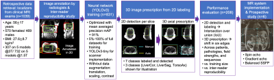 |
Automated MR Image Prescription of the Liver using Deep
Learning: Development, Evaluation & Prospective Implementation
Ruiqi Geng1,
Collin J. Buelo1,
Mahalakshmi Sundaresan2,
Jitka Starekova3,
Nikolaos Panagiotopoulos3,4,
Thekla Helene Oechtering3,4,
Edward M. Lawrence3,
Marcin Ignaciuk3,
Scott B Reeder5,
and Diego Hernando6
1Departments of Medical Physics, Radiology, University of Wisconsin, Madison, Madison, WI, United States, 2Department of Electrical and Computer Engineering, University of Wisconsin, Madison, Madison, WI, United States, 3Department of Radiology, University of Wisconsin, Madison, Madison, WI, United States, 4Department of Radiology and Nuclear Medicine, Universität zu Lübeck, Lübeck, Germany, 5Departments of Medical Physics, Radiology, Medicine, Emergency Medicine, Biomedical Engineering, University of Wisconsin, Madison, Madison, WI, United States, 6Departments of Medical Physics, Radiology, Electrical and Computer Engineering, Biomedical Engineering, University of Wisconsin, Madison, Madison, WI, United States Keywords: YIA, Liver This work developed a novel automated AI-based method for liver image prescription from a localizer and evaluated it in a large retrospective patient cohort (1,039 patients for training/testing), across pathologies, field strengths, and against radiologists’ inter-reader reproducibility performance. AI-based 3D axial prescription achieved a S/I shift of <2.3 cm compared to manual prescription for 99.5% of test dataset. The AI method performed well across all sub-cohorts and better in 3D axial prescription than radiologists’ inter-reader reproducibility performance. We successfully implemented the AI method on a clinical MR system, which demonstrated robust performance across localizer sequences. |
| 09:00 |
0004.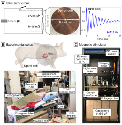 |
Measurement of Magnetostimulation Thresholds in the Porcine
Heart
Valerie Klein1,2,3,
Jaume Coll-Font2,3,4,
Livia Vendramini2,
Donald Staney2,
Mathias Davids2,3,
Natalie G. Ferris2,5,
Lothar R. Schad1,6,
David E. Sosnovik2,3,4,5,
Christopher Nguyen7,8,9,
Lawrence L Wald2,3,5,
and Bastien Guérin2,3
1Computer Assisted Clinical Medicine, Medical Faculty Mannheim, Heidelberg University, Heidelberg, Germany, 2A. A. Martinos Center for Biomedical Imaging, Department of Radiology, Massachusetts General Hospital, Charlestown, MA, United States, 3Harvard Medical School, Boston, MA, United States, 4Cardiovascular Research Center, Cardiology Division, Massachusetts General Hospital, Charlestown, MA, United States, 5Harvard-MIT Division of Health Sciences and Technology, Cambridge, MA, United States, 6Mannheim Institute for Intelligent Systems in Medicine, Medical Faculty Mannheim, Heidelberg University, Heidelberg, Germany, 7Cardiovascular Innovation Research Center, Heart Vascular & Thoracic Institute, Cleveland Clinic, Cleveland, OH, United States, 8Department of Radiology, Imaging Institute, Cleveland Clinic, Cleveland, OH, United States, 9Department of Biomedical Engineering, Lerner Research Institute, Cleveland Clinic, Cleveland, OH, United States Keywords: YIA, Heart High-amplitude gradient systems can surpass the IEC regulatory limit for cardiac stimulation (CS), which is based on animal electrostimulation data and simplified electromagnetic modeling. We assess CS in MRI by performing the first cardiac magnetostimulation threshold measurements in pigs, the primary animal model of the human cardiovascular system. We use the measurements to validate a detailed CS modeling pipeline applicable to both pigs and humans. Experimental thresholds and predictions in porcine-specific models agree within 19% NRMSE. CS modeling in a detailed human body model indicates that CS thresholds of the Connectome gradient are ~25-fold greater than the IEC CS limit. |
| 09:15 |
0005.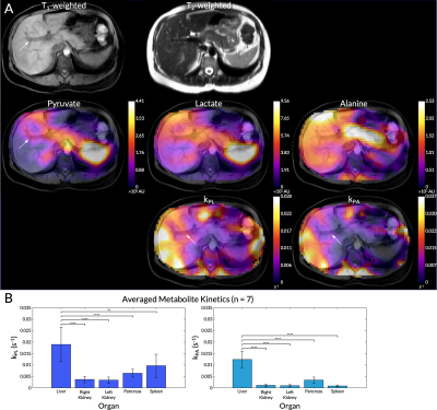 |
Whole-Abdomen Metabolic Imaging of Health Volunteers Using
Hyperpolarized [1-13C]pyruvate MRI
Philip Meng-en Lee1,
Hsin-Yu Chen2,
Jeremy W. Gordon2,
Zhen J Wang2,
Robert Bok2,
Ralph Hashoian3,
Yaewon Kim2,
Xiaoxi Liu2,
Tanner Nickles1,
Kiersten Cheung2,
Francesca De Las Alas2,
Heather Daniel2,
Peder EZ Larson1,
Cornelius von Morze4,
Daniel B Vigneron1,
and Michael A Ohliger2,5
1UC Berkeley-UCSF Graduate Program in Bioengineering; Dept. of Radiology & Biomedical Imaging, University of California, San Francisco, San Francisco, CA, United States, 2Department of Radiology & Biomedical Imaging, University of California, San Francisco, San Francisco, CA, United States, 3Clinical MR Solutions, Brookfield, WI, United States, 4Mallinckrodt Institute of Radiology, Washington University in St. Louis, St. Louis, MO, United States, 5Department of Radiology and Biomedical Imaging, Zuckerberg San Francisco General Hospital and Trauma Center, San Francisco, CA, United States Keywords: YIA, Hyperpolarized MR (Non-Gas), Body - Liver Whole-abdomen imaging with hyperpolarized 13C is challenging due to B0 and B1 inhomogeneities, respiratory motion, and broad spatial coverage. There is also little baseline data about healthy metabolism in abdominal organs. We developed and describe here a reliable imaging method to overcome these challenges, enabling metabolic imaging of the entire abdomen in a series of healthy volunteers. We present observed conversation rates of HP [1-13C]pyruvate to lactate and alanine in key organs such as the liver, kidneys, pancreas, and spleen. Methods established here set a firm foundation for investigating a broad spectrum of metabolic and neoplastic abnormalities in the liver. |
| 09:30 |
0006.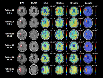 |
Predicting the Onset of Ischemic Stroke with Fast
High-Resolution 3D MR Spectroscopic Imaging
Zengping Lin1,
Ziyu Meng1,
Tianyao Wang2,
Rong Guo3,4,
Yibo Zhao3,5,
Yudu Li3,5,
Bin Bo1,
Yue Guan1,
Jun Liu2,
Hong Zhou6,
Xin Yu7,
David J Lin8,
Zhi-Pei Liang3,5,
Parashkev Nachev9,
and Yao Li1
1School of Biomedical Engineering, Shanghai Jiao Tong University, Shanghai, China, 2Radiology Department, Shanghai Fifth People's Hospital, Fudan University, Shanghai, China, 3Beckman Institute for Advanced Science & Technology, University of Illinois at Urbana-Champaign, Urbana, IL, United States, 4Siemens Medical Solutions USA, Inc, Urbana, IL, United States, 5Department of Electrical & Computer Engineering, University of Illinois at Urbana-Champaign, Urbana, IL, United States, 6Department of Radiology, The First Affiliated Hospital of South China of University, South China of University, Hengyang, China, 7Department of Biomedical Engineering, Case Western Reserve University, Cleveland, OH, United States, 8Center for Neurotechnology and Neurorecovery, Department of Neurology, Massachusetts General Hospital, Harvard Medical School, Boston, MA, United States, 9Institute of Neurology, University College London, London, United Kingdom Keywords: YIA, Ischemia Neurometabolite concentrations provide a direct index of infarct progression in stroke, but their relationship with stroke onset time remains unclear. Using a fast high-resolution 3D MRSI technique, this study assessed the temporal dynamics of N-acetylaspartate (NAA), creatine, choline, and lactate and estimated their value in predicting early (<6 hours) vs late (6–24 hours) hyperacute ischemic stroke groups. We found that lesional NAA and creatine was reduced from acute to subacute stroke patients and NAA level was inversely related to onset time in hyperacute patients. The changes in neurometabolite levels provided good discrimination between patients for early & late hyperacute time windows. |
| 09:45 |
0007. |
Submillimeter T1 Atlas for Subject-Specific Abnormality
Detection at 7T
Gian Franco Piredda1,2,3,
Samuele Caneschi1,
Tom Hilbert1,4,5,
Gabriele Bonanno1,6,7,
Arun Joseph1,6,7,
Karl Egger8,
Jessica Peter9,
Stefan Klöppel9,
Elisabeth Jehli10,11,
Matthias Grieder10,
Johannes Slotboom12,
David Seiffge13,
Martina Goeldlin13,
Robert Hoepner13,
Tom Willems14,
Serge Vulliemoz15,
Margitta Seeck15,
Punith B. Venkategowda16,
Ricardo A. Corredor Jerez1,4,5,
Bénédicte Maréchal1,4,5,
Jean-Philippe Thiran4,5,
Roland Weist6,12,
Tobias Kober1,4,5,
and Piotr Radojewski6,12
1Advanced Clinical Imaging Technology, Siemens Healthineers International AG, Lausanne, Switzerland, 2Human Neuroscience Platform, Fondation Campus Biotech Geneva, Geneva, Switzerland, 3CIBM-AIT, École Polytechnique Fédérale de Lausanne (EPFL), Lausanne, Switzerland, 4Department of Radiology, Lausanne University Hospital and University of Lausanne, Lausanne, Switzerland, 5LTS5, École Polytechnique Fédérale de Lausanne (EPFL), Lausanne, Switzerland, 6Translational Imaging Center (TIC), Swiss Institute for Translational and Entrepreneurial Medicine, Bern, Switzerland, 7Magnetic Resonance Methodology, Institute of Diagnostic and Interventional Neuroradiology, University of Bern, Bern, Switzerland, 8Department of Neuroradiology, Faculty of Medicine, University of Freiburg, Freiburg, Germany, 9University Hospital of Old Age Psychiatry and Psychotherapy, University of Bern, Bern, Switzerland, 10Translational Research Center, University Hospital of Psychiatry and Psychotherapy, University of Bern, Bern, Switzerland, 11Department of Neurosurgery, University Hospital of Zurich, Zurich, Switzerland, 12Support Center for Advanced Neuroimaging, Institute for Diagnostic and Interventional Neuroradiology, Inselspital, University of Bern, Bern, Switzerland, 13Department of Neurology, University Hospital of Bern, University of Bern, Bern, Switzerland, 14Institute of Psychology, University of Bern, Geneva, Switzerland, 15EEG & Epilepsy Unit, Department of Clinical Neurosciences, Geneva University Hospitals & Faculty of Medicine, Geneva, Switzerland, 16Siemens Healthcare Pvt. Ltd., Bangalore, India Keywords: YIA, Tissue Characterization, Acquisition & Analysis, Quantitative Imaging, Ultra-high field MRI Databases of normative values from healthy tissues are needed to leverage the improved comparability and hardware independence of quantitative MRI, and thus enable patient-specific detection of abnormalities in visual interpretation. Here, we present normative atlases of T1 relaxation times in the brain at 3T (1 mm isotropic) and 7T (0.6 mm isotropic) from two large cohorts of healthy subjects. The atlases were tested in single‐subject comparisons for detection of abnormal relaxation times in patients scanned at both field strengths. The presented detection of subtle alterations not visible in conventional MRI showed the clinical potential of the method. |
| 10:00 |
0008.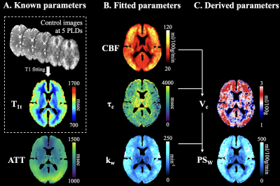 |
Quantification of Blood-Brain Barrier Water Exchange &
Permeability with Multi-Delay Diffusion Weighted pCASL
Xingfeng Shao1,
Chenyang Zhao1,
Qinyang Shou1,
Keith S St Lawrence2,3,
and Danny JJ Wang1
1Laboratory of FMRI Technology (LOFT), Mark & Mary Stevens Neuroimaging and Informatics Institute, Keck School of Medicine, University of Southern California, Los Angeles, CA, United States, 2Lawson Health Research Institute, London, ON, Canada, 3Department of Medical Biophysics, Western University, London, ON, Canada Keywords: YIA, Diffusion/other diffusion imaging techniques, Blood-brain barrier, water exchange, diffusion weighted perfusion imaging We developed an innovate pulse sequence and acquired diffusion weighted pCASL signals from a wide range of PLDs with improved SNR and spatial resolution. A 3-compartment single-pass approximation (SPA) model, which includes an additional venous compartment, was proposed to capture the full dynamics of the labeled blood bolus passing through capillaries while exchanging into tissue space before flowing into venules, and a LRL method was proposed for individual kw quantification. |
 Back to Meeting Home
 Back to the Program-at-a-Glance
The International Society for Magnetic Resonance in Medicine is accredited by the Accreditation Council for Continuing Medical Education to provide continuing medical education for physicians.
 Back to Meeting Home
 Back to the Program-at-a-Glance
 View Presentation Video