Power Pitch
Pitch: Novel Methods in Acquisition & Analysis
ISMRM & ISMRT Annual Meeting & Exhibition • 03-08 June 2023 • Toronto, ON, Canada

Power Pitch
Pitch: Novel Methods in Acquisition & Analysis
| 16:00 |
0347.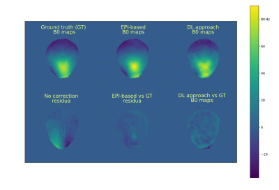 |
Comparison of dynamic B0 mapping approaches at 7T –
AI-based prediction via U-net vs measurement via
dual-echo EPI navigator
Stanislav Motyka1,
Paul Weiser2,
Bernhard Strasser1,
Dario Goranovic1,
Lukas Hingerl1,
Gilbert Hangel1,3,
Eva Niess1,
Stephan Wampl4,
Fabian Niess1,
Simon Robinson1,
Georg Langs2,
Siegfried Trattnig1,
and Wolfgang Bogner5
1High Field MR Center, Department of Biomedical Imaging and Image-guided Therapy, Medical University of Vienna, Vienna, Austria, 2Computational Imaging Research Lab, Department of Biomedical Imaging and Image-guided Therapy, Medical University of Vienna, Vienna, Austria, 3Department of Neurosurgery, Medical University of Vienna, Vienna, Austria, 4High Field MR Center, Center for Medical Physics and Biomedical Engineering, Medical University of Vienna, Vienna, Austria, 5Medical University of Vienna, Vienna, Austria Keywords: Motion Correction, Brain The abstract compares two approaches to dynamically estimate B0 maps at 7T. The U-net predicts B0 maps based on the initial B0 map and the movement information. The dual-echo EPI-based navigator directly measures B0 maps. Both methods yield comparable results. However, the deep learning approach could overcome the major disadvantage of the EPI-based navigator, thus the need for the dead time in the parent sequence, if the motion can be mapped externally. |
| 16:00 |
0348.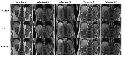 |
Comparison of k-space center, FID and Pilot Tone
navigators in abdominal motion tracking for optimal
XD-GRASP reconstruction
Cemre Ariyurek1,
Tess E. Wallace1,
Tobias Kober2,3,4,
Sila Kurugol1,
and Onur Afacan1
1Quantitative Intelligent Imaging Lab (QUIN), Department of Radiology, Boston Children's Hospital and Harvard Medical School, Boston, MA, United States, 2Advanced Clinical Imaging Technology, Siemens Healthineers International AG, Lausanne, Switzerland, 3Department of Radiology, Lausanne University Hospital and University of Lausanne, Lausanne, Switzerland, 4LTS5, École Polytechnique Fédérale de Lausanne (EPFL), Lausanne, Switzerland Keywords: Motion Correction, Body Abdominal MRI scans often require breath-holding to prevent image quality degradation, which can be challenging for patients. XD-GRASP enables generation of motion-robust images for free-breathing abdominal MRI by binning the data into respiratory phases. In this study, we compared three navigation techniques, namely k-space center, free induction decay navigators (FIDnavs) and pilot tone (PT), for XD-GRASP reconstruction. FIDnavs and PT have advantages over k-space center navigation since they are insensitive to gradient delays, independent of the imaging plane and acquired more frequently. The image quality ranking means (1=best,2=moderate,3=worst) were 1.4, 1.6 and 2 for FIDnavs, PT and k-space center, respectively. |
| 16:00 |
0349.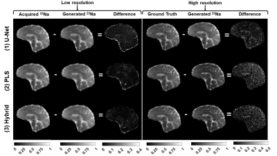 |
Super-resolution algorithms for simultaneous 1H
MRF/23Na MRI: Comparison between U-Net,
PLS-regression, and hybrid methods
Gonzalo Gabriel Rodriguez1,
Hector Lise de Moura1,
Lauren O'Donnell1,
Ravinder Regatte1,2,
and Guillaume Madelin1,2
1Center for Biomedical Imaging, Radiology Department, NYU School of Medicine, New York, NY, United States, 2Vilcek Institute of Graduate Biomedical Sciences, NYU Langone Health, New York, NY, United States Keywords: Data Processing, Non-Proton, Super-Resolution We compared three algorithms to generate a high-resolution (HR) 23Na image from simultaneously-acquired low-resolution (HR) 23Na density-weighted MRI and HR 1H density, T1 and T2 maps from MRF in brain at 7 T: U-net, PLS-regression, and hybrid. The multi-scale structural similarity index between generated HR 23Na images and HR ground truth was higher than 0.95 for the three methods. Overall, the hybrid method showed better results, generating the sharpest HR image while keeping the highest similarity between the acquired and generated LR images. |
| 16:00 |
0350.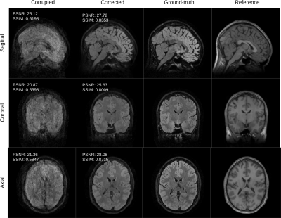 |
Retrospective motion correction and reconstruction
for clinical 3D brain MRI protocols with a reference
contrast
Gabrio Rizzuti1,
Tim Schakel1,
Niek Huttinga1,
Tristan van Leeuwen2,
and Alessandro Sbrizzi1
1Universitair Medisch Centrum Utrecht, Utrecht, Netherlands, 2Centrum Wiskunde & Informatica, Amsterdam, Netherlands Keywords: Motion Correction, Brain, brain, clinical protocols, motion correction, retrospective correction We discuss the application of a novel 3D retrospective rigid motion correction and reconstruction scheme to in-vivo data for clinical 3D brain protocols. The correction scheme leverages multiple scans contained in a typical MR session, of which some are corrupted by motion but at least one scan may be devoid of motion artifacts. The uncorrupted scan is then used as a reference to regularize a generalized rigid-motion registration problem. We discuss the potential of the proposed algorithm with a prospective in-vivo study. |
| 16:00 |
0351.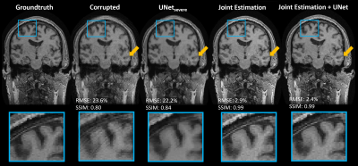 |
Stacked UNet-Assisted Joint Estimation for Robust 3D
Motion Correction
Brian Nghiem1,2,
Zhe Wu2,
Lars Kasper2,
and Kâmil Uludağ1,2
1Dpt. of Medical Biophysics, University of Toronto, Toronto, ON, Canada, 2University Health Network, Toronto, ON, Canada Keywords: Motion Correction, Motion Correction We showed that accurate 3D retrospective motion correction of T1w MPRAGE data can be achieved with a UNet-assisted joint estimation algorithm. We compared the proposed method to using the UNet on its own and the standard joint estimation algorithm. Joint estimation (with and without the UNet) outperformed using the stand-alone UNet. The UNet-assisted joint estimation algorithm converged faster than its UNet-free counterpart. We demonstrated the importance of adapting to the changing levels of artifacts over the course of the joint estimation algorithm by sequentially employing different UNets trained for correcting different levels of motion corruption. |
| 16:00 |
0352.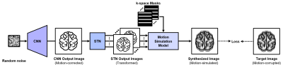 |
Motion Correction in Brain MRI using Deep Image
Prior
Jongyeon Lee1,
Wonil Lee1,
and HyunWook Park1
1Korea Advanced Institute of Science and Technology, Daejeon, Korea, Republic of Keywords: Motion Correction, Motion Correction, Brain Motion Correction Motion correction in MRI has been successful with deep learning techniques to reduce motion artifacts. However, these methods were based on supervised learning, which have required a large dataset to train neural networks. To overcome this issue, we propose a new technique to correct motion artifacts without any training dataset, which optimizes a neural network only with a single motion-corrupted image. We adopt Deep Image Prior framework to capture image priors from a convolutional neural network using motion simulation based on MR physics. Using the deep image prior, our method finely reduces motion artifacts without any dataset. |
| 16:00 |
0353.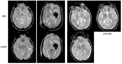 |
Motion-corrected multi-shot EPI: Viable alternative
to GRE for blood sensitive imaging with improved
image quality and reduced scan time
Zhiqiang Li1,
James Murchison1,
Dori Shoshan1,
Melvyn B Ooi2,
and John P Karis1
1Barrow Neurological Institute, Phoenix, AZ, United States, 2Philips Healthcare, Houston, TX, United States Keywords: Motion Correction, Motion Correction, artifact, rapid scan, clinical study, high value A motion-corrected 2D multi-shot EPI (msEPI) technique was compared to conventional 2D GRE to determine if it can be used as a faster technique for blood sensitive imaging in the emergency department (ED) setting. msEPI was found to be superior to GRE (p < 0.001) in motion artifact, overall image quality, and lesion detection. These results and reduced scan time make the motion-corrected 2D msEPI a viable alternative for blood sensitive imaging in the ED setting. |
| 16:00 | 0354. | WITHDRAWN |
| 16:00 |
0355.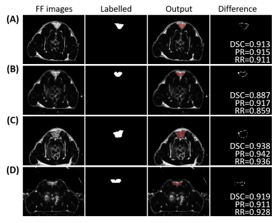 |
Automatic segmentation of the interscapular BAT in
rats by the dynamic magnetic resonance fat fraction
images
Chuanli Cheng1,
Lei Zhang2,
Hao Peng1,
Hairong Zheng1,
Xin Liu1,
Huimao Zhang2,
and Chao Zou1
1Shenzhen institutes of advanced technology, Chinese Academy of Sciences, Shenzhen, China, 2Radiology Department, Bethune First Hospital of Jilin University, Changchun, China Keywords: Segmentation, Fat, brown adipose tissue The noninvasive assessment of BAT volume is fundamental for characterizing the longitudinal effects in rodents. In this work, dynamic fat fraction images of 34 rats fed under different conditions were acquired before and after noradrenaline injection for 2.5 hours. The iBAT regions were recognized and labelled automatically by identifying the regions with significant changes in FF images. Then a deep learning network was built up by training the FF images and the automatically identified mask images in all rats. The dice similarity coefficient, precision rate and recall rate of the network were found to be 0.897±0.061, 0.901±0.068 and 0.889±0.086, respectively. |
| 16:00 |
0356.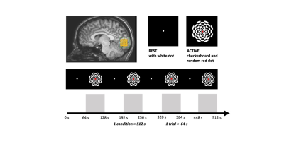 |
Multi-echo functional MRS Glutamate measurements in
visual cortex at 3T
Polina Emeliyanova1,2,
Laura M. Parkes1,2,
Stephen R. Williams3,
and Caroline Lea-Carnall1,2
1Division of psychology, communication and human neuroscience, Faculty of biology, medicine and health, University of Manchester, Manchester, United Kingdom, 2Geoffrey Jefferson Brain Research Centre, Manchester Academic Health Science Centre, Manchester, United Kingdom, 3Division of Informatics, Imaging and Data Science, University of Manchester, Manchester, United Kingdom Keywords: Data Acquisition, Spectroscopy, Functional MRS, multi-echo MRS, 3T, glutamate, creatine (Cr), N-Asetyl-Aspartate (NAA) fMRS studies have shown dynamic change in glutamate (Glu) concentrations during visual stimulation. Here, we examine the effect of TE from 9.3-280ms on the signal during fMRS acquisition and whether T2 relaxation parameters change during a visual task compared to rest. We observed a short Glu T2-compartment between 2.5-4.5ms arising from 48% (rest) and 16% (active) of the total signal with significant increase in Glu signal at TE =140ms during stimulation. These preliminary results indicate bi-exponential decay of Glu in the visual cortex at 3T encouraging future work with more robust fMRS paradigms. |
| 16:00 |
0357.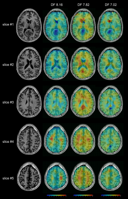 |
In vivo Whole-brain Downfield Metabolites Mapping
using 2π-CASP-EPSI at 7T
Guodong Weng1,2,
Piotr Radojewski1,2,
Federico Turco1,2,
and Johannes Slotboom1,2
1Support Center for Advanced Neuroimaging (SCAN), University of Bern, Bern, Switzerland, 2Translational Imaging Center, sitem-insel, Bern, Switzerland Keywords: Pulse Sequence Design, Spectroscopy, Downfield MRSI Many interesting metabolites, for instance adenosine triphosphate (ATP), glutathione (GSH) and homocarnosine (hCs), have resonances in the downfield range (>4.7 ppm) in 1H-MRS. Previous studies have studied the downfield spectra of metabolites in human brain using single-voxel spectroscopy (SVS) and, most recently, 2D one-slice MRSI. The study presents a 3D whole brain mapping sequence suited for downfield metabolites which does not require water suppression pulses. |
| 16:00 |
0358.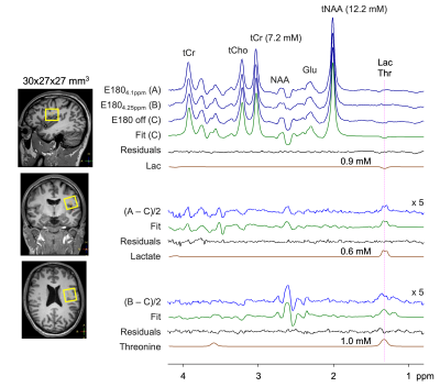 |
J-difference Editing of Lactate and Threonine in the
Human Brain at 3T
Ryan K Robison1,2,3,
Justin R Haynes2,3,
Sandeep Ganji4,5,
Wellington Pham2,3,
Seth A Smith2,3,
Victoria L Morgan2,3,
Reid C Thompson6,
Reed A Omary3,
John C Gore2,3,
and Changho Choi2,3
1Philips, Nashville, TN, United States, 2Vanderbilt University Institute of Imaging Science, Vanderbilt University Medical Center, Nashville, TN, United States, 3Radiology and Radiological Sciences, Vanderbilt University Medical Center, Nashville, TN, United States, 4Philips, Rochester, MN, United States, 5Mayo Clinic, Rochester, MN, United States, 6Neurological Surgery, Vanderbilt University Medical Center, Nashville, TN, United States Keywords: Data Acquisition, Spectroscopy Lactate is important as a source of energy and a product of metabolism, so the capability to measure lactate accurately and noninvasively by MRS is of high value. We report a new lactate editing approach that enables detection of the lactate 1.3 ppm resonance with minimal contaminations from threonine and other compounds. Narrow-band editing 180° RF pulses were implemented in 1H MR MEGA-PRESS and the sequence was optimized with numerical and phantom analyses. With data from six healthy adult subjects, the lactate and threonine concentrations in healthy brain were estimated to be 0.5±0.1 and 1.0±0.1 mM, respectively. |
| 16:00 |
0359.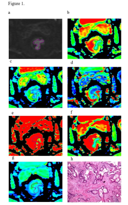 |
IVIM-DWI for the preoperative prediction of
perineural invasion status in rectal cancer: A
feasibility study with multivariate model analysis
Jia Yuping1 and
Dou Weiqiang2
1Lixia District, The First Affiliated Hospital of Shandong First Medical University & Shandong Provincial Qianfoshan Hospital, Jinan, China, 2MR Research, GE Healthcare, Beijing, China Keywords: Data Analysis, Cancer This study aimed to determine the clinical potential of intravoxel incoherent motion (IVIM) DWI in predicting perineural invasion (PNI) of rectal cancer (RC). IVIM parameters derived from different mathematical models, including apparent diffusion coefficient from mono-exponential model, true diffusion coefficientand and perfusion fraction from bi-exponential model, and the distributed diffusion coefficient from stretched-exponential model showed significant differences between 72 with PNI and 76 without PNI. With these findings, IVIM may thus be considered effective in preoperatively predicting PNI status in RC and further help clinicians make individual treatment plans. |
| 16:00 |
0360.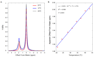 |
Temperature Mapping Based on Chemical Exchange
Saturation Transfer of Creatine
Siqi Cai1,2,
Yang Zhou1,
Chao Zou1,
Linhai Jiang3,
Qian Wan1,
Chunxiang Jiang1,
Changjun Tie1,
and Lijuan Zhang1,2
1Shenzhen Institute of Advanced Technology, Chinese Academy of Sciences, Shenzhen, China, 2University of Chinese Academy of Science, Beijing, China, 3The Instrumental Analysis Center of Shenzhen University, Shenzhen University, Shenzhen, China Keywords: Data Analysis, CEST & MT Chemical exchange between endogenous labile guanidinium (Guan) protons of creatine and free water and its thermal dependency were investigated with CEST imaging. The apparent offsets of Cr-CEST were linearly correlated to the experiment temperatures and was relatively insensitive to Cr concentration and pH variations at a range corresponding to physiological and pathological conditions, suggesting a potential utility of Cr-CEST for in vivo thermometry based on endogeneous creatine of the biological tissue. |
| 16:00 |
0361.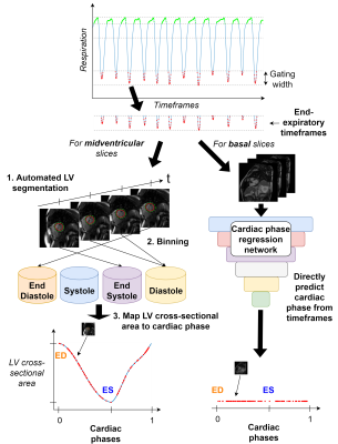 |
Respiratory controlled ECG-free cine construction
from real-time cardiac magnetic resonance images
acquired during free-breathing exercise
Julius Åkesson1,
Katarina Steding-Ehrenborg1,
Johannes Töger1,
Björn Östenson1,
Pia Sjöberg1,
and Einar Heiberg1
1Clinical Physiology, Department of Clinical Sciences Lund, Lund University, Skåne University Hospital, Lund University, Lund, Sweden Keywords: Data Processing, Software Tools, Exercise, real-time This study aimed to enable ECG-free construction of respiratory controlled 3D cardiac magnetic resonance cine series from short-axis 2D real-time images through automated detection of cardiac phases. This is important for imaging during exercise, since obtaining a reliable ECG-signal can be difficult. Visually coherent cines could be constructed for midventricular slice positions through detecting cardiac phases from automated left ventricular segmentations in real-time timeframes, but not for apical and basal slice positions. Deep learning-based regression of cardiac phases directly from images was investigated to handle this problem, but apical and basal cine construction remains an open problem. |
| 16:00 |
0362.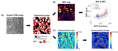 |
Effects of myelin structures on R2 values in the
aged rat corpus callosum
Hwapyeong Cho1 and
Hyung Joon Cho1
1Ulsan National Institute of Science and Technology, Ulsan, Korea, Republic of Keywords: Data Analysis, Relaxometry, Myelin, Magnetic field perturbation In this study, the effects of myelin structures such as myelin volume fraction (MVF) and myelin sheath thickness (MST) were investigated by measuring the intra/extracellular water signals in post-mortem aging rat brains. R2 values calculated by intra/extracellular water signals obtained using Carr-Purcell-Meiboom-Gill (CPMG) sequence are theoretically verified by comparison with transmission electron microscopy (TEM) and simulation results. It was confirmed that MVF affects the increase of R2 and MST affects the decrease of R2, and the analysis of R2 through this relationship was statistically significant. These results suggest that effects of myelin structures may affect CPMG-based intra/extracellular water signals. |
| 16:00 |
0363.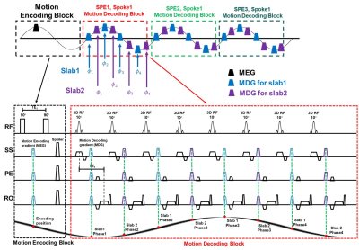 |
A 3D Fast Multiphase DENSE MRE Sequence with
Stack-of-Star Radial Sampling, Interleaved
Multi-Slab Acquisition and Hadamard Encoding
Yu Chen1,
Runke Wang1,
Ruokun Li2,
Fuhua Yan2,
and Yuan Feng1,2,3
1Shanghai JiaoTong University, School of Biomedical Engineering, Shanghai, China, 2Ruijin Hospital, Shanghai Jiaotong University School of Medicine, Shanghai, China, 3Institute of Medical Robotics, Shanghai Jiao Tong University, Shanghai, China Keywords: Elastography, Brain Conventional gradient echo (GRE) based Magnetic Resonance Elastography (MRE) suffers from long TE and scan time at low mechanical excitation (<25Hz). Displacement Encoding with a Stimulated Echo (DENSE) can record displacement with short TR, useful for low-frequency excitation. Here, 3D excitation was used to compensate for potential signal loss. A multiphase acquisition scheme with stack-of-star radial sampling was also used along with interleaved multi-slab acquisition and Hadamard encoding for MRE acceleration. Phantom experiments were conducted for validation. 32 slices brain MRE scan at 20Hz can be achieved in 11min30s, 5 folds faster than conventional GRE-based MRE. |
| 16:00 |
0364.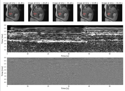 |
Detecting bladder and pelvic floor motion during MRI
using a small ultrasound-based sensor
Radhika Tibrewala1,
Mahesh Keerthivasan1,2,
Mary Bruno3,
Christopher Collins1,
Bruno Madore4,
and Daniel K Sodickson1
1Center for Advanced Imaging Innovation and Research, NYU Grossman School of Medicine, New York, NY, United States, 2Siemens Medical Solutions USA, New York, NY, United States, 3NYU Grossman School of Medicine, New York, NY, United States, 4Brigham and Women’s Hospital, Harvard Medical School, Boston, MA, United States Keywords: Motion Correction, Hybrid & Novel Systems Technology Deep-seated motion due to digestion and bladder filling can be difficult to monitor during MR acquisition. In this study, we use a small ultrasound-based sensor to detect bladder and pelvic floor motion, with a real-time MRI acquisition and a pilot-tone motion-sensitive signal as a reference. A neural network is used to predict the bladder area at various times during the MRI scan by using the US signal features. Our results indicate that the correlations between the ultrasound sensor and the MRI are beneficial for detecting bladder motion over time. |
| 16:00 |
0365.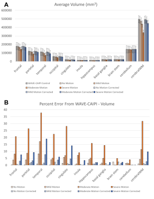 |
Evaluation of Scout Accelerated Motion Estimation
and Reduction (SAMER) MPRAGE for Visual Grading and
Volumetric Measurement of Brain Tissue
Nelson Homero Gil1,
Wei-Ching Lo2,
Bryan Clifford2,
Min Lang1,
Komal Awan1,
Daniel Nicolas Splitthoff3,
Daniel Polak3,
Stephen Cauley1,
and Susie Huang1
1Radiology, Massachusetts General Hospital, Boston, MA, United States, 2Siemens Medical Solutions USA, Boston, MA, United States, 3Siemens Healthcare GmbH, Erlangen, Germany Keywords: Motion Correction, Neurodegeneration Volumetric brain MRI using the T1-weighted-MPRAGE sequence is an important component of the clinical evaluation of dementia. However, MPRAGE has a long acquisition time and is especially prone to patient motion artifact. A recent development to address this issue is a technique called Scout Accelerated Motion Estimation and Reduction (SAMER). In this work, we used a set of 90 MPRAGE scans derived from 10 healthy volunteers to demonstrate that SAMER is effective at correcting various degrees of motion, including severe motion with non-diagnostic image quality, and greatly increases the accuracy of volumetric brain measurements. |
| 16:00 |
0366.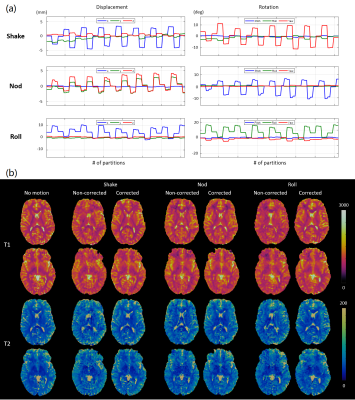 |
3D MR Fingerprinting with Fat-navigator-based Motion
Correction for Pediatric Imaging
Siyuan Hu1,
Yong Chen1,
Mark Griswold1,
and Dan Ma1
1Case Western Reserve University, Cleveland, OH, United States Keywords: Motion Correction, MR Fingerprinting MR Fingerprinting (MRF) simultaneously measures multiple tissue properties within a single scan. Although 2D MRF has been shown to be less sensitivie to motion than conventional imaging, bulk motion is still problematic for longer 3D MR Fingerprinting scans. We propose to integrate 3D fat navigators with 3D MRF acquisitions for motion correction in neuroimaging, especially for non-sedated pediatric imaging. The proposed method was validated in in vivo scans of healthy subjects with various types of motions and in non-sedated infants. We showed that the fat-navigated 3D MRF framework could resolve and correct bulk motions of both healthy volunteers and infants. |
 Back
to Meeting Home
Back
to Meeting Home
 Back
to the Program-at-a-Glance
Back
to the Program-at-a-Glance
The International Society for Magnetic Resonance in Medicine is accredited by the Accreditation Council for Continuing Medical Education to provide continuing medical education for physicians.
 Back
to Meeting Home
Back
to Meeting Home Back
to the Program-at-a-Glance
Back
to the Program-at-a-Glance Full Session Video