Power Pitch
Pitch: Data Analysis & Processing
ISMRM & ISMRT Annual Meeting & Exhibition • 03-08 June 2023 • Toronto, ON, Canada

08:15 |
0470.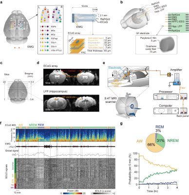 |
Mouse sleep fMRI with simultaneous electrophysiology at 9.4T
Yalin Yu1,
Yue Qiu2,
Chuanjun Tong3,
and Zhifeng Liang1
1Institute of Neuroscience, CAS Key Laboratory of Primate Neurobiology, Center for Excellence in Brain Science and Intelligence Technology, Chinese Academy of Sciences, Shanghai, China, Shanghai, China, 2Department of Anesthesia, Zhongshan Hospital, Fudan University, Shanghai, China, Shanghai, China, 3School of Biomedical Engineering, Southern Medical University, Guangzhou, China, Shanghai, China Keywords: Data Analysis, Brain Simultaneous electrophysiology and fMRI could provide both macroscopic and microscopic observations, but it is highly technically challenging and not widely used in sleep research. We developed mouse sleep fMRI based on simultaneous electrophysiology at 9.4T and allowed manifestation of the full sleep cycle (NREM/REM) during fMRI. The results revealed global state-dependent patterns. Rich state transition epochs demonstrated that state transitions were global, irreversible and sequential phenomenon, which can be predicted using LSTM RNN models. Importantly, simultaneous hippocampal recording revealed enhanced sharp-wave ripple triggered global patterns during NREM than awake state, which may attribute to co-occurrence of spindle events. |
| 08:15 |
0471.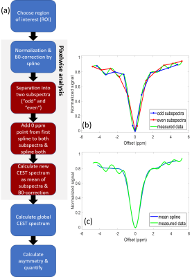 |
Cardiac CEST MRI under the influence of very short
saturation pulses – an analysis approach
Daniel Schache1,
Ajay Peddi1,
Ali Nahardani2,
Cornelius Faber1,
and Verena Hoerr1,2
1Translational Research Imaging Center, Clinic of Radiology, University of Münster, Münster, Germany, 2Heart Center Bonn, Department of Internal Medicine II, University Hospital Bonn, Bonn, Germany Keywords: Data Analysis, CEST & MT Cardiac CEST MRI in mice rely on the application of very short saturation pulses. These pulses cause disturbing signal modulation in the CEST spectrum preventing a consistent data analysis. To correct for this, an additional approach named DOSE was established in the data analysis pipeline. The DOSE-corrected analysis was validated in vitro on a glucose concentration series as well as in vivo on cardiac glucoCEST experiments of nine healthy mice. Hereby, oscillations in the CEST spectra could be reduced substantially ensuring a consistent quantification. |
| 08:15 |
0472.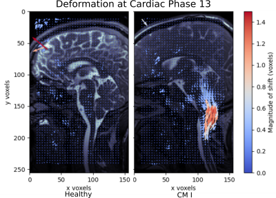 |
Phase-Based Registration for Visualisation of Pulsatile
Brain Motion
Robin Laven1,
Sam Richardson1,
Nadine Hidalgo2,
Dale Justin Sasis2,
Eryn Kwon3,4,
Samantha Holdsworth3,4,
and Poul Nielsen1,2
1Auckland Bioengineering Institute, University of Auckland, Auckland, New Zealand, 2Department of Engineering Science, University of Auckland, Auckland, New Zealand, 3Department of Anatomy and Medical Imaging & Center for Brain Research, University of Auckland, Auckland, New Zealand, 4Mātai Medical Research Institute, Tairāwhiti-Gisborne, New Zealand Keywords: Data Processing, Brain, Chiari 1 Malformation Pulsatile brain motion is a valuable tool in understanding injury and congenital disorders. These pulsatile motions have a typical magnitude of < 1 pixel. Amplified MRI (aMRI) amplifies the pulsatile motion to the point of visibility, enabling qualitative examination. However, the amplification in frequency space results in nonlinear distortion of motion preventing the recovery of unaugmented displacements. Here, we propose using phase-based subpixel registration. This methodology enables the recovery of displacement quantification of pulsatile brain motion. |
08:15 |
0473.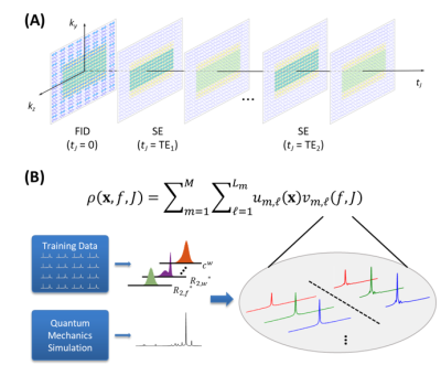 |
Removal of Water and Lipid Signals from J-resolved 1H-MRSI
Data Using an FID Reference and Physics-Based Subspaces
Wen Jin1,2,
Yibo Zhao1,2,
Yudu Li1,3,
Rong Guo1,4,
Ruihao Liu1,5,
Jie Luo5,
Yao Li5,
and Zhi-Pei Liang1,2
1Beckman Institute for Advanced Science and Technology, University of Illinois at Urbana-Champaign, Urbana, IL, United States, 2Department of Electrical and Computer Engineering, University of Illinois at Urbana-Champaign, Urbana, IL, United States, 3National Center for Supercomputing Applications, University of Illinois at Urbana-Champaign, Urbana, IL, United States, 4Siemens Medical Solutions USA, Inc., Urbana, IL, United States, 5School of Biomedical Engineering, Shanghai Jiao Tong University, Shanghai, China Keywords: Data Processing, Spectroscopy J-resolved 1H-MRSI provides a unique capability to quantify both brain neurotransmitters and metabolites. This paper presents a novel method to effectively remove water and lipid signals from J-resolved 1H-MRSI data with limited spatial encodings and without lipid suppression. The proposed method has been validated using data from a healthy subject and a brain tumor patient, producing impressive results. |
| 08:15 |
0474.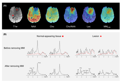 |
Separation of Macromolecules and Metabolites in
Ultrashort-TE MRSI Data with Learned Probabilistic Subspaces
Yibo Zhao1,2,
Yudu Li1,3,
Wen Jin1,2,
Rong Guo1,4,
Wenli Li5,
Yao Li5,
Jie Luo5,
and Zhi-Pei Liang1,2
1Beckman Institute for Advanced Science and Technology, University of Illinois at Urbana-Champaign, Urbana, IL, United States, 2Department of Electrical and Computer Engineering, University of Illinois at Urbana-Champaign, Urbana, IL, United States, 3National Center for Supercomputing Applications, University of Illinois at Urbana-Champaign, Urbana, IL, United States, 4Siemens Medical Solutions USA, Inc., Urbana, IL, United States, 5School of Biomedical Engineering, Shanghai Jiao Tong University, Shanghai, China Keywords: Data Analysis, Spectroscopy, Macromolecule Separation of macromolecules and metabolites in ultrashort-TE MRSI data has been very difficult due to limited SNR and strong spectral overlap. In this work, we proposed a new solution to the problem using a subspace-based approach aided with long-TE navigator signals. Physics-based prior information was incorporated through pre-learned spectral bases and probability distributions of spatial coefficients. The proposed method has been validated using experimental data from healthy and brain tumor subjects, producing impressive results. |
| 08:15 |
0475.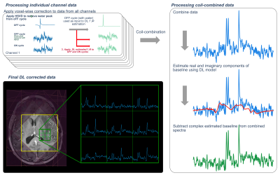 |
Evaluation of Deep Learning Models for Processing
Lactate-Edited MR Spectroscopic Imaging in Patients with
Glioma
Sana Vaziri1,
Adam W Autry1,
Marisa Lafontaine1,
Janine Lupo1,
Susan Chang2,
and Yan Li1
1Radiology and Biomedical Imaging, University of California, San Francisco, San Francisco, CA, United States, 2Neurological Surgery, University of California, San Francisco, San Francisco, CA, United States Keywords: Data Processing, Spectroscopy The use of deep learning models for frequency and phase correction of spectral data was evaluated to reduce processing time of 3D MRSI datasets acquired from patients with glioma. Models for frequency and phase offset estimation were trained for data prior to coil-combination. Separate models for baseline removal of coil-combined data were similarly trained and evaluated. The voxel-wise peaks for spectra processed using the standard approach were evaluated and compared to the spectra processed using the proposed deep learning approach. Compared to standard processing, deep learning processing produced spectra with comparable SNR and linewidths in a shorter processing time. |
| 08:15 |
0476. |
The value of Cinematic Volume Rendering Technique: magnetic
resonance imaging in diagnosing tumors around the brachial
plexus
Rui Chen1,
Yuncai Ran1,
Junxia Niu1,
Yanglei Wu2,
Yong Zhang1,
and Jingliang Cheng1
1Department of Magnetic Resonance, The First Affiliated Hospital of Zhengzhou University, Zhengzhou, China, 2MR Collaborations, Siemens Healthineers Ltd., Beijing, China Keywords: Visualization, Nerves, Cinematic Volume Rendering Technique (cVRT);Brachial plexus nerve Cinematic Volume Rendering Technique (cVRT) can clearly display the contours of the tumor, brachial plexus, and peripheral blood vessels, and the extent of their involvement while simultaneously imaging them. The three-dimensional anatomical effect is more realistic, playing a direct role in guiding the surgical plan. |
| 08:15 |
0477.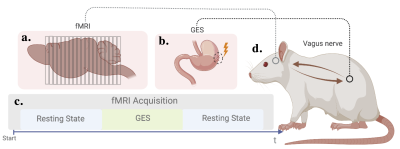 |
Targeted Gastric Electrical Stimulation Modulates Functional
Connectivity of the Interoceptive Network in the Rat Brain
Fatimah M. Alkaabi1,2,
Jiayue Cao1,
Angela Yee1,
Xiaokai Wang1,
Ulrich Scheven1,
Chih Hsuan Tsai1,
Ana C. Saavedra1,
and Zhongming Liu1,3
1Biomedical Engineering, University of Michigan, Ann Arbor, MI, United States, 2Biomedical Engineering, Imam Abdulrahman Bin Faisal University, Dammam, Saudi Arabia, 3Electrical Engineering and Computer Science, University of Michigan, Ann Arbor, MI, United States Keywords: Data Analysis, Animals Gastric Electrical Stimulation (GES) is an FDA approved therapy for gastroparesis with unspecified working mechanisms. One plausible mechanism is that it activates the vagal afferents to engage the brain’s interoceptive network in regulating the stomach. Orientation and location-specific stimulation that targets the vagal-gastric receptors can effectively activate the brainstem. We asked whether and how this type of GES can engage interoceptive regions and modulate their interactions, especially the anterior cingulate (ACC) and insular cortices (IC), the primary visceromotor and viscerosensory processing regions, respectively. Therefore, we evaluated the functional connectivity in the rat brain before, during, and after targeted GES. |
| 08:15 |
0478.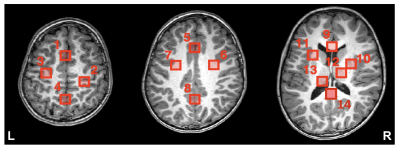 |
Reproducibility of conventional and GABA-edited multi slice
MRSI in the human brain at 3T
Dillip K. Senapati1,2,
Helge J. Zöllner1,2,
Ipek Özdemir 1,2,
Georg Oeltzschner1,2,
Doris D.M. Lin1,2,
and Peter B. Barker1,2
1Russell H. Morgan Department of Radiology and Radiological Science, The Johns Hopkins University School of Medicine, Baltimore, MD, United States, 2F. M. Kirby Research Center for Functional Brain Imaging, Kennedy Krieger Institute, Baltimore, MD, United States Keywords: Data Analysis, Spectroscopy, GABA, MR Spectroscopy, MRSI, Reproducibility, Brain Reproducibility of both conventional and GABA-edited multi-slice MRSI of the human brain recorded at 3T was investigated in 11 healthy adult volunteers. GABA-edited data were post-processed using a retrospective motion compensation (MoCo) scheme. Test-retest coefficients of variation(CV) were calculated. Overall reproducibility was good for conventional MRSI for selected brain regions, but edited-MRSI data showed significant variations. Edited-MRSI scans may benefit from both improved post-doc correction schemes to minimize subtraction artifacts, as well as better B0 field homogeneity over the brain, and possibly also prospective MoCo schemes. |
| 08:15 |
0479.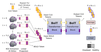 |
Multivariate Classification of fMRI Time Series with Fused
Window Transformers
Hasan Atakan Bedel1,2,
Irmak Şıvgın1,2,
Onat Dalmaz1,2,
Salman Ul Hassan Dar1,2,
and Tolga Çukur1,2,3
1Department of Electrical and Electronics Engineering, Bilkent University, Ankara, Turkey, 2National Magnetic Resonance Research Center (UMRAM), Bilkent University, Ankara, Turkey, 3Neuroscience Program, Bilkent University, Ankara, Turkey Keywords: Data Analysis, fMRI (resting state) Functional MRI (fMRI) experiments serve a key role in advancing our understanding of human brain function during normal and disease states. Analysis of high-dimensional fMRI data can significantly benefit from recent deep learning approaches, yet existing methods are insufficiently sensitive to the contextual representations in fMRI data across diverse time scales. Here, we present a novel transformer model for fMRI analysis that effectively captures local and global dependencies in fMRI data. Comprehensive demonstrations are provided that show the superior performance of BolT in gender and disease detection against state-of-the-art learning-based methods. |
| 08:15 |
0480.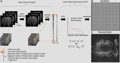 |
Improved NORDIC denoising for submillimetre BOLD fMRI using
patch formation via non-local pixels similarity -
pixel-matching (PM) NORDIC
Alessandro Nigi1 and
Jeroen C.W. Siero1
1Radiology, UMCU, Utrecht, Netherlands Keywords: Data Processing, Data Processing, denoising, NORDIC, BOLD fMRI, thermal noise Submillimetre BOLD fMRI enables studying brain function at the mesoscopic level but is limited by low SNR. The NORDIC PCA algorithm reduces thermal noise levels in fMRI in a patch-wise manner via singular value thresholding (SVT). However, the NORDIC patch formation uses adjacent pixels that often contain signals from multiple tissues, which can degrade the denoising performance. We propose an alternative patch formation using the similarity between non-local pixels, dubbed pixel-matching (PM) NORDIC. PM-NORDIC outperforms standard NORDIC in terms of temporal SNR and spatial smoothness estimates. |
| 08:15 |
0481.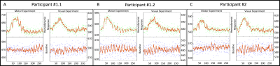 |
fMRI with whole-brain coverage, 75ms temporal resolution and
high SNR by combining HiHi reshuffling and Multiband imaging
Tim Schmidt1,2,
Johanna Vannesjo3,
Stefan Sommer4,5,6,
and Zoltan Nagy1
1Laboratory for Social and Neural Systems Research (SNS Lab), University of Zürich, Zürich, Switzerland, 2Institute for Biomedical Engineering, ETH Zürich and University of Zürich, Zürich, Switzerland, 3Norwegian University of Science and Technology, Trondheim, Norway, 4Siemens Healthineers International AG, Zürich, Switzerland, 5Swiss Center for Musculoskeletal Imaging (SCMI), Balgrist Campus, Zürich, Switzerland, 6Advanced Clinical Imaging Technology (ACIT), Siemens Healthineers International AG, Lausanne, Switzerland Keywords: Data Processing, fMRI By combining an fMRI data shuffling method and multiband accelerated acquisition, we were able to measure the hemodynamic response in the primary motor and visual cortices with 75ms temporal resolution while maintaining high SNR. With the addition of multiband imaging, we were able to achieve whole-brain coverage in a feasible scan time, and use appropriate event-related stimulus paradigm to map the BOLD response in the primary motor and visual cortices in a combined experiment. |
| 08:15 |
0482.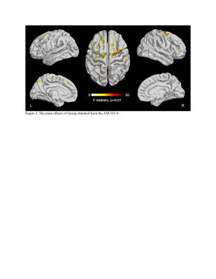 |
Multiscale Entropy Analysis of Resting State fMRI in
Pre-Adolescents with ADHD
Ru Zhang1,
Kay Jann1,
Danny J.J. Wang1,
Christina J. Duval2,
and Stuart B. Murray2
1Mark and Mary Stevens Neuroimaging and Informatics Institute, University of Southern California, Los Angeles, CA, United States, 2Department of Psychiatry and Behavioral Sciences, University of Southern California, Los Angeles, CA, United States Keywords: Data Analysis, fMRI (resting state) The current study used resting state functional magnetic resonance imaging to measure the complexity of pre-adolescents with ADHD but no other comorbidities. We ran a factorial 2 (Group: ADHD, Control) by 2 (Sex: Male, Female) analyses of covariance on the multiscale entropy (MSE) maps with the pubertal development status as a covariate. It revealed MSE values were significantly lower in the ADHD group than the control group in various regions, including bilateral superior frontal gyrus, bilateral precuneus, right inferior/middle frontal gyrus/postcentral gyrus/insular, and left precentral gyrus/middle cingulate gyrus (t = -3.131 to -2.445, p < 0.015). |
| 08:15 |
0483.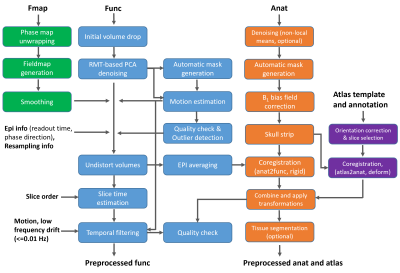 |
Optimized fMRI preprocessing pipeline enables robust
functional connectivity analysis of mouse brain at laminar
level
Wei Zhu1,
Guangle Zhang1,
Xiao-Hong Zhu1,
and Wei Chen1
1University of Minnesota, Minneapolis, MN, United States Keywords: Data Processing, fMRI High-resolution BOLD fMRI has become an essential tool for studying neural circuit and hemodynamic changes at mesoscopic scale. Nevertheless, it is more prone to the poor sensitivity and non-neural signal contamination as the spatial resolution increases. Group-level analysis also imposes new requirements on the subject alignment accuracy. To deal with these challenges, we developed a fMRI preprocessing pipeline featured in random matrix theory-based PCA denoising, one-time image voxel shift correction, and enhanced subject-level alignment. We applied this pipeline to the high-resolution mouse resting state fMRI and achieved high-quality hierarchical connectomes from large brain regions to thin cortical layers. |
| 08:15 |
0484.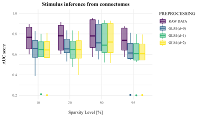 |
Pseudo resting-state by task-evoked functional connectivity.
Alice Giubergia1,
Sara Mascheretti2,
Valentina Lampis3,
Tommaso Ciceri1,
Martina Villa4,5,6,
Chiara Andreola7,
Filippo Arrigoni8,
and Denis Peruzzo1
1Neuroimaging Unit, IRCCS Eugenio Medea, Bosisio Parini (LC), Italy, 2Department of Brain and Behavioral Sciences, University of Pavia, Pavia (PV), Italy, 3Child Psychopathology Unit, IRCCS Eugenio Medea, Bosisio Parini (LC), Italy, 4Department of Psychological Sciences, University of Connecticut, Storrs, CT, United States, 5Institute for the Brain and Cognitive Sciences (IBACS), University of Connecticut, Storrs, CT, United States, 6Haskins Laboratories, New Haven, CT, United States, 7Laboratoire de Psychologie de Développement et de l’Éducation de l’Enfant (LaPsyDÉ), Université Paris Cité, Paris, France, 8Paediatric Radiology and Neuroradiology Department, V. Buzzi Children’s Hospital, Milano (MI), Italy Keywords: Data Processing, Brain Connectivity Functional connectomics investigates how brain regions are functionally associated. Studies in literature seek to infer a “pseudo-resting” state from task data, however it should be tested whether resting-state-like connectivity can be inferred by task-fMRI data. This work investigates several preprocessings of task-evoked connectivity performing classification experiments, to test their ability to reproduce a real “resting-state” connectivity and their impact on a clinical context, namely the comparison of Typical Readers and Developmental Dyslexics. Our results suggest that a task-free “pseudo-resting” connectivity cannot be inferred from task-fMRI data and that signal preprocessing does not influence the way the classification rules are inferred. |
| 08:15 |
0485.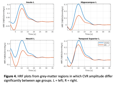 |
Age-related differences in dynamic cerebrovascular
reactivity based on CO2-response modelling
Colette Clare Milbourn1,
Seyedmohammad J Shams2,
and Jean J Chen2,3,4,5
1School of Life Sciences, University of Nottingham, Nottingham, United Kingdom, 2Research, Rotman Research Institute, Toronto, ON, Canada, 3Research, Baycrest Health Sciences Centre, Toronto, ON, Canada, 4Department of Medical Biophysics, University of Toronto, Toronto, ON, Canada, 5Institute of Biomedical Engineering, University of Toronto, Toronto, ON, Canada Keywords: Data Analysis, Neuroscience, blood vessels, cerebrovascular reactivity, deconvolution Secondary category: contrast mechanism Cerebrovascular Reactivity (CVR) was measured in healthy, young and older adults. Specifically, dynamic CVR (dCVR) was measured as the carbon dioxide (CO2)-response function. This was estimated by deconvolving a sinusoidal hypercapnic time course from simultaneously acquired blood oxygen level dependent (BOLD) fMRI data using a canonical-correlation (CCA) based approach. The results demonstrate differences in dCVR amplitude and rise time between age groups. This proof-of-concept study establishes the feasibility of dCVR modelling for the study of aging. |
| 08:15 |
0486.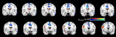 |
Detecting the role of hippocampal subfields in pattern
separation and completion task using 7T object memory task
fMRI
Zhengshi Yang1,
Xiaowei Zhuang2,
Mark Lowe3,
and Cordes Dietmar1
1Lou Ruvo Center for Brain Health, Cleveland Clinic, Las Vegas, NV, United States, 2Cleveland Clinic, Las Vegas, NV, United States, 3Cleveland Clinic, Cleveland, OH, United States Keywords: Data Analysis, fMRI, Task fMRI; reigstration In this study, we have established a reliable hippocampal subfield segmentation and activation analysis pipeline to probe the role of hippocampal subfields in pattern separation task by using 7T fMRI data, which could be able to identify the role of subfield dysfunction in cognitive impairment in prodromal AD. |
| 08:15 |
0487.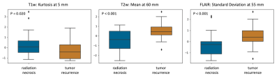 |
Biophysical deformation in brain-around-tumor on MRI can
distinguish radionecrosis vs recurrent tumor in brain
metastases: A feasibility study
Hyemin Um1,
Virginia Hill2,
Marwa Ismail3,
Sushant Puri4,
Ameya Nayate5,
Prateek Prasanna6,
Lisa Rogers5,
Jennifer Yu7,
and Pallavi Tiwari3
1Case Western Reserve University, Cleveland, OH, United States, 2Northwestern Medicine, Chicago, IL, United States, 3University of Wisconsin-Madison, Madison, WI, United States, 4Johns Hopkins Medicine, Baltimore, MD, United States, 5University Hospitals Cleveland Medical Center, Cleveland, OH, United States, 6Stony Brook University, Stony Brook, NY, United States, 7Cleveland Clinic, Cleveland, OH, United States Keywords: Quantitative Imaging, Machine Learning/Artificial Intelligence A significant challenge in the management of metastatic brain tumors following radiation therapy is distinguishing radiation necrosis from tumor recurrence. Differential diagnosis is difficult on routine MRI and patients are subject to invasive procedures to confirm the absence of disease. We explored the feasibility of deformation features from the normal parenchyma to identify disease recurrence versus radiation effects. Our results suggest that measurements of the subtle tissue deformations in the normal-appearing brain regions may elucidate differences in the tissue microarchitecture of radionecrosis and tumor recurrence and may serve as surrogate markers to non-invasively characterize treatment response in brain metastases. |
| 08:15 |
0488.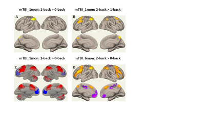 |
Functional Activation Pattern to varied working memory loads
in Mild Traumatic Brain Injury Patients
Li Jiang1,
Jiachen Zhuo1,
Steven Roys1,
Chandler Sours Rhodes2,
Prashant Raghavan Raghavan1,
and Rao Gullapalli1
1Department of Diagnostic Radiology and Nuclear Medicine, University of Maryland Baltimore, Baltimore, MD, United States, 22National Intrepid Center of Excellence, Walter Read National Military Medical Center, Rockville, MD, United States Keywords: Data Analysis, Traumatic brain injury Mild traumatic brain injury (mTBI) account for 85% of all TBIs. Working memory impairment is one of the most common symptoms in mTBI patients and may persist years post-mTBI. In this study, we used N-back fMRI to investigate the varying N-back task loads on the functional activation pattern in patients with mild TBI and age-matched healthy controls (HCs) as well as the longitudinal changes within mTBI group. Our results suggested that appropriate task load would be more sensitive in detecting the subtle brain functional changes, even though differences in behavioral performance between mTBI and HCs were absent. |
 Back to Meeting Home
 Back to the Program-at-a-Glance
The International Society for Magnetic Resonance in Medicine is accredited by the Accreditation Council for Continuing Medical Education to provide continuing medical education for physicians.
 Back to Meeting Home
 Back to the Program-at-a-Glance
 Full Session Video