Power Pitch
Pitch: Novel Acquisition Strategies
ISMRM & ISMRT Annual Meeting & Exhibition • 03-08 June 2023 • Toronto, ON, Canada

Power Pitch
Pitch: Novel Acquisition Strategies
| 08:15 |
0104.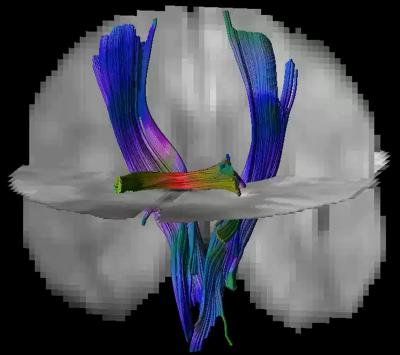 |
Tensors and Tracts at 64 mT
Alix Plumley1,
Francesco Padormo2,
Mara Cercignani1,
Rafael O'Halloran2,
Rui Teixeira2,
Álvaro Planchuelo-Gómez1,3,
Antoine Legouhy4,
Tianrui Luo2,
and Derek K Jones1
1Cardiff University, Cardiff, United Kingdom, 2Hyperfine Inc., Guilford, CT, United States, 3University of Valladolid, Valladolid, Spain, 4University College London, London, United Kingdom Keywords: Data Analysis, Low-Field MRI, Diffusion Tensor Imaging We present the first ever demonstration of Diffusion Tensor Magnetic Resonance Imaging (DT-MRI) including quantitative measures of mean diffusivity, fractional anisotropy, and successful tractographic reconstruction of projection and commissural pathways on a portable system operating at 64 mT. |
| 08:15 |
0105.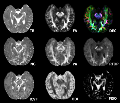 |
Migration of Potential Microstructural Markers of AD by
High-Resolution Hippocampal Diffusion MRI
Courtney Joy Comrie1,
Samantha Schatz1,
Kevin Johnson1,
Scott Squire1,
Nan-kuei Chen1,
and Elizabeth Hutchinson1
1Biomedical Engineering, University of Arizona, Tucson, AZ, United States Keywords: Data Acquisition, Alzheimer's Disease Diffusion MRI has been identified as a promising tool for identifying novel and early makers of AD and comorbid pathology. This study worked towards translating advanced microstructural techniques to clinical acquisitions for the purpose of potential AD diagnosis. High resolution diffusion human data was acquired using a 4-way phase-encoding and specialized hippocampal FOV in healthy subjects for method development. High quality maps were achieved that revealed more hippocampal microstructural information than what is acquired in database protocols according to preliminary results. |
| 08:15 |
0106. |
High Resolution Diffusion Tensor Imaging using Rapid
Single-Slab 3D EPI Encoding
Hyunkyung Maeng1 and
Jaeseok Park1,2
1Department of Biomedical Engineering, Sungkyunkwan University, Suwon, Korea, Republic of, 2Department of Intelligent Precision Healthcare Convergence, Sungkyunkwan University, Suwon, Korea, Republic of Keywords: Data Acquisition, Diffusion Tensor Imaging, High resolution 3D DTI To propose an accelerated high resolution whole brain DTI and investigate its feasibility (1.0$$$mm^3$$$ isotropic spatial resolution, imaging time ~ 15 min) |
| 08:15 |
0107.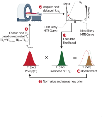 |
Adaptive Magnetic Resonance
Assaf Tal1,
Inbal Beracha1,
and Amir Seginer2
1Chemical and Biological Physics, Weizmann Institute of Science, Rehovot, Israel, 2Siemens Healthcare Ltd., Rosh Haayeen, Israel Keywords: Hybrid & Novel Systems Technology, Pulse Sequence Design, Accelerating data acquisition We present a completely general framework for adaptively changing the radiofrequency pulses and delays in real time in response to incoming data from the subject within the scanner. This "personalized-radiology" framework is shown to increase the precision of T2 estimation for n-acetyl-aspartate in-vivo by a factor of 1.7, and accelerate its acquisition 2.5-fold.
|
08:15 |
0108.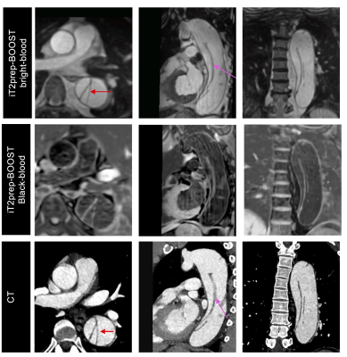 |
Efficient iT2prep-BOOST for simultaneous contrast-free 3D
aortic lumen and vessel wall imaging for the assessment of
thoracic aortopathy
Anastasia Fotaki1,
Camila Munoz1,
Alina Hua1,
Karl P Kunze1,2,
Radhouene Neji3,
Kuberan Pushparajah1,
Rene M Botnar4,5,6,7,
and Claudia Prieto4,5,6,7 1King's College London, London, United Kingdom, 2MR Research Collaborations, Siemens Healthcare Limited, Camberley, United Kingdom, 3MR Research Collaborations, Siemens Healthcare Limited, Camberely, United Kingdom, 4Biomedical Engineering, King's College London, London, United Kingdom, 5Institute for Biological and Medical Engineering, Pontificia Universidad Católica de Chile, Santiago, Chile, 6Millennium Institute for Intelligent Healthcare Engineering, Santiago, Chile, 7School of Engineering, Pontificia Universidad Católica de Chile, Santiago, Chile Keywords: Data Acquisition, Cardiovascular, aorta Bright- and black-blood MRI sequences are clinically acquired sequentially, for aortic lumen and vessel wall imaging respectively, in patients with thoracic aortic disease. A novel, free-breathing, 3D sequence (iT2prep-BOOST) is proposed for contrast-free simultaneous depiction of lumen and vessel wall. We clinically validated the iT2prep-BOOST in a cohort of 25 patients with thoracic aortic disease against the conventional bright-blood T2-prep bSSFP sequence and black-blood 2D HASTE. Quantitative and qualitative image quality assessment demonstrated that iT2Prep-BOOST enabled time-efficient, bright- and black-blood aortic imaging, with improved image quality compared to conventional approaches, and comparable measurements for aortic wall and lumen dimensions. |
| 08:15 |
0109.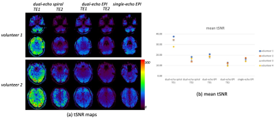 |
A 3D dual-echo spiral sequence for dynamic susceptibility
contrast imaging
Zhiqiang Li1,
Poonam Choudhary1,
Dinghui Wang2,
Sudarshan Ragunathan1,
Melvyn B Ooi3,
John P Karis1,
James G Pipe2,
Ashley M Stokes1,
and C Chad Quarles1,4
1Barrow Neurological Institute, Phoenix, AZ, United States, 2Mayo Clinic, Rochester, MN, United States, 3Philips Healthcare, Houston, TX, United States, 4The University of Texas MD Anderson Cancer Center, Houston, TX, United States Keywords: Pulse Sequence Design, DSC & DCE Perfusion, spiral, perfusion, quantitative imaging Dynamic susceptibility contrast (DSC)-MRI is an important tool to assess brain tumor status for diagnosis, surgical planning, and surveillance. Dual-echo EPI based DSC-MRI enables accurate perfusion measurements using a single-dose of contrast agent, simultaneous DSC- and dynamic contrast enhanced (DCE)-MRI measures and greater parameter flexibility. However, EPI-related distortion artifacts impair its clinical utility. In this work, we proposed a 3D dual-echo spiral acquisition to mitigate EPI-related artifacts, and improve the fidelity of DCE-MRI derived parameters and arterial input function estimation. Preliminary data from volunteers and a glioma tumor patient showed reduced geometric distortion, increased temporal SNR, and accurate perfusion measurements. |
| 08:15 |
0110.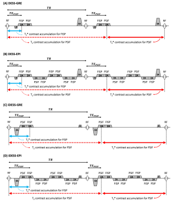 |
Multi-contrast Imaging using Dual-pathway Echo-planar
Imaging Sequence
Silu Han1,
Sudhir Ramanna1,
and Nan-kuei Chen1,2
1Department of Biomedical Engineering, The University of Arizona, Tucson, AZ, United States, 2BIO5 Institute, The University of Arizona, Tucson, AZ, United States Keywords: Pulse Sequence Design, Brain Multi-contrast imaging, commonly acquired with spoiled gradient recalled echo (SPGR) in clinical routine examinations, has less-than-optimal scan efficiency1. Dual-pathway sequences, such as double-echo steady-state (DESS) and inverse double-echo steady-state (iDESS), have been developed to improve signal-to-noise ratio (SNR) and scan efficiency as compared with SPGR2. Here we propose to further combine DESS/iDESS sequences and echo-planar imaging (EPI) to enhance scan efficiency, with significant implications to temperature mapping and parametric mapping (e.g., T1-, T2-, T2*-mapping and B0-field mapping, quantitative susceptibility mapping (QSM)3, 4). |
| 08:15 |
0111.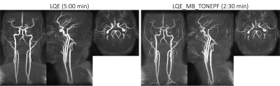 |
Simultaneous Brain and Neck Time of Flight using Spiral
Multiband with Localized Quadratic Encoding
Xi Peng1,
Dinghui Wang1,
Guruprasad Krishnamoorthy1,2,
Daniel D. Borup1,2,
and James G. Pipe1
1Mayo Clinic, Rochester, MN, United States, 2Philips Healthcare, Rochester, MN, United States Keywords: New Trajectories & Spatial Encoding Methods, Blood vessels A spiral multiband localized quadratic encoding (LQE) method is proposed to achieve simultaneous brain and neck time of flight MRA within a single 2.5-minute scan. LQE acquisition is efficient for both SNR and in-flow enhancement. Multi-band LQE enables increased coverage with no increase in scan time, but can introduce venous signal contamination. We propose a multi-band LQE approach with TONE and partial-Fourier slice selection to reduce the venous signal and achieve simultaneous time-of-flight MRA of intracranial and carotid arteries. |
| 08:15 |
0112.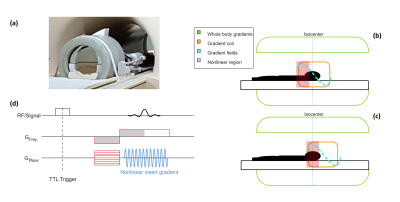 |
High-fidelity PSF reconstruction for fast and silent MRI
using nonlinear spatially encoding gradient fields switched
at 20kHz
Michael JB McGrory1,
Edwin Versteeg1,
Alessando Sbrizzi1,
Cornelis AT van den Berg1,
Dennis WJ Klomp1,
and Jeroen CW Siero1,2
1Radiology, University Medical Center Utrecht, Utrecht, Netherlands, 2Spinoza Center for Neuroimaging, Amsterdam, Netherlands Keywords: Gradients, Gradients Acoustic noise in MRI scans can be reduced by utilising gradient switching frequencies at 20kHz. High slew rates required to achieve such high-frequency switching can lead to peripheral nerve stimulation (PNS), adding difficulty for application to whole-body gradients. Instead, nonlinear encoding gradients have been shown to limit PNS while achieving desired slew rates. In this work, we show the feasibility of using a nonlinear silent gradient for spatial encoding by employing a PSF-based reconstruction and investigate image fidelity on a 4-fold accelerated in-vivo scan. This method could potentially be utilised for a whole-body gradient design for silent and fast MRI. |
| 08:15 |
0113.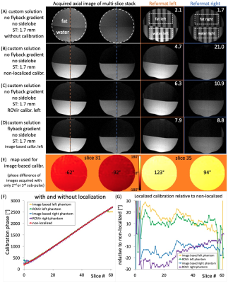 |
Fast localized calibration for spatial-spectral excitation
without fly-back gradients
Michael Schär1,
Robert G Weiss2,
and Allison G Hays2
1Department of Radiology, Johns Hopkins University School of Medicine, Baltimore, MD, United States, 2Department of Cardiology, Johns Hopkins University School of Medicine, Baltimore, MD, United States Keywords: Pulse Sequence Design, RF Pulse Design & Fields, calibration, localized calibration, spectral-spatial, fat suppression Spiral MRI requires fat suppression because fat and other off-resonant spins are blurred. For fast cardiac spiral MRI at 3T, spectral-spatial water-only excitation is often used. Standard spectral-spatial pulses apply a fly-back gradient, limiting how thin the slices can be (>4mm). Without fly-back gradients slices can be as thin as ~1.7mm, but require a phase calibration for the radiofrequency sub-pulses with inverted gradients due to system imperfections. Here we propose and test a fast (<1s), localized pre scan enabling thin-slice water only excitation. As a potential application we show spiral multi-slice coronary angiography images acquired in a single breath-hold. |
| 08:15 |
0114.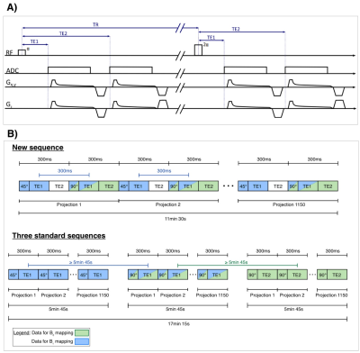 |
Development of a Pulse Sequence with Alternating Excitation
for Respiratory-Sorted B1+ and B0 Field Mapping in 23Na
Human Torso MRI
Jana Felz1,2,
Fabian J. Kratzer1,
Armin M. Nagel1,3,
Peter Bachert1,2,
Mark E. Ladd1,2,4,
and Tanja Platt1
1Medical Physics in Radiology, German Cancer Research Center, Heidelberg, Germany, 2Faculty of Physics and Astronomy, Heidelberg University, Heidelberg, Germany, 3Institute of Radiology, University Hospital Erlangen, Erlangen, Germany, 4Faculty of Medicine, Heidelberg University, Heidelberg, Germany Keywords: Pulse Sequence Design, Non-Proton, High-Field MRI, Body, 23Na MRI, Field Mapping 23Na MRI is a promising imaging method, but suffers from lower SNR, hence from longer acquisition times and lower image resolution than 1H MRI. Especially in the torso, motion and magnetic field inhomogeneities impede quantitative analysis of 23Na concentrations. To tackle this, a new pulse sequence is presented that yields self-gated respiratory-sorted B1+ and B0 maps from a single measurement. B1+ mapping with the new sequence is less prone to motion artefacts, since k-space projections of two flip angles are acquired in an interleaved manner. This reduces the influence of varying respiration, which can lead to artefacts in conventional mapping. |
| 08:15 |
0115.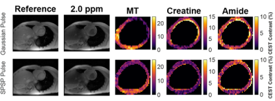 |
Advanced Spatial-Spectral Pulse Design for Metabolite
Specific Filtered CEST in the Human Heart
Cindy Ayala1,
Huiwen Luo2,
Kevin Godines1,
William Grissom2,
and Moriel Vandsburger1
1University of California, Berkeley, Berkeley, CA, United States, 2Vanderbilt University, Nashville, TN, United States Keywords: Pulse Sequence Design, CEST & MT Chemical Exchange Saturation Transfer (CEST) MRI has been used to probe metabolism via total creatine contrast using conventional contrast generation with Gaussian saturation. We developed a spatial-spectral (SPSP) saturation pulsed CEST protocol to separate the contrast generated by metabolic subcomponents creatine and phosphocreatine (PCr) in phantoms and the human heart (n=10). Phantom studies revealed uniform CEST contrast for creatine and PCr following saturation with Gaussian pulses, and selective PCr contrast following saturation with SPSP pulses. Human studies revealed both enhanced B1-uniformity and selective PCr contrast with SPSP saturation, enabling quantitation of the PCr/total creatine ratio in the myocardium. |
| 08:15 |
0116.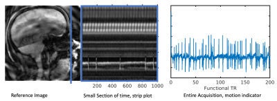 |
Accelerating Dynamic Imaging in SimulScan: Simultaneous
Functional MRI and Dynamic Imaging of Tongue Motion
Bradley P. Sutton1,2,3,
Georgia A. Malandraki4,
Riwei Jin1,3,
Charles Marchini1,3,
Aaron Anderson3,
Paul Arnold2,3,5,
and Zhi-Pei Liang2,6
1Bioengineering, University of Illinois Urbana Champaign, Urbana, IL, United States, 2Carle Illinois College of Medicine, Urbana, IL, United States, 3Beckman Institute, University of Illinois Urbana Champaign, Urbana, IL, United States, 4Speech, Language, and Hearing Sciences, Purdue University, West Lafayette, IN, United States, 5Neuroscience Institute, Carle Foundation Hospital, Urbana, IL, United States, 6Electrical and Computer Engineering, University of Illinois Urbana Champaign, Urbana, IL, United States Keywords: Data Acquisition, fMRI (task based) Leveraging low rank and Partial Separability models, the SimulScan sequence and reconstruction was updated to drastically improve the quality and speed of dynamic imaging. This enables simultaneous functional MRI and dynamic imaging of oropharyngeal motions to be examined despite the many air/tissue interfaces. We demonstrate the improvement on a healthy adult performing a self-paced tongue tapping during the scan. The improved imaging will enable the examination of neural control of fine scale articulatory and oral movements critical for speech, feeding, and swallowing events. |
| 08:15 |
0117.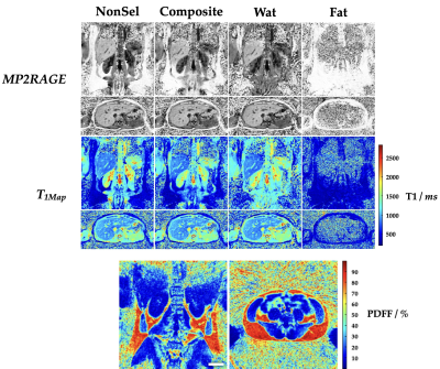 |
Radial MP2RAGE combined with binomial pulses for
multi-parametric (T1 and PDFF) abdominal mapping.
François E Maingault1,
William Lefrançois1,
Nadège Corbin1,
Aurélien J Trotier1,
Laurence Dallet1,
Eric Thiaudière1,
Sylvain Miraux1,
and Emeline EJ Ribot1
1Centre de Résonance Magnétique des Systèmes Biologiques, UMR5536, CNRS/Université de Bordeaux, Bordeaux, France Keywords: Pulse Sequence Design, Fat, Water, pdff, Abdomen A 3D radial-encoding MP2RAGE sequence was developed with binomial pulses within the GRE trains. These give the possibility to obtain water-specific, fat- specific or composite 3D T1-maps of the abdominal cavity during free-breathing in less than 10 minutes. A 3D Proton Density Fat Fraction (PDFF) map can also be determined with the same scan. The T1 values are similar with the standard MP2RAGE and the PDFF correspond to the ones obtained with MR spectroscopy. A test-retest on healthy volunteers shows the good reproducibility of the method. |
| 08:15 |
0118.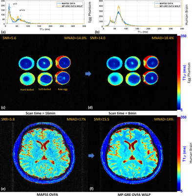 |
Improving 3D-T1rho Mapping of the Human Brain Using
Optimized Variable Flip-Angles and Weighted Spin-Lock Pulses
Marcelo V. W. Zibetti1,
Rajiv Menon1,
Hector L. De Moura1,
Mahesh B. Keerthivasan2,
and Ravinder R. Regatte1
1Radiology, NYU Grossman School of Medicine, New York, NY, United States, 2Siemens Medical Solutions, Malvern, PA, United States Keywords: Data Acquisition, Pulse Sequence Design, optimization This study shows 3D-T1rho mapping of the human brain using optimized variable flip-angles (OVFAs) and weighted spin-lock pulses (WSLP). Our preliminary results suggest that the proposed sequence based on OVFAs and WSLP can improve SNR by almost 3X in brain T1rho mapping, reduces data acquisition time by half, and improve the mean of normalized absolute deviation (MNAD) compared to MAPSS sequence for the same application. |
| 08:15 |
0119.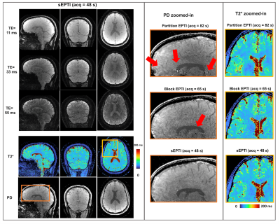 |
Spherical Echo-Planar Time-resolved Imaging (sEPTI) for 3D
highly-accelerated, distortion-free, time-resolved
whole-brain T2* mapping
Nan Wang1,
Yannick WE Brackenier1,
Congyu Liao1,
Siddharth Srinivasan Iyer1,2,
Xiaozhi Cao1,
Justin Haldar3,
and Kawin Setsompop1,4
1Department of Radiology, Stanford University, Stanford, CA, United States, 2Department of Electrical Engineering and Computer Science, Massachusetts Institute of Technology, Cambridge, MA, United States, 3Ming Hsieh Department of Electrical and Computer Engineering, University of Southern California, Los Angeles, CA, United States, 4Department of Electrical Engineering, Stanford University, Stanford, CA, United States Keywords: Data Acquisition, Data Acquisition, Echo-planar imaging, time-resolved imaging EPTI is a rapid time-resolved quantitative imaging method. In this work, we developed a spherical EPTI sampling trajectory (sEPTI) to improve its speed. To achieve fast imaging, sEPTI traverses a tight 3D spherical k-space using full ramp-sampling and variable echo-spacing, which also desirably increases its spatiotemporal incoherency. sEPTI was demonstrated in vivo to provide improved imaging performance over conventional block 3D-EPTI that requires 1.4x longer scan; achieving high-quality 1mm isotropic whole-brain proton-density and T2* maps in 48s. As part of this work, a novel and effective reconstruction approach to mitigate high-spatial-order ghosts in echo-planar acquisitions was also developed. |
| 08:15 |
0120.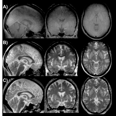 |
Ultrashort-echo time magnetization transfer (UTE-MT) for
brain iron imaging
Humberto Monsivais1,
Gianna Nossa1,
Seokkyoon Hong2,
Taewoong Park2,
Fethi Sila Erdil1,
Xin Shen3,
Antonia Susnjar2,
Ali Özen4,
Serhat Ilbey4,
Mark Chiew5,
Jessica Huber6,
Ulrike Dydak1,7,
and Uzay Emir1,2
1School of Health Sciences, Purdue University, West Lafayette, IN, United States, 2Weldon School of Biomedical Engineering, Purdue University, West Lafayette, IN, United States, 3Radiology, University of California San Francisco, San Francisco, CA, United States, 4Department of Radiology, Medical Physics, University of Freiburg, Freiburg, Germany, 5Welcome Centre for Integrative Neuroimaging, University of Oxford, England, United Kingdom, 6Department of Audiology and Speech Sciences, Purdue University, West Lafayette, IN, United States, 7Department of Radiology and Imaging Sciences, Indiana University School of Medicine, Indianapolis, IN, United States Keywords: Contrast Mechanisms, Brain We have established a novel 3D dual-echo UTE-MT imaging method to assess hyperintense T1w signal in iron-rich brain areas by eliminating the ultra-short T2 constituents of the myelin signal via the magnetization transfer (MT) technique. Our preliminary results show improved positive image contrast in deep brain areas such as the substantia nigra (SN) and the LC. Other iron-rich areas in the basal ganglia (globus pallidus and putamen) also show improved contrast. |
| 08:15 |
0121.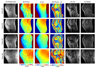 |
Ultrashort Echo Time Single Point Dixon Using 3D Phase
Modeling
Nathan Newbury1,
Sam Sedaghat1,
Jiyo Athertya1,
Michael Carl2,
Jiang Du1,
and Hyungseok Jang1
1Department of Radiology, University of California San Diego, La Jolla, CA, United States, 2GE Healthcare, La Jolla, CA, United States Keywords: Fat, MSK Fat signals can obscure morphological structures and influence quantitative parameter mapping in ultrashort echo time (UTE) musculoskeletal MRI. However, conventional fat suppression methods have challenges in UTE imaging of short T2 species. For example, the chemical-shift based fat saturation can significantly attenuate short T2 signals with broad spectra, and water excitation with long composite pulses may yield a long minimum echo time (TE). Alternatively, the feasibility of single-point Dixon (1p-Dixon) has been demonstrated for UTE imaging. In this study, we investigate the feasibility of 1p-Dixon based on the 3D phase modeling approach, which achieves fat suppression without additional data acquisition. |
| 08:15 |
0122.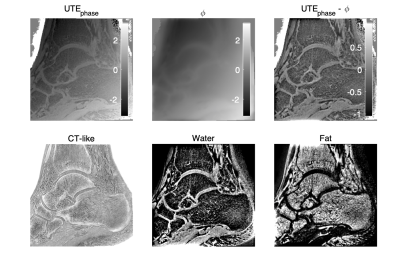 |
Accelerated single UTE-Dixon for simultaneous short T2*water
and fat imaging using a FLORET trajectory
Anh T. Van1,
Kilian Weiss2,
Georg C. Feuerriegel1,
Philipp Braun1,
Guruprasad Krishnamoorthy3,4,
Alexandra S. Gersing1,
James G. Pipe4,
and Dimitrios C. Karampinos1
1Department of Diagnostic and Interventional Radiology, School of Medicine, Technical University of Munich, Munich, Germany, 2Philips GmbH Market DACH, Hamburg, Germany, 3Philips Healthcare, Rochester, MN, United States, 4Department of Radiology, Mayo Clinic College of Medicine and Science, Rochester, MN, United States Keywords: Bone, Skeletal Ultra-short echo time (UTE) imaging enables the depiction of short-T2* tissues and is being increasingly used for the generation of CT-like bone images. UTE imaging has been recently combined with single-echo Dixon processing to enable the separation of water and fat signals from a single echo UTE image, but was primarily previously employed in radial stack-of-stars UTE acquisitions with prolonged scan durations. The present work combines single UTE-Dixon processing with a Fermat looped, orthogonally encoded trajectory (FLORET) to enable accelerated simultaneous short T2* water- and fat-separated imaging at sub-millimeter isotropic resolution. The technique is applied in the ankle. |
| 08:15 |
0123.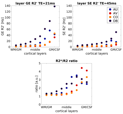 |
Characterization of the R2*/R2 ratio of the BOLD
laminar-specificity through biophysical simulations using
realistic vascular architectures
Mario Gilberto Báez-Yáñez1,
Vanja Curcic1,
Jeroen Siero1,
Matthias J.P. van Osch2,
and Natalia Petridou1
1Department of Radiology, Center for Image Sciences, UMC Utrecht, Utrecht, Netherlands, 2C.J. Gorter MRI Center, LUMC, Leiden, Netherlands Keywords: Signal Modeling, fMRI GE-BOLD fMRI is the most used technique to measure functional hyperemia across cortical depth. GE-BOLD is sensitive towards all vascular contributions, while SE-BOLD increases the specificity towards small vessels– being more specific to neuronal activity. However, SE-BOLD fMRI measurements may not completely remove the macrovascular contribution. Using realistic vascular models, we simulated specific oxygenation states in different vascular compartments to characterize the GE-and SE-BOLD specificity across cortical depth quantified through the R2*/R2-ratio. Simulations show 1)similar ratio values at deeper layers indicating comparable specificity to microvessels for both sequences, 2)layer-fMRI SE-BOLD cannot completely eliminate the macrovascular contribution towards the pial surface. |
 Back to Meeting Home
 Back to the Program-at-a-Glance
The International Society for Magnetic Resonance in Medicine is accredited by the Accreditation Council for Continuing Medical Education to provide continuing medical education for physicians.
 Back to Meeting Home
 Back to the Program-at-a-Glance
 Full Session Video