Power Pitch
Pitch: What's New in Body Imaging: Emerging Techniques & AI
ISMRM & ISMRT Annual Meeting & Exhibition • 03-08 June 2023 • Toronto, ON, Canada

Power Pitch
Pitch: What's New in Body Imaging: Emerging Techniques & AI
| 08:15 |
1208.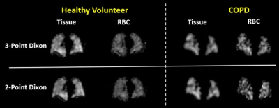 |
Hyperpolarized xenon-129 dissolved MRI based on two-point
Dixon method
Hengjie Chen1,
Jaime F Mata2,
Y. Michael Shim2,
John P. Mugler2,
Xiaoping Hu1,
Li Zhao3,
An Liu4,
and Kun Qing1,4
1University of California, Riverside, Riverside, CA, United States, 2University of Virginia, Charlottesville, VA, United States, 3Zhejiang University, Hangzhou, China, 4City of Hope National Medical Center, Duarte, CA, United States Keywords: Lung, Hyperpolarized MR (Gas) In this work, a 2-point Dixon-based method is developed to provide separation of the hyperpolarized xenon-129 dissolved-phase magnetic resonance imaging (MRI) components for pulmonary applications. Based on analysis of data from 6 healthy volunteers and 20 patients with lung disease, the separated tissue and red blood cell (RBC) components obtained from this method are highly consistent with those from the previously-described 3-point Dixon method. The 2-point Dixon based method has the potential to greatly improve the signal-to-noise ratio for dissolved-phase MRI while still maintaining accurate tissue/RBC separation. |
| 08:15 |
1209.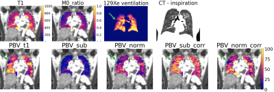 |
Lung perfusion imaging: influence of signal-to-concentration
transformation method and comparison with 129Xe biomarkers
Marta Tibiletti1,
Paul JC Hughes2,
James A Eaden2,
Josephine H Naish1,3,
Helen A Marshall4,
John C Waterton1,5,
Stephen A Bianchi6,
Jim M Wild4,7,
and Geoff JM Parker1,8
1Bioxydyn Ltd, Manchester, United Kingdom, 2POLARIS, University of Sheffield MRI unit, The University of Sheffield, Sheffield, United Kingdom, 3MCMR, Manchester University NHS Foundation Trust, Wythenshawe, United Kingdom, 4POLARIS, The University of Sheffield, Sheffield, United Kingdom, 5Centre for Imaging Sciences, University of Manchester, Manchester, United Kingdom, 6Academic Department of Respiratory Medicine, Sheffield Teaching Hospitals NHS Foundation Trust, Sheffield, United Kingdom, 7Insigneo Insititute for in silico medicine, Sheffield, United Kingdom, 8Centre for Medical Image Computing, Department of Medical Physics and Biomedical Engineering, University College London, London, United Kingdom Keywords: Lung, Perfusion Quantitative pulmonary perfusion can be derived from MR imaging with injection of contrast agent (CA). Tracer-kinetic theory yields physiological parameters such as pulmonary blood flow (PBF), blood volume (PBV) and mean transit time (MTT) but relies on accurate calculation of CA concentration. Previous studies have used ‘subtraction’ or ‘normalisation’ methods without T1-mapping. Using both simulations and data from interstitial lung disease patients, we demonstrate how the ‘subtraction’ method weights PBV by local lung density. This causes likely spurious correlations with hyperpolarised 129Xe biomarkers linked to lung ventilation, microstructure and density, potentially obscuring information of diagnostic interest. |
| 08:15 |
1210.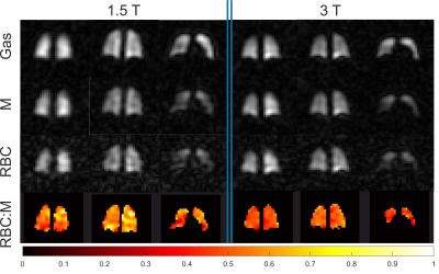 |
Implementation of dissolved 129Xe lung MRI with 4-echo 3D
radial spectroscopic imaging at 3T: comparing with results
at 1.5T in healthy volunteers
Guilhem Jean Collier1,
Ho-Fung Chan1,
Graham Norquay1,
Neil J. Stewart1,
Ryan S. Munro1,
Oliver Rodgers1,
Rolf F. Schulte2,
and Jim M. Wild1
1POLARIS, University of Sheffield, Sheffield, United Kingdom, 2GE Healthcare, Munich, Germany Keywords: Lung, Hyperpolarized MR (Gas) Four-echo 3D radial spectroscopic imaging of hyperpolarised 129Xe in the lung is implemented at 3T to measure ratios of xenon gas and dissolved xenon in blood (RBC) and lung tissues (M). Thanks to an interleaved acquisition of the echo times and the choice of a 0.32ms echo time spacing, data acquisition is possible within the short relaxation time of dissolved 129Xe (T2*~1ms). 11 healthy volunteers have been imaged at both 1.5 and 3T after the inhalation of a 1L dose of hyperpolarised 129Xe. Results show comparable images at both field strengths and a significantly reduced (p=0.02) RBC:M at 3T. |
| 08:15 |
1211.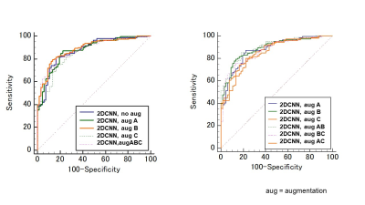 |
Usefulness of Deep Learning Models of DWI in Direct
Differentiating Malignant and Benign Breast Tumors Without
Lesion Segmentation
Mami Iima1,2,
Kazuki Tsuji3,
Ryosuke Mizuno4,
Toshiki Yamazaki3,
Masako Kataoka1,
Maya Honda1,5,
Rie Ota1,6,
Aika Okazawa7,
Keiho Imanishi8,
Masakazu Toi9,
and Yuji Nakamoto1
1Kyoto University Graduate School of Medicine, Kyoto, Japan, 2Kyoto University Hospital, Institute for Advancement of Clinical and Translational Science (iACT), Kyoto University Hospital, Kyoto, Japan, 3Kyoto University Faculty of Medicine, Kyoto, Japan, 4A.I.System Research CO.,Ltd., Kyoto, Japan, 5Diagnostic Radiology, Kansai Electric Power Hospital, Osaka, Japan, 6Diagnostic Radiology, Tenri Hospital, Nara, Japan, 7Kyoto University Graduate School of Medi, Kyoto, Japan, 8e-Growth Co., Ltd., Kyoto, Japan, 9Breast Surgery, Kyoto University Graduate School of Medi, Kyoto, Japan Keywords: Breast, Breast 348 women suspected of breast tumors were enrolled, and 206 breast lesions (139 malignant, 67 benign) were further analyzed. Breast 5b-value-DWI was performed, and a deep learning model dedicated to this breast DWI dataset was established. Several comparative experiments were performed; comparison of data augmentations, small network VS. large network, 2DCNN VS. 3DCNN, and analysis in 5b-DWI VS. ADC maps. Augmentations using elastic deformation, affine transform, and Gaussian noise improved diagnostic performance up to AUC=0.90. The use of small CNN and without ADC map also showed higher diagnostic performance (AUC=0.88-0.90), showing AI potential to improve breast DWI diagnostic performance. |
| 08:15 |
1212.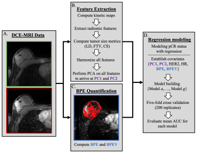 |
Combining radiomic features with background parenchymal
enhancement from DCE-MRI data for predicting treatment
response in breast cancer
Kalina Polet Slavkova1,
Eric A Cohen1,
Rhea Chitalia2,
Snekha Thakran1,
Walter C Mankowski1,
Alex Nguyen2,
Hannah Horng2,
Elizabeth S McDonald3,
Michael D Feldman4,
Angela DeMichele5,6,
and Despina Kontos7
1Department of Radiology, University of Pennsylvania, Philadelphia, PA, United States, 2Department of Bioengineering, University of Pennsylvania, Philadelphia, PA, United States, 3Department of Radiology, Hospital of the University of Pennsylvania, Philadelphia, PA, United States, 4Division of Surgical Pathology, Hospital of the University of Pennsylvania, Philadelphia, PA, United States, 5Division of Hematology Oncology, Perelman Center for Advanced Medicine, Philadelphia, PA, United States, 6Abramson Cancer Center, University of Pennsylvania, Philadelphia, PA, United States, 7Radiology, University of Pennsylvania, Philadelphia, PA, United States Keywords: Breast, Breast, radiomics, DCE-MRI Our objective is to predict pathological complete response (pCR) outcome to neoadjuvent chemotherapy in breast cancer patients. We combine radiomic features with background parenchymal enhancement -- computed from standard-of DCE-MRI data from the ISPY-2 trial -- and model treatment outcome via multivariable logistic regression. During training and testing, we demonstrate that models including BPE alongside radiomic and clinical covariates yielded the highest AUC values among all tested regression models, thus improving the prediction of pCR outcome. |
| 08:15 |
1213.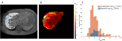 |
3D MR Elastography: Exploring Quality Assurance Measures
Shan Cai1,2,
Jens Tellman1,2,
Christian Simonsson1,2,3,
Wolf Bartholomä1,4,
Jonatan Eriksson1,2,
Nils Dahlström1,4,
Stergios Kechagias5,
Patrik Nasr5,
Mattias Ekstedt5,
Ralph Sinkus6,
and Peter Lundberg1,2
1Center for Medical Imaging Science and Visualization, Linköping University, Linköping, Sweden, 2Department of Medical Radiation Physics, and Department of Health, Medicine and Caring Sciences, Linköping University, Linköping, Sweden, 3Department of Biomedical Engineering, Linköping University, Linköping, Sweden, 4Department of Radiology in Linköping, and Department of Health, Medicine and Caring Sciences, Linköping University, Linköping, Sweden, 5Department of Gastroenterology and Hepatology, and Department of Health, Medicine and Caring Sciences, Linköping University, Linköping, Sweden, 6School of Biomedical Engineering and Imaging Science, King’s College London, London, United Kingdom Keywords: Liver, Elastography, 3D MRE Magnetic resonance elastography (MRE) is a powerful tool that can grade liver fibrosis non-invasively. In comparison with 2D MRE, 3D MRE can provide additional biomechanical tissue parameters for evaluating liver fibrosis. However, there is a great need to develop a quality assurance protocol for 3D MRE measurements. In this study, we have investigated quality assurance parameters for a 3D MRE research system and identified the cut-off values of the quality parameters to assign the 3D MRE data into three quality categories. A confidence map was constructed based on the evaluation of these quality parameters. |
| 08:15 |
1214.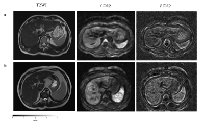 |
A Noninvasive Method with Three-Dimensional MR elastography
in Assessing of Liver Fibrosis and Inflammation
Yikun Wang1,
Jiahao Zhou1,
Huimin Lin1,
Fuhua Yan1,
and Ruokun Li1
1Ruijin Hospital, Shanghai Jiaotong University School of Medicine, Shanghai, China Keywords: Liver, Liver, MR elastography Differentiating fibrosis and inflammation is vital important clinical distinction in patients with chronic liver disease (CLD). Liver histopathology is the worldwide reference standard for staging fibrosis and inflammation. Therefore, there is urgent need to develop non-invasive and low risk methods for staging liver fibrosis and inflammation. Current researches has been demonstrated that three-dimensional MRE allows showed higher diagnosis accuracy for staging liver fibrosis even inflammation than 2D MRE. Here, we prospectively evaluate the diagnosis efficacy of the parameters 3D MRE-derived for staging liver fibrosis and grade of inflammation in real clinical situations. |
| 08:15 |
1215.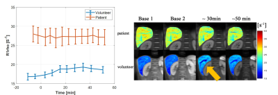 |
Dynamic glucose-enhanced imaging of the liver using
breath-hold black blood quantitative T1rho MRI
Yurui Qian1,
Vincent Wai-Sun Wong2,
Jian Hou1,
Baiyan Jiang1,3,
Xinrong Zhang2,
Grace Lai-Hung Wong2,
Zhigang Wu4,
Queenie Chan5,
Simon Chun Ho Yu1,
Winnie Chiu-Wing Chu1,
and Weitian Chen1
1Imaging and Interventional Radiology, The Chinese University of Hong Kong, Hong Kong, Hong Kong, 2Medical Data Analytics Centre (MDAC), Department of Medicine & Therapeutics, The Chinese University of Hong Kong, Hong Kong, Hong Kong, 3Illuminatio Medical Technology Limited, Hong Kong, Hong Kong, 4Philips Healthcare, Shenzhen, China, 5Philips Healthcare, Hong Kong, Hong Kong Keywords: Liver, Liver Non-alcoholic fatty liver disease (NAFLD) is often associated with abnormal metabolic syndrome. In this study, we investigated dynamic glucose enhanced imaging of the liver using T1rho MRI after glucose ingestion. We hypothesize this approach can be used to assess metabolic activities in the liver. Sixteen young volunteers and four patients with NAFLD were recruited in this study. The preliminary results suggest that the proposed approach has the potential to detect metabolic variations between normal subjects and subjects with fatty liver. |
08:15 |
1216.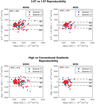 |
Reproducibility of M1-optimized Liver DWI Across Field
Strengths and Gradient Performance
Timothy J Allen1,
Srijyotsna Volety1,
Rianne A van der Heijden2,
Greg Simchick2,
and Diego Hernando1,2
1Medical Physics, University of Wisconsin-Madison, Madison, WI, United States, 2Radiology, University of Wisconsin-Madison, Madison, WI, United States Keywords: Liver, Diffusion/other diffusion imaging techniques Liver DWI using M1-optimized diffusion imaging (MODI) minimizes motion-induced signal dropout while suppressing lesion-mimicking vessel signal. However, early MODI implementations have been confined to 3.0T MR systems with high-performance gradients. The feasibility and reproducibility of MODI DWI on 1.5T systems and systems with conventional gradient performance remains untested. In this work, liver DWI was acquired using conventional and MODI acquisitions in 8 healthy human subjects on 3 separate MR systems with different field strength and gradient performance. ADC estimation with MODI achieved smaller coefficients of variation and reproducibility coefficients across MR systems in comparison to conventional Stejskal–Tanner (monopolar) DWI. |
| 08:15 |
1217.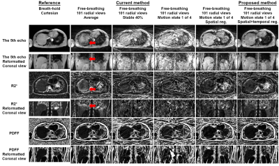 |
Improved Free-Breathing Volumetric Liver Fat and R2*
Quantification Using 3D Stack-of-Radial GRE Dixon MRI and
XD-GRASP Reconstruction
Xiaodong Zhong1,
Marcel D Nickel2,
Stephan A.R. Kannengiesser2,
Brian M Dale3,
Holden H Wu4,
and Vibhas Deshpande5
1MR R&D Collaborations, Siemens Medical Solutions USA, Inc., Los Angeles, CA, United States, 2MR Application Predevelopment, Siemens Healthcare GmbH, Erlangen, Germany, 3MR R&D Collaborations, Siemens Medical Solutions USA, Inc., Cary, NC, United States, 4Department of Radiological Sciences, David Geffen School of Medicine, University of California Los Angeles, Los Angeles, CA, United States, 5MR R&D Collaborations, Siemens Medical Solutions USA, Inc., Austin, TX, United States Keywords: Liver, Quantitative Imaging, Fat, R2*, Iron Respiratory motion compensation is necessary for free-breathing stack-of-radial liver fat and R2* quantification. While self-gating is a valid approach, it may lead to degraded quality of images and quantitative maps and possible prolonged acquisition. A 3D XD-GRASP stack-of-radial technique was developed and evaluated in a motion phantom and in vivo subjects. Results demonstrated PDFF and R2* agreement of the proposed method compared to reference methods. Improved image and map quality and PDFF and R2* quantification agreement of the proposed method using an acceleration factor of 4 (equivalent to 105 seconds of time saving) were observed compared to the self-gating method. |
| 08:15 |
1218.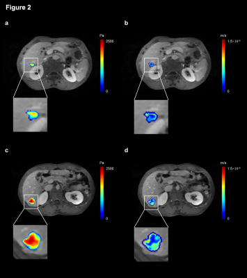 |
Predicting microvascular invasion of HCC using whole-lesion
histogram analysis with the interstitial fluid pressure and
velocity model
Liyun Zheng1,2,3,
Chun Yang1,2,
Yongming Dai4,
and Mengsu Zeng1,2
1Shanghai Institute of Medical Imaging, Shanghai, China, 2Department of Radiology, Zhongshan Hospital, Fudan University, Shanghai, China, 3Shenzhen United Imaging Research Institute of Innovative Medical Equipment, Shenzhen, China, 4MR Collaboration, Central Research Institute, United Imaging Healthcare, Shanghai, China Keywords: Liver, Liver Most solid tumors have increased interstitial fluid pressure (IFP), and the increased IFP is an obstacle to treatment. This study applied a non-invasive dynamic contrast-enhanced magnetic resonance imaging-based model to acquire IFP and interstitial fluid velocity (IFV). Based on the IFP and IFV maps, histogram analysis was applied to evaluate the spatial distributions of pixel gray levels with reduced sampling bias. IFP- and IFV-derived parameters have proven to be significant predictors of microvascular invasion status. Therefore, this optimal model, together with histogram analysis, offers a new way for in vivo and non-invasive assessment of hepatocellular carcinoma. |
| 08:15 |
1219.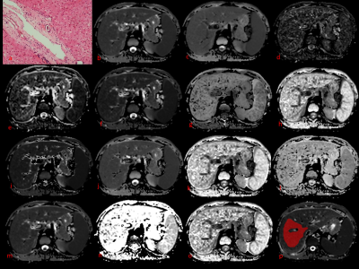 |
Feasibility of integrating 6 diffusion models within a
single acquisition and their performance comparison in liver
fibrosis staging
Yanli Jiang1,2,3,
Fengxian Fan1,3,
Jie Zou1,2,3,
Pin Yang1,3,
Pengfei Wang1,3,
Jing Zhang1,3,
and Shaoyu Wang4
1magnetic resonance imaging department, Lanzho u university second hospital, LANZHOU, China, 2Lanzhou university, LANZHOU, China, 3Gansu Province Clinical Research Center for Functional and Molecular Imaging, LANZHOU, China, 4Siemens Healthineer, ShangHai, China Keywords: Liver, Diffusion/other diffusion imaging techniques DWI has been broadly used in clinical practice, which can provide quantitative parameters. Several DWI models have been used for liver fibrosis staging but diagnostic performance variance in different research because their acquisitions are different and normally difficult to be compared in one study. In this study, we compared 6 DWI models, including 14 parameters in detecting significant liver fibrosis using date from one acquisition. The results showed that the DWI models were similarly valuable in detecting significant liver fibrosis in patients with liver disease. The combined index with the three parameters of CTRW model had the highest AUC. |
| 08:15 |
1220.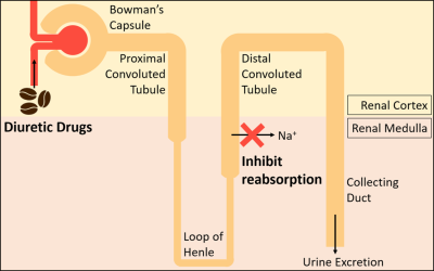 |
Quantitative measurement of renal oxygenation in human using
QSM and pCASL
Yujin Jung1,
Hyun-Seo Ahn1,
and Sung-Hong Park1
1Department of Bio and Brain Engineering, Korea Advanced Institute of Science and Technology, Daejeon, Korea, Republic of Keywords: Kidney, Oxygenation Measuring the renal oxygenation level is important because it is related to the progress of kidney diseases such as chronic kidney disease. In this study, we noninvasively quantified and mapped the renal metabolic rate of oxygen (RMRO2) using quantitative susceptibility mapping and pseudo-continuous arterial spin labeling and evaluated the results using the caffeine challenge. The results demonstrated higher renal oxygenation in the medulla than the cortex, and lower renal oxygenation on the caffeine day compared to the control day, as expected. This technique can be applied to identifying patients at risk of kidney diseases with no contrast agent. |
| 08:15 |
1221.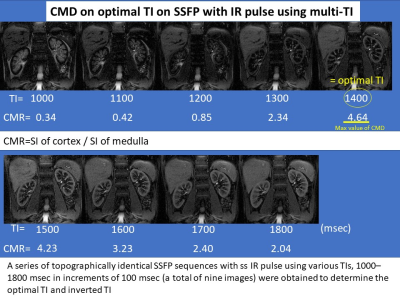 |
Multiparametric MR imaging to identify the changes in
normoalbuminuric diabetic kidney disease (NADKD)
noninvasively
Akira Yamamoto1,
Tsutomu Tamada1,
Yu Ueda2,
Ayumu Kido1,
Atsushi Higaki1,
Akihiko Kanki1,
and Yoshihiko Fukukura1
1Kawasaki Medical School, Kurashiki, Japan, 2Philips Japan, Tokyo, Japan Keywords: Kidney, fMRI This study suggests the possibility that MRI using the values of CMD on optimal TI on SSFP with IR pulse with multi TI, which can sensitively capture fibrotic changes in the renal cortex of NADKD, can be a new parameter to evaluate NADKD non-invasively and in a short period of time. |
| 08:15 |
1222.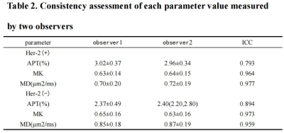 |
The value of amide proton-weighted imaging and diffusion
kurtosis imaging in predicting Her-2 expression in
endometrial cancer
Xiwei Li1,
Shifeng Tian1,
Changjun Ma1,
Nan Wang1,
Jiazheng Wang2,
and Ailian Liu1
1The First Affiliated Hospital of Dalian Medical University, Dalian, China, 2Philips Healthcare,Beijing, Beijing, China Keywords: Cancer, Body Endometrial cancer (EC) is one of the three major malignant tumors in women, and its commonly used treatment is mainly surgery, radiotherapy, chemotherapy and hormone therapy . However, some patients still have low response to the above treatments. However, molecular therapy targeting Her-2 gene has significant value in the clinical diagnosis and improving the prognosis. Amide proton weighted imaging (APTw) and diffusion kurtosis imaging (DKI)can be used for the diagnosis of diseases by reflecting the level of molecular metabolism and cellular microstructure. This study explored the value of the above two sequences in quantitative prediction of HER-2 gene expression levels. |
| 08:15 |
1223.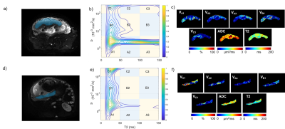 |
Probing Possible Composition Changes of Placenta over
Gestation Age Using Diffusion‐Relaxometry Correlated
Spectroscopy Imaging
Xilong Liu1,
Chantao Huang1,
Wentao Hu2,
Jie Feng1,
Wenjun Qiao1,
Sijin Chen1,
Bo Liu1,
Yongming Dai2,
and Yikai Xu1
1Department of Medical Imaging Center, Nanfang Hospital, Southern Medical University, Guangzhou, China, 2MR Collaboration, Central Research Institute, United Imaging Healthcare, Shanghai, China Keywords: Placenta, Diffusion/other diffusion imaging techniques Currently, antenatal noninvasive imaging techniques to directly evaluate placental function remains challenge. The objective of this study was using diffusion-relaxometry correlated spectrum imaging (DR-CSI) as a tool to probe the possible placental composition changes over gestation in normal pregnancy. Four to five peaks were visible in the D-T2 spectrum for most subjects. DR-CSI compartments were defined according to peak distribution. Significant correlation between DR-CSI compartment volume fractions and GA was found. We suggested that the DR-CSI technique could be used to reveal features of placental composition. |
| 08:15 |
1224.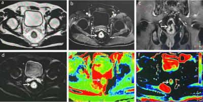 |
The value of glucose-chemical exchange saturation transfer
and amide proton transfer in predicting tumor grading and
staging in rectal cancer
Han Jiang1,
Nan Meng2,
Ziqiang Li1,
Bo Dai2,
Pengyang Feng3,
Yu Luo2,
Zhiwei Shen4,
and Meiyun Wang*2
1Department of Medical Imaging, XinxiangMedical University & Henan Provincial People's Hospital, Zhengzhou, China, 2Department of Medical Imaging, Zhengzhou University People's Hospital & Henan Province People's Hospital, Zhengzhou, China, 3Department of Medical Imaging, Henan University People’s Hospital & Henan Provincial People’s Hospital, Zhengzhou, China, 4Philips healthcare, Beijing, China Keywords: Pelvis, CEST & MT, rectal cancer What are the values of glucose-chemical exchange saturation transfer (glucoCEST) and amide proton transfer (APT) imaging in WHO grading and T staging of rectal cancer? We performed 3D-glucoCEST, 3D-APT, and DWI scans in 21 rectal adenocarcinoma cases. GlucoCEST signal intensity (SI) and 3D-APT SI could distinguish high-grade from low-grade, and T3 stage from T2 stage rectal adenocarcinoma, and both had comparable diagnostic efficacy. In addition, 3D-glucoCEST SI and 3D-APT SI were positively correlated with tumor grade. |
| 08:15 |
1225.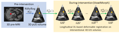 |
3D MRI-US multimodal alignment for real-time intervention -
a tradeoff between accuracy and computation time
Jhimli Mitra1,
Chitresh Bhushan1,
Soumya Ghose1,
David Mills1,
Heather Chan1,
Matthew Tarasek1,
Shane Wells2,
Sydney Jupitz3,
Chris Brace4,
Bryan Bednarz3,
Thomas Foo1,
James Holmes5,
and Desmond Yeo1
1GE Research, Niskayuna, NY, United States, 2Depts. of Radiology and Urology, University of Wisconsin-Madison, Madison, WI, United States, 3Dept. of Medical Physics, University of Wisconsin-Madison, Madison, WI, United States, 4Depts. of Radiology and Biomedical Engineering, University of Wisconsin-Madison, Madison, WI, United States, 5Dept. of Radiology, University of Iowa, Iowa City, IA, United States Keywords: Liver, Multimodal, Real-time multimodal alignment, image-guided intervention, MRI-US deformable fusion Multimodal MRI-US image fusion in image-guided therapy such as liver microwave ablation aids in correct placement of the applicator device. Significant tissue deformation due to breathing motion requires alignment of pre-interventional MRI on real-time interventional US to compensate for the motion. In this work, we present a hybrid framework (conventional and deep learning) for multimodal deformable registration to provide higher accuracy and retain the low latency required for real-time interventions. |
| 08:15 |
1226.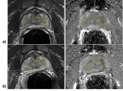 |
Quantitative and Automated MRI Cancer Risk Maps used for
Identification of Prostate Cancer Progression
Matthew Gibbons1,
Janet E Cowan2,
Peter R Carroll2,
Matthew R Cooperberg2,
and Susan M Noworolski1
1Department of Radiology and Biomedical Imaging, University of California, San Francisco, San Francisco, CA, United States, 2Department of Urology, University of California, San Francisco, San Francisco, CA, United States Keywords: Prostate, Cancer This study’s objective was to determine whether automated mpMRI cancer risk maps could identify prostate cancer progression during active surveillance. Derived lesion masks were used to analyze factors for progression. A decision tree model for progression was generated with sensitivity = 0.84, specificity = 0.56, and ROC AUC 0.75. The identification results indicate the potential of mpMRI and MRI cancer risk maps to assist in identifying progression during prostate cancer active surveillance. |
| 08:15 |
1227.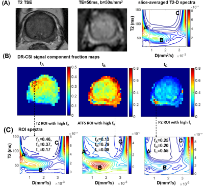 |
Rapid In Vivo Prostate Microstructure MRI using
Diffusion-Relaxation Correlation Spectrum Imaging with
Random Matrix Theory-Based Denoising
Zhaohuan Zhang1,
Shu-Fu Shih1,
Kyunghyun Sung1,
Steven Raman1,
and Holden H. Wu1
1Department of Radiology, University of California, Los Angeles, Los Angeles, CA, United States Keywords: Prostate, Microstructure Diffusion-Relaxation Correlation Spectroscopic Imaging (DR-CSI) has shown promises for quantifying prostate microscopic tissue compartments for prostate cancer characterization, but in vivo DR-CSI faces challenges such as lower signal-to-noise ratio (SNR), and signal averages could lead to prolonged scan time. This work investigated the combination of DR-CSI with random matrix theory-based denoising to take advantage of the large number of TE-b values contrast encodings to improve SNR, and enables rapid in vivo prostate microstructure MRI in <6min at 3T. |
 Back to Meeting Home
 Back to the Program-at-a-Glance
The International Society for Magnetic Resonance in Medicine is accredited by the Accreditation Council for Continuing Medical Education to provide continuing medical education for physicians.
 Back to Meeting Home
 Back to the Program-at-a-Glance
 Full Session Video