Power Pitch
Pitch: Cardiovascular Liquid Dynamics: Advanced Flow & Angiography
ISMRM & ISMRT Annual Meeting & Exhibition • 03-08 June 2023 • Toronto, ON, Canada

Power Pitch
Pitch: Cardiovascular Liquid Dynamics: Advanced Flow & Angiography
15:45 |
1069.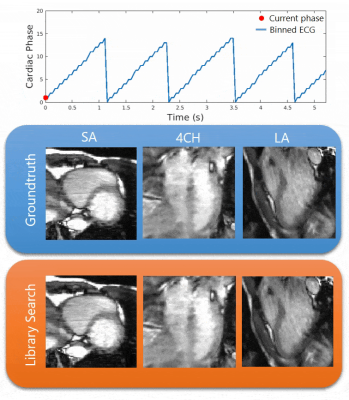 |
Cardiac Library Based Real-time Imaging (CALIBRI): towards
3D cardiac visualization during MR-guided cardiac
interventions
Anne Spakman1,
Eric M Schrauben1,
Renske Merton1,
Gustav J Strijkers2,
Aart J Nederveen1,
Marco JW Götte3,
and Bram F Coolen2
1Department of Radiology & Nuclear Medicine, Amsterdam UMC, Amsterdam, Netherlands, 2Department of Biomedical Engineering & Physics, Amsterdam UMC, Amsterdam, Netherlands, 3Department of Cardiology, Amsterdam UMC, Amsterdam, Netherlands Keywords: Arrhythmia, MR-Guided Interventions, Real-time imaging We propose Cardiac Library Based Real-time Imaging (CALIBRI), which allows the use of the same data for both catheter tracking and real-time visualization of cardiac motion states. This is achieved by matching the acquired k-lines with a library of pre-acquired cardio/respiratory gated 3D CINE data. In this study, we implemented our novel method on a 3T MRI system and showed the feasibility of this approach in multiple healthy volunteers. |
| 15:45 |
1070.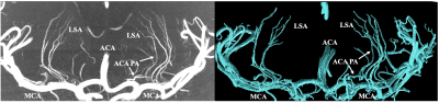 |
Automatic 3D Segmentation of perforating arteries from
ultra-high resolution 7T Compressed Sensing MRA images.
Qingle Kong1,
Zhe Zhang2,3,
and Jing Jing2,3,4
1MR Collaboration, Siemens Healthineers Ltd., Beijing, China, 2Tiantan Neuroimaging Center of Excellence, Beijing, China, 3Beijing Tiantan Hospital, Capital Medical University, China National Clinical Research Centre for Neurological Diseases, Beijing, China, 4Beijing Tiantan Hospital, Capital Medical University, Neurology Department, Beijing, China Keywords: Vessels, Segmentation, perforating arteries segmentation The evaluation of perforating arteries is important for the diagnosis of small vessel disease. It’s challenging to image perforating arteries because of their small caliber size, which requires an extremely high resolution. Moreover, to determine pathological changes, the vessel trees need to be segmented and quantified. In this study, an automatic 3D Segmentation method was introduced and perforating arteries around the Circle of Willis were segmented and quantified. The number of stems of perforators was counted and compared based on segmentation results and MIPs images. The results revealed that the segmentation method robustly achieved the segmentation of perforators. |
| 15:45 |
1071.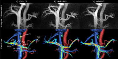 |
Abbreviated Three Minute 4D Flow MRI of the Portal
Circulation: Prospective Evaluation in Obese Subjects
Thekla Helene Oechtering1,2,
AMK Muntasir Shamim3,
David Harris4,
Nikolaos Panagiotopoulos1,2,
Alma Spahic5,
Oliver Wieben1,6,
Kevin M Johnson1,3,
Alejandro Roldán-Alzate1,7,8,
and Scott B Reeder1,6,7,8,9
1Department of Radiology, University of Wisconsin-Madison, Madison, USA, WI, United States, 2Department of Radiology and Nuclear Medicine, Universität zu Lübeck, Lübeck, Germany, 3Department of Electrical and Computer Engineering, University of Wisconsin-Madison, Madison, USA, WI, United States, 4University of Wisconsin-Madison, Madison, USA, WI, United States, 5Department of Medical Physics, University of Wisconsin-Madison, Madison, USA, WI, United States, 6Department of Medical Physics, University of Wisconsin-Madison, Madison, WI, United States, 7Department of Mechanical Engineering, University of Wisconsin-Madison, Madison, WI, United States, 8Department of Biomedical Engineering, University of Wisconsin-Madison, Madison, WI, United States, 9Department of Emergency Medicine, University of Wisconsin-Madison, Madison, WI, United States Keywords: Flow, Liver Constant, non-pulsatile flow in the portal venous system offers the possibility of time-averaged reconstruction for shortening the acquisition time. We acquired both a 10min and 3min radial 4D flow MRI (PCVIPR) in 10 subjects in a test-retest paradigm. Time-averaged 10min and 3min exams were compared to the reference standard, i.e., cardiac-phase resolved 10min exam (14 timeframes). All datasets were analyzable with quality rated as “good” for all 3min exams. Small vessels were best visualized in the time-averaged 10min data. Conservation of mass analysis and test-retest repeatability yielded excellent results for all reconstructions. Quantitative results agreed well between reconstructions. |
| 15:45 |
1072.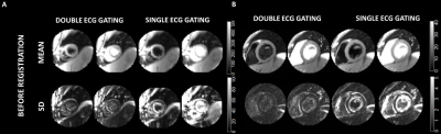 |
Effects of double-ECG gating in synchronized breathing
myocardial arterial spin labeling
Verónica Aramendía-Vidaurreta1,2,
Sergio M. Solís-Barquero1,2,
Marta Vidorreta3,
Ana Ezponda1,2,
Gorka Bastarrika1,2,
and María A. Fernández-Seara1,2
1Radiology, Clínica Universidad de Navarra, Pamplona, Spain, 2IdiSNA, Instituto de Investigación Sanitaria de Navarra, Pamplona, Spain, 3Siemens Healthineers, Madrid, Spain Keywords: Heart, Perfusion Myocardial perfusion can be quantitatively measured noninvasively using arterial spin labeling (ASL). Motion due to the cardiac cycle is tackled with the use of single- or double-ECG gating. The goals of this study were to investigate the performance of double-gating, in synchronized breathing myocardial ASL with presaturation pulses by comparison with single-gating, and to compare the different quantification strategies for double-gated data. This study showed that double-gating is more robust to heart rate variability than single-gating in synchronized breathing ASL sequences with presaturation pulses, but accurate saturation-recovery fitting requires the acquisition of several baseline images. |
| 15:45 |
1073.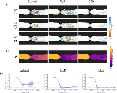 |
Joint Velocity and Acceleration Encoding for Improved
Pressure Gradient Mapping in Flow Imaging
Michael Loecher1,2,
Priya J Nair3,
and Daniel B Ennis1,2
1Radiological Sciences Laboratory, Stanford University, Stanford, CA, United States, 2Radiology, Veterans Administration Health Care System, Palo Alto, CA, United States, 3Bioengineering, Stanford University, Stanford, CA, United States Keywords: Flow, Velocity & Flow In this work, we introduce an optimized motion encoding strategy for flow measurements that simultaneously and non-colinearly encodes velocity and acceleration into data phase. This enables velocity-based flow measurements and improves pressure gradient mapping derived from the acceleration data. The method is tested in a stenotic flow phantom, where the proposed method shows good velocity agreement with conventional 4D-flow methods, while also producing improved pressure gradient maps when compared to both 4D-flow and a CFD reference. |
| 15:45 |
1074.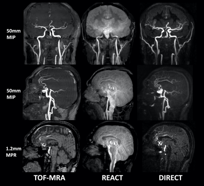 |
DIRECT: non-contrast-enhanced relaxation-based
flow-independent intracranial MR angiography using modified
REACT
Masami Yoneyama1,
Takayuki Sakai2,
Yasuhiro Goto3,
Masanobu Nakamura1,
Daichi Murayama2,
Michinobu Nagao4,
Shuo Zhang5,
Kayoko Abe4,
Yutaka Hamatani3,
Kazuo Kodaira3,
Takumi Ogawa3,
Mana Kato3,
Isao Shiina3,
and Marc Van Cauteren6
1Philips Japan, Tokyo, Japan, 2Department of Radiology, Eastern Chiba Medical Center, Chiba, Japan, 3Department of Radiological Services, Tokyo Women's Medical University, Tokyo, Japan, 4Department of Diagnostic Imaging and Nuclear Medicine, Tokyo Women’s Medical University, Tokyo, Japan, 5Philips Healthcare, Hamburg, Germany, 6Philips Healthcare, Best, Netherlands Keywords: Vessels, Blood vessels A new non-contrast-enhanced, relaxation-based, flow-independent MRA method, called Relaxation-Enhanced Angiography without ContrasT (REACT) has recently been proposed for vascular imaging. One of the major limitations of REACT is that bright signal of long-T1 fluids such as CSF is visible, which can obscure vascular structure in the intracranial vessels. We developed a new sequence, called DIRECT (Dual-Inversion REaCT) with modified magnetization preparation scheme of REACT to suppress CSF signals while maintain the high signal intensity from vessels. DIRECT enables robust and high-quality whole-brain intracranial MR angiography with uniform fat, CSF and brain parenchyma suppression within clinically feasible scan time. |
| 15:45 |
1075.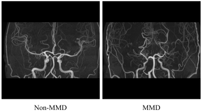 |
Automatic identification of Moyamoya disease based on
time-of-flight MR angiography
Zheng Tan1,
Mingming Lu2,
Shuai Liu1,
Shitong Liu2,
Hongtao Zhang2,
Xiaoying Tang1,
Jianming Cai2,
and Fei Shang1
1Department of Biomedical Engineering, School of Life Science, Beijing Institute of Technology, Beijing, China, 2Department of Radiology, The Fifth Medical Center of PLA General Hospital, Beijing, China Keywords: Vessels, Stroke Moyamoya disease (MMD) is a rare chronic progressive cerebrovascular disease that causes strokes. For the diagnosis of MMD, time-of-flight magnetic resonance angiography (TOF-MRA) can be an alternative to digital subtraction angiography (gold standard) owing to its non-invasive and radiation-free attributes. In this study, the deep learning method (ResNet-50) was used for MMD automatic diagnosis on the maximum intensity projection images from 3D TOF-MRA, and five-fold cross-validation was used for validation. The method exhibits the accurate ability (AUC: 0.990 ± 0.008, accuracy: 0.933 ± 0.063) to identify MMD and has the potential to improve the clinical management of MMD. |
| 15:45 |
1076.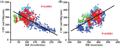 |
Toward accurate cerebral blood flow estimation in mice after
accounting for anesthesia
Zhiliang Wei1,2,
Yuguo Li1,2,
Adnan Bibic2,
Jiadi Xu1,2,
and Hanzhang Lu1,2,3
1Russell H. Morgan Department of Radiology and Radiological Science, Johns Hopkins University School of Medicine, Baltimore, MD, United States, 2F. M. Kirby Research Center for Functional Brain Imaging, Kennedy Krieger Research Institute, Baltimore, MD, United States, 3Department of Biomedical Engineering, Johns Hopkins University School of Medicine, Baltimore, MD, United States Keywords: Flow, Animals CBF measurements in mice were often confounded by the utilizations of anesthesia, which are generally vasoactive. Here, we aimed to systematically understand the relationships between CBF and related physiological factors, not only the anesthesia dose but also respiratory rate (RR), heart rate (HR), and exposure time to anesthesia. We found that CBF measurements in mice were affected by anesthesia dose and time, but can be corrected by using respiratory rate and heart rate. The correction scheme will facilitate applications of CBF measurements in mechanistic understanding of vascular diseases and preclinical therapeutic trials by providing anesthesia-independent CBF measurements. |
| 15:45 |
1077.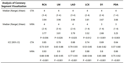 |
Comparative analysis of image quality by 3.0 T Non-contrast
Free-breathing Whole-heart CMRA with CCTA in patients with
coronary artery stenosis
Ping Tian1,
Ying Liu1,
Jianxiu Lian2,
Minwen Zheng1,
Jingji Xu1,
and Jianmin Zheng1
1Department of Radiology, The First Affiliated Hospital of Air Force Medical University, Xi’an, China, 2Philips Healthcare, Beijing, China Keywords: Vessels, Myocardium, Coronary magnetic resonance angiography Atherosclerotic lesions in coronary vessels cause vascular lumen stenosis or obstruction, resulting in myocardial ischemia, hypoxia or necrosis, which is an important cause of coronary heart disease. This study was designed to evaluate the performance of coronary magnetic resonance angiography(CMRA) sequence in volunteer subjects. The ability visualization of coronary Main Branch segments were no statistically significant difference in MRA coronary quality scores compared with CTA(p>0.05) with sensitivity, specificity for per patient were 86%, 85%, respectively. The coronary artery visualization by CMRA can be used to detect clinically significant coronary artery stenosis in patients with suspected coronary heart disease. |
| 15:45 |
1078.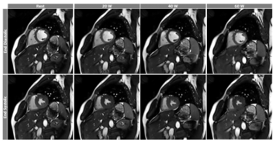 |
Biventricular and Hemodynamic Assessment under Multi-stage
Exercise using Real-time CMR
Preethi S Chandrasekaran1,
Chong Chen2,
Yingmin Liu1,
Christopher Crabtree3,
Syed Murtaza Arshad4,
Matthew Tong5,
Yuchi Han5,
and Rizwan Ahmad2
1Davis Heart and Lung Research Institute, Ohio State University, Columbus, OH, United States, 2Biomedical Engineering, Ohio State University, Columbus, OH, United States, 3Department of Human Sciences, Ohio State University, Columbus, OH, United States, 4Electrical and Computer Engineering, Ohio State University, Columbus, OH, United States, 5Cardiovascular Medicine, Ohio State University, Columbus, OH, United States Keywords: Heart, Velocity & Flow, Exercise CMR A comprehensive exercise stress cardiovascular MRI (Ex-CMR) was performed in twelve healthy subjects. Biventricular quantification was performed from real-time cine, while hemodynamic parameters in the ascending aorta and main pulmonary artery were estimated using real-time flow. The acquisition process was repeated two to three times at increasing exercise intensities. The highly accelerated (R = 7-8 for cine and R = 16 for flow) real-time data were reconstructed inline using Gadgetron-based compressed sensing reconstruction. In agreement with the literature, the ejection fraction and cardiac output correlated positively with exercise intensity. |
| 15:45 |
1079.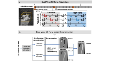 |
Dual Venc 5D flow MRI with Increased Velocity Dynamic Range:
An in-vitro and in-vivo Validation and Feasibility Study
Elizabeth Weiss1,
Justin Baraboo1,
Liliana Ma1,
Mariana B. L. Falcão2,
Christopher W. Roy2,
Matthias Stuber2,
and Michael Markl1
1Radiology, Northwestern University, Chicago, IL, United States, 2University of Lausanne (CHUV), Lausanne, Switzerland Keywords: Flow, Pulse Sequence Design We illustrate a pilot study of the first implementation of dual-venc 5D flow MRI. We found excellent voxel-wise agreement with single-venc 5D flow in a pulsatile phantom. In two healthy controls, we find good agreement, but identify unexpected aliasing in the dual venc 5D flow which requires further investigation. |
| 15:45 |
1080.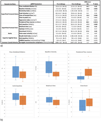 |
Acute Effects of Electronic and Tobacco Cigarette Aerosol
Inhalation on Vascular Function Detected at Quantitative MRI
Marianne Nabbout1,
Michael C Langham1,
Alessandra S Caporale1,2,3,
Wensheng Guo4,
and Felix W Wehrli1
1Department of Radiology, University of Pennsylvania, Philadelphia, PA, United States, 2Department of Neurosciences, Imaging and Clinical Sciences, G. d’Annunzio University’ of Chieti-Pescara, Chieti, Italy, 3Institute for Advanced Biomedical Technologies (ITAB), G. d’Annunzio University’ of Chieti-Pescara, Chieti, Italy, 4Department of Biostatistics, Epidemiology and Informatics, University of Pennsylvania, Philadelphia, PA, United States Keywords: Vessels, Blood vessels, Vascular Reactivity, Oxygenation, Blood Velocity To assess the acute effects of tobacco and electronic cigarette inhalation on vascular function, multiple MRI markers were analyzed in a study targeting peripheral, central and neurovascular beds of healthy young subjects. Smokers and vapers (9 subjects for a total N=15 study visits), ages 22 to 45 years, underwent two MRI scans, with a smoking or vaping challenge in between. Data from smoking/vaping challenges were combined to assess for their pooled effect. Smoking/vaping had significant acute effects on peripheral reactivity, notably on the superficial femoral artery flow-mediated dilation, baseline velocity and superficial femoral vein baseline oxygen saturation, among others. |
| 15:45 |
1081.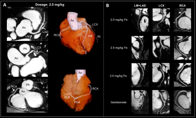 |
3.0T Whole-Heart Contrast‑Enhanced Coronary Magnetic
Resonance Angiography Using Ferumoxytol: A Feasibility Study
Yuehong Liu1,
Bin Sun2,
Xinyu Wang1,
Chen Zhang3,
Xuewei Fan4,
Zhenyu Li4,
Jing An5,
and Qi Yang1
1Beijing Chaoyang Hospital, Capital Medical University, Beijing, China, Beijing, China, 2Fujian Medical University Union Hospital, Fuzhou, China, 3MR Scientific Marketing, Siemens Healthcare, Beijing, China, 4Central Hospital Affiliated to Xinxiang Medical University, Xinxiang, China, 5Siemens Shenzhen Magnetic Resonance Ltd., Shenzhen, China Keywords: Vessels, Blood vessels, Ferumoxytol This study suggested the feasibility of combining low-dose ferumoxytol (2.0-3.0 mg/kg) with ECG-triggered, navigator-gated, inversion-recovery prepared, segmented gradient-echo sequence to obtain high-quality sub-millimeter 3D whole-heart CMRA images under free-breathing for approximately 7 mins on MR 3.0T scanner. Compared to gadobenate, ferumoxytol produced more intense and prolonged cardiac vessel enhancement (24-36 hours) Ferumoxytol could help to reduce acquisition time of CMRA while maintaining a high signal-to-noise ratio, great image quality, and clear delineation of the coronary arteries. This study of healthy volunteers also suggested that ferumoxytol-enhanced CMRA has the potential to identify patients without coronary artery disease. |
| 15:45 |
1082.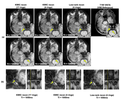 |
Improved Simultaneous Intracranial Vessel Wall Imaging and
dynamic MRA Using Low-rank Reconstruction
Kaiyu Zhang1,
Zhensen Chen2,
Yin Guo1,
Xin Wang3,
Gador Canton4,
Niranjan Balu4,
Thomas Hatsukami5,
Chun Yuan4,6,
and Xiaodong Ma6
1Department of Bioengineering, University of Washington, Seattle, WA, United States, 2Institute of Science and Technology for Brain-Inspired Intelligence, Fudan University, Shanghai, China, 3Department of Electrical Engineering, University of Washington, Seattle, WA, United States, 4Department of Radiology, University of Washington, Seattle, WA, United States, 5Department of Surgery, University of Washington, Seattle, WA, United States, 6Department of Radiology and Imaging Sciences, University of Utah, Salt Lake City, UT, United States Keywords: Vessel Wall, Blood vessels Acquiring multi-contrast images of intracranial vessels from one single MRI sequence is beneficial for comprehensive neurovascular disease diagnosis. Vessel wall (VW) images and dynamic MRA (dMRA) can be obtained from a time-efficient multi-contrast sequence named iSNAP. However, the previously adopted k-space sharing reconstruction method generated high-quality dMRA but low-quality VW images. In this study, we aim to develop a novel low-rank-based image reconstruction method to reconstruct VW and dMRA from highly undersampled 4D MRI. High quality VW images with sharper wall delineation are obtained, while hemodynamic information from dMRA is preserved. |
| 15:45 |
1083.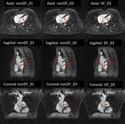 |
Free-running 3D-CINE MRI of patients with congenital heart
disease using inter-bin compensation of cardiac motion
Bastien Milani1,
Christopher Roy1,
Jean-Baptist Ledoux1,
David C. Rotzinger1,
Salim Si-mohamed1,2,3,
Ambra Masi1,
Jerome Yerly1,4,
Tobias Rutz1,
Milan Prsa1,
Jurg Schwitter1,
and Matthias Stuber1,4
1Lausanne University Hospital (CHUV) and University of Lausanne (UNIL), Lausanne, Switzerland, 2INSA-Lyon, CNRS, Inserm, CREATIS, Université de Lyon,, Villeurbanne, France, 3Louis Pradel Hospital, Hospices Civils de Lyon, Bron, France, 4Center for Biomedical Imaging (CIBM), Lausanne, Switzerland Keywords: Heart, Cardiovascular, ejection fraction We present in this work a 3D-CINE whole-heart reconstruction that we developed for free-running 3D‑radial fully self-gated acquisitions. The reconstruction is compressed-sensing-based with temporal-total-variation (tTV) regularization, which is known to corrupt or compress motion and to blur moving structures. In order to solve these drawbacks, we regularize by an improved tTV equal to the one-norm of the sum of the motion-corrected-residuals between adjacent frames. While this strategy has already been applied for various trajectories, it has never been applied for 3D-radial in free-running. In this study, we demonstrate quantitatively and qualitatively that this strategy in fact improves image quality. |
| 15:45 |
1084.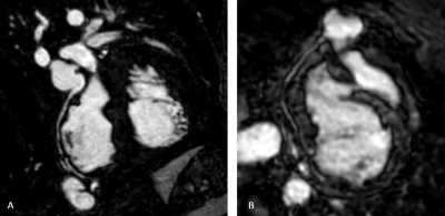 |
Ferumoxytol Dose Optimization for 3D Whole-Heart Congenital
Heart Disease Imaging
Sanja Dzelebdzic1,
Maher Abadeer1,
Gerald Greil1,
and Tarique Hussain1
1Cardiac MRI, Children's Medical Center, UT Southwestern, Dallas, TX, United States Keywords: Heart, Cardiovascular, image quality, ferumoxytol, contrast With its concomitant ability to shorten the T1 relaxivity of blood and long intravascular half-life, ferumoxytol has become a popular “blood pool” contrast agent. The optimum contrast dose is not known. Reducing the ferumoxytol dose to 2 mg/kg is an efficient method to optimize the image quality, diagnostic performance, and achieve reduction in Gibbs’ truncation artefact. |
| 15:45 |
1085.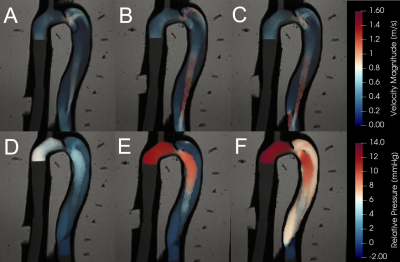 |
Validation of Non-Invasive Relative Pressure Mapping by 4D
Flow MRI in Aortic Dissection
Brandon K. Hardy1,
Judith Zimmermann2,
Nicholas S. Burris1,3,
Daniel B. Ennis4,
David Marlevi5,6,
and David A. Nordsletten1
1Dept. of Biomedical Engineering, University of Michigan, Ann Arbor, MI, United States, 2Dept. of Radiology and Biomedical Imaging, University of California San Francisco, San Francisco, CA, United States, 3Dept. of Radiology, University of Michigan, Ann Arbor, MI, United States, 4Dept. of Radiology, Stanford University, Stanford, CA, United States, 5Dept. Molecular Medicine and Surgery, Karolinska Institutet, Stockholm, Sweden, 6Institute for Medical Engineering and Science, Massachusetts Institute of Technology, Cambridge, MA, United States Keywords: Quantitative Imaging, Blood vessels, Pressure Estimation False lumen (FL) growth rate in type B aortic dissection (TBAD) is correlated with FL pressurization. 4D flow MRI allows for quantitative assessment of three-dimensional flow, including spatial mapping of relative pressure. Despite its promise, 4D flow relative pressure mapping has not yet been validated in a TBAD-specific context. Here, we validate a state-of-the-art proposed Stokes Estimator (STE) of relative pressure against catheter measurements using three physiologically accurate TBAD flow phantoms and subsequently analyze the method’s sensitivity to image noise. STE pressure estimates closely matched catheter data and were robust to noise, indicating STE’s potential for TBAD treatment planning. |
| 15:45 |
1086.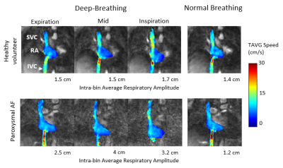 |
Investigation of the effect of deep-breathing on the caval
circulation in patients with cardiac rhythm disorders using
5D radial Flow MRI
Sara Boccalini1,
Marta Beghella2,
Loic Boussel1,2,
Philippe Douek2,3,
Philippe Chevalier4,
Claudia Prieto5,
and Monica Sigovan6
1Department of Radiology, HCL, Lyon, France, 2CREATIS, Lyon, France, 3Departement of Radiology, HCL, Lyon, France, 4Department of Cardiac Rythm, HCL, Lyon, France, 5School of Biomedical Engineering and Imaging Sciences, King’s College London, London, United Kingdom, 6CNRS, CREATIS Lab, Lyon, France Keywords: Quantitative Imaging, Velocity & Flow Respiratory motion effects thoracic blood flow mainly by intrathoracic pressure changes. In addition, deep-inspirations strongly increase the systemic venous return with immediate repercussions on right ventricular stroke volume. Simultaneously, pulmonary resistance is increased leading to decreased inflow in the left chambers, which is compensated in the following heartbeats. In patients with atrial myopathy (ex. atrial fibrillation), these adjustments might be more difficult. Our aim was to assess the impact of deep breathing on hemodynamic parameters in patients with atrial fibrillation, paroxysmal and permanent, and in a healthy population as compared to normal breathing. |
| 15:45 |
1087.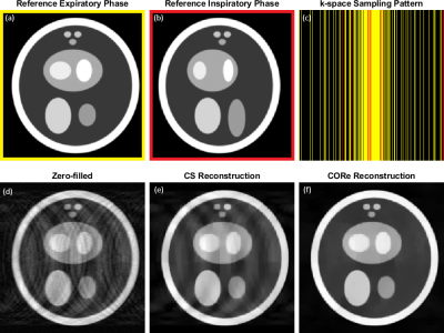 |
Motion artifact reduction in self-gated CMR 4D flow imaging
under exercise stress
Syed Murtaza Arshad1,
Chong Chen2,
Yingmin Liu3,
Preethi Chandrasekaran3,
Christopher Crabtree4,
Ning Jin5,
and Rizwan Ahmad2
1Department of Electrical & Computer Engineering, The Ohio State University, Columbus, OH, United States, 2Department of Biomedical Engineering, The Ohio State University, Columbus, OH, United States, 3Davis Heart and Lung Research Institute, The Ohio State University, Columbus, OH, United States, 4Department of Human Sciences, The Ohio State University, Columbus, OH, United States, 5Cardiovascular MR R&D, Siemens Medical Solutions Inc, Columbus, OH, United States Keywords: Flow, Image Reconstruction, 4D Flow Free-breathing self-gated CMR 4D flow imaging using traditional Compressed Sensing (CS) methods invariably contains motion artifacts due to the inaccuracy of self-gating signal. Self-gating signal degrades even further in the case of exercise stress imaging due to excessive movement of the subject. We propose Compressive recovery with Outlier Rejection (CORe) to reduce the motion artifacts. Using data from a 2D digital phantom and 4D flow data under rest and stress conditions, we demonstrate that CORe is effective in suppressing motion artifacts while maintaining agreement with 2D-PC based flow quantification. |
| 15:45 |
1088.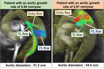 |
Evaluation of hemodynamic parameters for prediction of
aortic growth in patients with chronic Stanford type B
aortic dissection using 4D flow MRI.
Satoshi Higuchi1,
Hideki Ota1,
Ryuichi Mori2,
Yuki Ichinoseki2,
Hiroki Kamada1,
and Kei Takase1
1Diagnostic Radiology, Tohoku University Hospital, Sendai, Japan, 2Radiology, Tohoku University Hospital, Sendai, Japan Keywords: Flow, Vessels, Aortic Dissection Uncomplicated type B aortic dissection (AD) has a poor long-term outcome and further optimization of predictors for aortic expansion is required. Seventeen patients with chronic type B AD who underwent 4D flow MRI were included and divided into two groups based on the aortic growth rate. The morphological and hemodynamic parameters in each group were retrospectively analyzed. The forward flow and volume in true lumen and ratio of these parameters at entry and false lumen to true lumen were significantly higher in the fast growth-rate group than in the slow one; no morphological parameters showed a significant difference. |
 Back to Meeting Home
 Back to the Program-at-a-Glance
The International Society for Magnetic Resonance in Medicine is accredited by the Accreditation Council for Continuing Medical Education to provide continuing medical education for physicians.
 Back to Meeting Home
 Back to the Program-at-a-Glance
 Full Session Video