Power Pitch
Pitch: Relaxometry & Multicontrast MRI: From Cancer to White Matter & More
ISMRM & ISMRT Annual Meeting & Exhibition • 03-08 June 2023 • Toronto, ON, Canada

Power Pitch
Pitch: Relaxometry & Multicontrast MRI: From Cancer to White Matter & More
13:45 |
1348.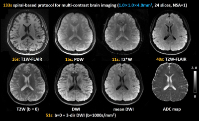 |
Spiral-based multi-contrast imaging protocol for a full
brain MR exam in 2 minutes
Guangqi Li1,
Yajing Zhang2,
and Hua Guo1
1Center for Biomedical Imaging Research, Department of Biomedical Engineering, School of Medicine, Tsinghua University, Beijing, China, 2MR Clinical Science, Philips Health Technology (China), Beijing, China Keywords: Multi-Contrast, Brain Fast multi-contrast brain exams are highly desirable in clinical practice. Spiral sampling has high efficiency in terms of spatial encoding. Thus it holds great potential for many MRI applications. In this work, we optimized a fast multi-contrast brain imaging protocol based on multi-shot spiral acquisitions. Six contrasts (T1W-FLAIR, T2W, PDW, T2*W, T2W-FLAIR, DWI) and apparent diffusion coefficient (ADC) maps with an in-plane resolution of 1.0 mm2 for a full brain MR exam can be obtained in about 2 minutes. |
| 13:45 |
1349. |
Decoding the phase-cycled bSSFP signal for maximized
parameter quantification - T1, T2, proton density and
magnetic field inhomogeneity
Nils Marc Joel Plähn1,
Adèle Mackowiak2,
Berk Açikgöz3,
Eva Peper3,
Giulia Rossi2,
and Jessica Bastiaansen3
1Department of Diagnostic, Interventional and Pediatric Radiology (DIPR), Inselspital, Bern Universit, Bern, Switzerland, 2Department of Radiology, Lausanne University Hospital (CHUV) and University of Lausanne (UNIL), Lausanne, Switzerland, 3Department of Diagnostic, Interventional and Pediatric Radiology (DIPR), Inselspital, Bern University Hospital, University of Bern, Bern, Switzerland Keywords: Relaxometry, Data Analysis, phase-cycled bSSFP, T1-quantification, T2-quantification, B0 quantification, proton-density quantification, multi-parameter quantification A novel analytical method for off-resonance encoded parameter quantification of voxel-wise phase-cycled bSSFP signal profiles was developed. The approach conserves both magnitude and phase information in a complete, linearized, and compact way. A framework for ultra-rapid and simultaneous $$$T_1$$$, $$$T_2$$$, $$$B_0$$$ inhomogeneity and proton density quantification were developed. The approach was validated in simulations, phantom and in vivo knee experiments and compared with gold-standard reference measurements when available. Simulations and experiments validated the proposed method for multi-parameter quantification with high accuracy and precision, providing the first step towards novel analytical quantification possibilities with phase-cycled bSSFP. |
| 13:45 |
1350.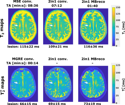 |
Rapid PD, T2, and T2* Mapping with 2in1-RARE-EPI and
Model-Based Reconstruction in Multiple Sclerosis
Jose Raul Velasquez Vides1,
Carl J. J. Herrmann1,2,
Ludger Starke1,3,
Hampus Olsson1,
and Thoralf Niendorf1,4
1Berlin Ultrahigh Field Facility (B.U.F.F), Max Delbrück Center for Molecular Medicine in the Helmholtz Association, Berlin, Germany, 2Department of Physics, Humboldt University of Berlin, Berlin, Germany, 3Digital Health - Machine Learning Research Group, Digital Health Center, Hasso Plattner Institute, University of Potsdam, Potsdam, Germany, 4Experimental and Clinical Research Center (ECRC), a joint cooperation between the Charité Medical Faculty and the Max Delbrück Center for Molecular Medicine in the Helmholtz Association, Berlin, Germany Keywords: Relaxometry, Multiple Sclerosis One obstacle of established quantitative resonance imaging methods is the excessive scan time. This study examines the use of radially-sampled 2in1-RARE-EPI in conjunction with model-based reconstruction methods for accelerated and simultaneous PD, T2, and $$$T_{2}^{*}$$$ mapping. We demonstrate that this approach facilitates a substantial decrease in acquisition time (01:40 vs. 07:12 min) of 2in1-RARE-EPI without impairing the quality of the parametric maps. Our patient data demonstrate the applicability of model-based reconstructed 2in1-RARE-EPI for T2 and $$$T_{2}^{*}$$$ mapping of multiple sclerosis lesions.
|
| 13:45 |
1351. |
Delta-relaxometry with contrast-enhanced MR Fingerprinting:
phantom validation and application to tumor imaging
Shengwen Deng1,
Walter Zhao2,3,
David W. Jordan1,
Chaitra Badve 1,4,
and Dan Ma2
1Department of Radiology, Case Western Reserve University and University Hospitals Cleveland Medical Center, Cleveland, OH, United States, 2Department of Biomedical Engineering, Case Western Reserve University, Cleveland, OH, United States, 3Case Western Reserve University School of Medicine, Cleveland, OH, United States, 4Seidman Cancer Center and Case Comprehensive Cancer Center,, Cleveland, OH, United States Keywords: Relaxometry, MR Fingerprinting, Delta Relaxometry; Tumor Imaging The influence of hemodynamics and contrast concentration can be eliminated with ratios between delta-relaxometry in contrast-enhanced MR Fingerprinting (MRF). This delta ratio can be used to characterize the in vivo contrast-specific tissue response, beyond the conventional T1/T2 shortening effect. In this abstract, we: 1) developed a MRF-based strategy to image the concentration-independent, contrast-specific tissue response using delta-relaxometry; 2) validated reproducibility and linearity of delta-relaxometry in phantom experiments; 3) reported the novel Delta-relaxometry image contrast distinct from current clinical image contrasts; and 4) illustrated the sensitivity of delta-relaxometry in brain tumor characterization and classification. |
| 13:45 |
1352.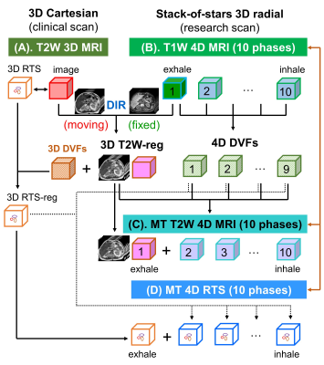 |
Real-time Multi-Contrast 4D MRI using Motion Transfer for
Low-Latency Volumetric Motion Tracking on a 1.5T MR-Linac
System
Can Wu1,
Victor Murray1,
Syed Siddiq1,
Neelam Tyagi1,
Marsha Reyngold2,
Christopher Crane2,
and Ricardo Otazo1,3
1Department of Medical Physics, Memorial Sloan Kettering Cancer Center, New York, NY, United States, 2Department of Radiation Oncology, Memorial Sloan Kettering Cancer Center, New York, NY, United States, 3Department of Radiology, Memorial Sloan Kettering Cancer Center, New York, NY, United States Keywords: Multi-Contrast, Cancer Real-time multi-contrast 4D MRI is proposed by transferring motion from 4D T1-weighted images to 3D T2-weighted images and performing fast signature matching based on T1-weighted 3D radial stack-of-stars acquisitions. The proposed approach exploits the anatomical correlations between T1-weighted and T2-weighted images and fast acquisition of T1-weighted data to generate real-time multi-contrast volumetric motion information for adaptation and monitoring of radiation treatment of tumors affected by respiratory motion on an MR-Linac system. The feasibility of the proposed method was demonstrated on patients with pancreatic cancer. |
| 13:45 |
1353.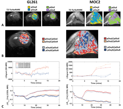 |
Oxygen-enhanced and glucose-enhanced MRI for dual mapping of
hypoxia and glucose uptake in tumours at high spatial
resolution
Ben Dickie1,2,
Thomas Kisby3,
Emily Rowling4,
Julius Chung5,
Mohammad Babur4,
Lidan Christie1,
Tao Jin5,
Kostas Kostarelos3,6,
Kaye Williams4,
and James O'Connor7
1Division of Informatics, Imaging and Data Sciences, Faculty of Biology, Medicine and Health, University of Manchester, UK, Manchester, United Kingdom, 2Geoffrey Jefferson Brain Research Centre, University of Manchester, Manchester Academic Health Science Centre, UK, Manchester, United Kingdom, 3Nanomedicine Lab, Faculty of Biology, Medicine and Health and National Graphene Institute, University of Manchester, UK, Manchester, United Kingdom, 4Division of Pharmacy and Optometry, School of Health Sciences, Faculty of Biology, Medicine and Health, University of Manchester, UK, Manchester, United Kingdom, 5Department of Radiology, University of Pittsburgh, Pittsburgh, Pennsylvania, USA, Pittsburgh, PA, United States, 6Catalan Institute of Nanoscience and Nanotechnology (ICN2), UAB Campus Bellaterra, Barcelona, Spain, Barcelona, Spain, 7Division of Cancer Sciences, School of Health Sciences, Faculty of Biology, Medicine and Health, University of Manchester, UK, Manchester, United Kingdom Keywords: Multi-Contrast, Cancer We have developed a non-invasive imaging protocol for dual assessment of hypoxia and glucose uptake in tumours using oxygen-enhanced MRI and glucoCESL MRI, and test feasibility in GL261 glioblastoma and MOC2 head and neck cancer mouse models. GL261 tumours had a lower non-perfused fraction (p = 0.0015), a trend to higher glucose enhancing fraction (p = 0.07) and a smaller hypoxic glucose refractory fraction (p= 0.021). The normoxic glucose enhancing fraction was significantly larger than the normoxic glucose refractory fraction (p < 0.0001) in both models. These imaging tools will be used to assess the effects of hypoxia modifying drugs. |
| 13:45 |
1354.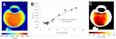 |
MRI relaxometry to measure the oxygen partial pressure and
viscosity of the vitreous humour of the aging eye
Xingzheng Pan1,
Alyssa Lie2,
Renita Martis1,
Beau Pontre3,
Julie Lim1,
Thomas White4,
and Paul Donaldson1
1Physiology, University of Auckland, Auckland, New Zealand, 2Optometry and Vision Science, University of Auckland, Auckland, New Zealand, 3Anatomy and Medical Imaging, University of Auckland, Auckland, New Zealand, 4Physiology and Biophysics, Stony Brook University, New York, NY, United States Keywords: Relaxometry, Oxygenation, Human eye The vitreous humour is a clear, gel-like fluid to provide structural support to the eye. Recently, it has been shown that the vitreous is important in regulating oxygen levels within the back of the eye. However, with ageing, the vitreous undergoes liquefaction, and as a result, oxygen is able to be move more freely throughout the vitreous, which increases the exposure of tissues such as the lens to oxygen. In this study, we developed MRI-based protocols to clinically monitor the oxygen levels and the fluid viscosity of the vitreous and applied these protocol in a cohort of elderly partipants. |
| 13:45 |
1355.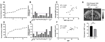 |
Towards a multi-parametric MRI-based myelin marker for
detecting enhanced myelination in the Gli1-/- mouse brain
Choong Heon Lee1,
Mara Holloman2,
Jacob Brady3,
James L. Salzer2,
and Jiangyang Zhang1
1Bernard and Irene Schwartz Center for Biomedical Imaging, Department of Radiology, New York University School of Medicine, New York, NY, United States, 2Department of Neuroscience and Physiology, Neuroscience Institute, New York University School of Medicine, New York, NY, United States, 3UConn Health, Farmington, CT, United States Keywords: Multi-Contrast, Microstructure Although multiple MRI-based myelin marker have been introduced, their sensitivity and specificity remained limited. Multi-parametric MRI can potentially enhance myelin mapping, but validation remains challenging. In this study, we compared myelin histology from the Gli1-/- mouse brain, which has enhanced myelination, with MRI markers based on relaxation, magnetization transfer, and diffusion properties from the same animals. We found varying degrees of correlation between MRI markers and myelin basic protein signals in multiple brain regions. Partial least square regression analysis demonstrated that multi-parametric MRI can indeed improve myelin mapping and provided information for further optimization. |
| 13:45 |
1356.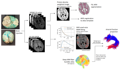 |
FibraVasc: towards ex vivo MRI mapping of cerebral vascular
territories, application to the vascularization of deep
white matter bundles
Simon Legeay1,
Mykyta Smirnov2,
Maëlig Chauvel1,
Bastien Herlin1,
Laurent Barantin2,3,
Ivy Uszynski1,
Igor Lima Maldonado2,3,
Christophe Destrieux2,3,
and Cyril Poupon1
1BAOBAB, NeuroSpin, Université Paris-Saclay, CNRS, CEA, Gif-sur-Yvette, France, 2UMR 1253, iBrain, Université de Tours, Inserm, Tours, France, 3CHRU de Tours, Tours, France Keywords: Relaxometry, Ex-Vivo Applications, Quantitative Imaging The arterial distribution territories of the brain remain poorly characterized and are of major importance for treating ischemic strokes. We propose a new quantitative MRI approach for ex vivo mapping of vascular territories using paramagnetic gelatin injection into cerebral arteries to enhance MRI contrast at the gelatin level. This method was applied on one healthy human brain injected into the middle cerebral artery and accurately quantified the volume fraction of blood using a dedicated protocol and a modeling of the injected tissue signal. We projected the resulting mapping along the deep white matter fiber bundles. |
| 13:45 |
1357.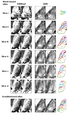 |
In-vivo imaging of the human thalamus: a comprehensive
evaluation of structural imaging approaches for thalamic
nuclei differentiation at 7T
Cristina Sainz Martinez1,2,
José P. Marques3,
Gabriele Bonanno4,5,6,
Tom Hilbert4,7,8,
Constantin Tuleasca8,9,10,
Meritxell Bach Cuadra2,7,
and João Jorge1
1CSEM - Swiss Center for Electronics and Microtechnology, Bern, Switzerland, 2CIBM Center for Biomedical Imaging, Lausanne, Switzerland, 3Donders Centre for Cognitive Neuroimaging, Radboud University, Nijmegen, Netherlands, 4Advanced Clinical Imaging Technology, Siemens Healthineers International AG, Bern, Switzerland, 5Translational Imaging Center (TIC), Swiss Institute for Translational and Entrepreneurial Medicine (SITEM), Bern, Switzerland, 6Magnetic Resonance Methodology, Institute of Diagnostic and Interventional Neuroradiology, University of Bern, Bern, Switzerland, 7Department of Radiology, Lausanne University Hospital (CHUV) and University of Lausanne (UNIL), Lausanne, Switzerland, 8Signal Processing Laboratory 5 (LTS-5), École Polytechnique Fédérale de Lausanne, Lausanne, Switzerland, 9Department of Clinical Neurosciences, Neurosurgery Service and Gamma Knife Center, Centre Hospitalier Universitaire Vaudois (CHUV), Lausanne, Switzerland, 10Faculty of Biology and Medicine, University of Lausanne (UNIL), Lausanne, Switzerland Keywords: Multi-Contrast, High-Field MRI, Thalamus, Nuclei, 7 Tesla, QSM The ability to non-invasively image the thalamus and its different nuclei would be highly valuable to neuroscience and neuroradiology, but has remained challenging. Here, we initiated a comprehensive practical review of recent thalamic imaging approaches at 7 Tesla, based on T1, T2, T2* and susceptibility properties. These were all acquired on the same in-vivo brain, to avoid anatomical variability confounds. The images were qualitatively compared to histological atlases. Upon systematic assessment, QSM and GM/WM-optimized MP2RAGE proved the most valuable to differentiate specific nuclei. To our knowledge, this is the most comprehensive evaluation to date of thalamic imaging modalities at 7T. |
| 13:45 |
1358. |
Investigations of T1 Anisotropy in ex-vivo White Matter
using a Tiltable Coil
Niklas Wallstein1,
André Pampel1,
Carsten Jäger1,
Roland Müller1,
and Harald E Möller1,2
1Max Planck Institute for Human Cognitive and Brain Sciences, Leipzig, Germany, 2Department of Physics, Leipzig University, Leipzig, Germany Keywords: Relaxometry, White Matter, T1 Anisotropy WM In general, a broad consensus exists that dipolar couplings between water protons and motional restricted macromolecules are prominent modulators of longitudinal relaxation in WM. Recent studies demonstrate anisotropic relaxation in human white matter (WM) in dependence of the fibre-to-field orientation. Our approach for further investigations of such effects overcomes previous limitations, in particular, sample reorientations for a direct demonstration of orientation effects, which were missing in earlier studies. Samples of ex-vivo WM tissue of pig brain yielded slightly reduced spin-lattice relaxation times by 1-2% around an angle of 40° between the main fibre direction and B0. |
| 13:45 |
1359.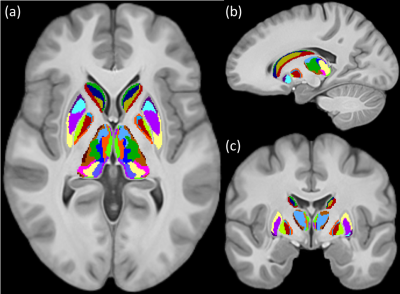 |
In-vivo parcellation of human subcortex by diffusion and
anatomical MRI
Tonima Ali1,2,
Jinglei Lv1,2,
Marshall Dalton2,3,
Steve Kassem4,
Arkiev D'Souza2,5,
and Fernando Calamante1,2,6
1School of Biomedical Engineering, The University of Sydney, Sydney, Australia, 2Brain and Mind Centre, The University of Sydney, Sydney, Australia, 3School of Psychology, The University of Sydney, Sydney, Australia, 4Neuroscience Research Australia, Sydney, Australia, 5Translational Research Collective, The University of Sydney, Sydney, Australia, 6Sydney Imaging, The University of Sydney, Sydney, Australia Keywords: Multi-Contrast, Diffusion/other diffusion imaging techniques, Brain, Gray matter, Segmentation, Neuro We integrated the information from structural and diffusion MRI from a group of healthy subjects, to develop a data-driven parcellation of the human subcortex. We first identified the data features that are sensitive to the micro-architectural variabilities within subcortex and then segregated specialised sub-regions with discernible properties. Our parcellated sub-regions demonstrated remarkable similarities to known nuclei and sub-structures within striatum, globus pallidus, and thalamus. Our parcellation has also identified regions which are known to have distinct anatomical and functional properties but that are yet to be explicitly added to extant human brain atlases. |
| 13:45 |
1360.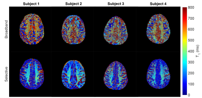 |
Spectrally Selective On-Resonance Inversion Recovery at 7T
for Multiple Component T1 Mapping
Paul S Jacobs1,
Neil Wilson1,
Ravi Prakash Reddy Nanga1,
Blake Benyard1,
Kosha Ruparel2,
Dushyant Kumar1,
Mark A Elliott1,
David Roalf2,
and Ravinder Reddy1
1Center for Advanced Metabolic Imaging in Precision Medicine, Department of Radiology, University of Pennsylvania, Philadelphia, PA, United States, 2Department of Psychiatry, University of Pennsylvania, Philadelphia, PA, United States Keywords: Contrast Mechanisms, White Matter, High-Field MRI, Inversion Recovery Two sets of images were acquired on four healthy subjects using separate on-resonance selective and broadband inversion recovery (IR) sequences at nine different inversion times. These images were fit to a bi-exponential T1 fitting model aiming to separate out specific long and short components. The selective IR data showed a substantial increase in white matter contrast compared to the broadband data with an average T1 of approximately 300ms. This agrees with reported literature values of the T1 of myelin and suggest it may be a potential driver of this selective short T1 component contrast. |
| 13:45 |
1361. |
A Rapid, Whole-brain Quantification of T1 Relaxation Time
Using Submillimetre Inversion-recovery EPI at 7T
Seong Dae Yun1 and
N. Jon Shah1,2,3,4
1Institute of Neuroscience and Medicine 4, INM-4, Forschungszentrum Juelich, Juelich, Germany, 2Institute of Neuroscience and Medicine 11, INM-11, JARA, Forschungszentrum Juelich, Juelich, Germany, 3JARA - BRAIN - Translational Medicine, Aachen, Germany, 4Department of Neurology, RWTH Aachen University, Aachen, Germany Keywords: Contrast Mechanisms, Quantitative Imaging, 7T, Inversion-recovery EPI, Rapid T1 mapping, Submillimetre and Whole-brain Knowledge of T1 relaxation time is of great interest for clinical diagnosis or MRI sequence optimisation. For the quantitative measurement of T1, the inversion-recovery method is widely used due to its relatively good accuracy or tolerance to B1 inhomogeneity. However, 2D multi-slice- or 3D segmentation-based readout methods often preclude the effect of different signal recovery modulation depending on slice locations. Therefore, this work presents a single-slice-based inversion-recovery 2D EPI method, combined with TR-external EPI phase correction at 7T, to provide rapid, whole-brain T1 mapping with a voxel size of 0.73 × 0.73 mm2. |
| 13:45 |
1362.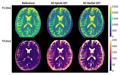 |
Simultaneous T1 and T2 relaxometry of the human brain at 7T
using Quantitative Transient-state Imaging
Matteo Cencini1,
Rolf F Schulte2,
Marta Lancione1,
Carolin M Pirkl2,
Laura Biagi1,
Graziella Donatelli3,4,
and Michela Tosetti1
1IRCCS Stella Maris, Pisa, Italy, 2GE Healthcare, Munich, Germany, 3IMAGO7 Foundation, Pisa, Italy, 4Azienda Ospedaliero-Universitaria Pisana, Pisa, Italy Keywords: High-Field MRI, High-Field MRI Fast quantitative MR methods enable repeatable and reproducible assessment of tissue properties improving diagnosis and follow-up. Here, we implemented two different 3D relaxometry methods based on quantitative transient-state imaging (QTI) for fast T1 and T2 mapping of the human brain at 7T. The two techniques were demonstrated both in-vitro and in-vivo and provided good quality parametric maps with low geometric distortion and blurring in clinically feasible acquisition and reconstruction times. |
| 13:45 |
1363.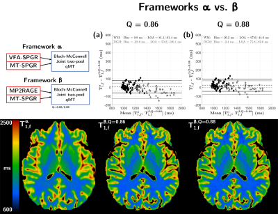 |
Disentangling T1 relaxation from MT effects in the MP2RAGE
sequence
Lucas Soustelle1,2,
Andreea Hertanu1,2,
Thomas Troalen3,
Maxime Guye1,2,
Jean-Philippe Ranjeva1,2,
Guillaume Duhamel1,2,
and Olivier M. Girard1,2
1Aix-Marseille Univ, CNRS, CRMBM, Marseille, France, 2APHM, Hôpital Universitaire Timone, CEMEREM, Marseille, France, 3Siemens Healthcare SAS, Marseille, France Keywords: Relaxometry, Modelling, Microstructure, Nervous System Common T1 mapping methods are dependent on the biophysical model assumptions. Previous work based on a qMT-SPGR framework demonstrated the influence of magnetization transfer effects and the importance of appropriate sequence design and signal modelling for T1 estimation. In this work, we expanded upon this optimized framework using an MP2RAGE sequence and demonstrated that quantitative T1 values in agreement with VFA-based experiments can be obtained in the human brain. |
| 13:45 |
1364.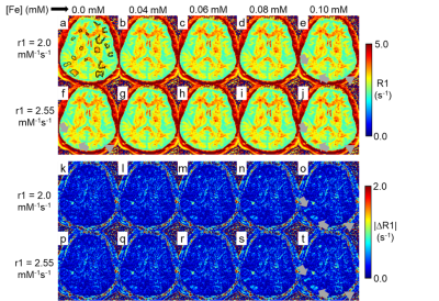 |
Reproducibility and sensitivity of in vivo human brain R1
map at ultralow field 64 mT
Joong Hee Kim1,
Govind Nair1,
Daniel Reich1,
and David L. Brody1
1National Institute of Health, Bethesda, MD, United States Keywords: Relaxometry, Low-Field MRI, Ultra low field MR parameter mapping provides objective biomarkers for living tissues. However, long scan times and high field MR imaging are typically required, whereas few ultralow field MR scanner studies have been performed. Here we propose a simplified R1 mapping protocol with only two inversion delay time points (R1approx), taking advantage of the fast R1 at ultralow field. The R1 characteristics of human brain were largely preserved in R1approx compared to standard inversion recovery R1 mapping. R1approx. required under 30 minutes vs. ~two hours for standard R1 mapping. In addition, R1approx. shows high reproducibility and good sensitivity to an R1 contrast agent. |
| 13:45 |
1365.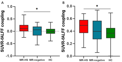 |
Elevated Metabolic-functional Coupling in Epileptogenic
Lesion and Network are Related with Surgical Outcomes of
Mesial Temporal Lobe Epilepsy
Siyu Yuan1,
Hui Huang1,
Miao Zhang2,
Wei Liu3,
Jiwei Li1,
Bingyang Cai1,
Ya Cui1,
and Jie Luo1
1School of Biomedical Engineering, Shanghai Jiao Tong University, Shanghai, China, 2Department of Nuclear Medicine, Ruijin Hospital, Shanghai Jiao Tong University School of Medicine, Shanghai, China, 3Department of Neurosurgery, Ruijin Hospital, Shanghai Jiao Tong University School of Medicine, Shanghai, China Keywords: fMRI, PET/MR It has been unclear whether metabolic-functional coupling could be disturbed in epileptogenic lesion and network by epileptic activities. In this study, we investigated metabolic changes, functional alterations and their couplings in epileptogenic lesion and network of mesial temporal lobe epilepsy (MTLE) using simultaneous PET/MR, and further evaluated the relationship between altered couplings and surgical outcome. MTLE patients showed abnormalities of standardized uptake value ratio (SUVR) and fractional amplitude of low-frequency fluctuation (fALFF) in default mode network (DMN). MTLE patients with hippocampal sclerosis (MR-HS) had higher SUVR-fALFF coupling in ipsilateral hippocampus and DMN, and the couplings were associated with surgical outcomes. |
| 13:45 |
1366.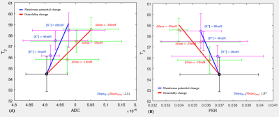 |
The respective contribution of cell swelling and changes in
hydrated water molecules to T2 changes due to changes in
membrane potential
Seong-min Kim1,
Kyeongseon Min2,
Jung Seung Lee1,3,
and Jang-Yeon Park1,3
1Department of Biomedical Engineering, Sungkyunkwan University, Suwon, Korea, Republic of, 2Laboratory for Imaging Science and Technology, Department of Electrical and Computer Engineering, Seoul National University, Seoul, Korea, Republic of, 3Department of Intelligent Precision Healthcare Convergence, Sungkyunkwan University, Suwon, Korea, Republic of Keywords: Contrast Mechanisms, Challenges, cell scan, qMR, ADC, T2, PSR Our group recently reported a method for direct imaging of neuronal activity (DIANA), suggesting that its contrast mechanism is T2 changes due to changes in membrane potential during neuronal activation, which accompanies cell swelling and changes in hydrating water modlecules of the cell membrane. In this study, by measuring apparent-diffusion-coefficient (ADC) and pool-size-ratio (PSR) versus T2, respectively, we verified the respective contribution of cell swelling and changes in hydrating water modlecules to T2 changes when only osmotic pressure or membrane potential was altered in T-lymphocyte cells in vitro. |
| 13:45 |
1367.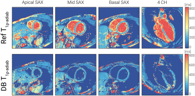 |
Suppressing blood signal in myocardial T1ρ mapping at 3T
through novel dark-blood adiabatic spin-lock preparations.
Chiara Coletti1,
Maša Bozic-Iven1,2,
Joao Tourais1,
Christal van de Steeg-Henzen3,
Mehmet Akcakaya4,
and Sebastian Weingärtner1
1TU Delft, Delft, Netherlands, 2Heidelberg University, Heidelberg, Germany, 3HollandPTC, Delft, Netherlands, 4University of Minnesota, Minneapolis, MN, United States Keywords: Relaxometry, New Signal Preparation Schemes, T1ρ, Dark-blood, adiabatic T1ρ-mapping is emerging as a promising, contrast-free alternative to LGE for assessment of myocardial viability. However, high blood T1ρ values can obfuscate endocardial scar. In this work, we propose a dark-blood adiabatic T1ρ preparation to suppress the blood signal and improve depiction of the blood-myocardium interface. A slice-selection gradient is added to an odd number of adiabatic full passage pulses to achieve blood inversion outside the imaging slab. Phantom results show that DBT1ρ yields unbiased estimation of T1ρ time. In vivo, thorough blood suppression is achieved for the trade-off against a moderate increase in DBT1ρ variance. |
 Back to Meeting Home
 Back to the Program-at-a-Glance
The International Society for Magnetic Resonance in Medicine is accredited by the Accreditation Council for Continuing Medical Education to provide continuing medical education for physicians.
 Back to Meeting Home
 Back to the Program-at-a-Glance
 Full Session Video