Power Pitch
Pitch: Getting the Best Out of Diffusion MRI
ISMRM & ISMRT Annual Meeting & Exhibition • 03-08 June 2023 • Toronto, ON, Canada

Power Pitch
Pitch: Getting the Best Out of Diffusion MRI
| 13:30 |
0976.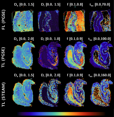 |
Quantifying features of human gray matter microstructure
postmortem using Neurite Exchange Imaging (NEXI) at
ultra-high field
Andreea Hertanu1,
Quentin Uhl1,
Tommaso Pavan1,
Christophe M. Lamy2,
and Ileana O. Jelescu1
1Dept. of Radiology, Lausanne University Hospital (CHUV) and University of Lausanne, Lausanne, Switzerland, 2Division of Anatomy, Faculty of Medicine, University of Geneva, Geneva, Switzerland Keywords: Microstructure, Modelling, Diffusion In this work, we characterized microstructure properties of cortical gray matter and hippocampus using diffusion MRI, on two samples of postmortem fixed healthy human brain. The weak-to-absent diffusivity time-dependence and marked kurtosis time-dependence supported the suitability of the Neurite Exchange Imaging (NEXI) model to probe postmortem human gray matter microstructure. NEXI parametric estimations revealed region-specific values, calling for further validation using histology and further clarifying the discrepancy between compartment diffusivities estimated here vs. the literature. This study offers an important window on human gray matter microstructure at a spatial resolution unachievable in vivo. |
| 13:30 |
0959.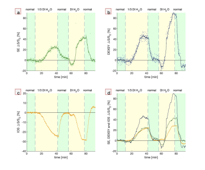 |
Diffusion-weighted signals and intrinsic optical signal
share a similar contrast mechanism
Nathan Hu Williamson1,
Rea Ravin1,2,
Teddy Xuke Cai1,3,
and Peter Joel Basser1
1NICHD, NIH, Potomac, MD, United States, 2Celoptics, Rockville, MD, United States, 3Wellcome Centre for Integrative Neuroimaging, Oxford, Oxford, United Kingdom Keywords: Diffusion/other diffusion imaging techniques, Neuroscience, Microstructure Diffusion-weighted imaging (DWI) and intrinsic optical signal (IOS) are both used to measure neural tissue structure and function. Here we demonstrate with simultaneous real-time low-field, high-gradient MR and optical microscopy that DW signals and IOS share a similar contrast mechanism, most likely the ratio between intracellular and extracellular volume. Signals monitor how cells are affected and respond to adverse conditions, providing novel insight into pathological mechanisms and links between structure and function. |
| 13:30 |
0960.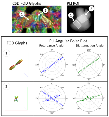 |
Diattenuation and retardance metrics from complete
polarimetry differentiate microscale and macroscale
anisotropy
Rhea Carlson1,
Courtney Comrie1,
Justina Bonaventura2,
Kellys Morara1,
Noelle Daigle2,
Elizabeth Hutchinson1,
and Travis W. Sawyer1,2
1Biomedical Engineering, University of Arizona, Tucson, AZ, United States, 2Wyant College of Optical Sciences, University of Arizona, Tucson, AZ, United States Keywords: Data Acquisition, Multimodal, Polarized Light Imaging, Optics, Ferret This study uses complete polarimetry to capture diattenuation and retardance in post-mortem ferret optic chiasm (OC) chosen for its well understood fiber structure. These methods are novel due to the limited analysis of diattenuation metrics on tissue. Our results suggest that retardance measurements are sensitive to microscale features rather than to macroscale geometry when compared to fractional anisotropy (FA) and propagator anisotropy (PA). In addition, we have shown fiber orientation distributions (FODs) created through direct optical measurement of the tissue on a 2D plane are comparable to diffusion FODs. |
| 13:30 |
0961.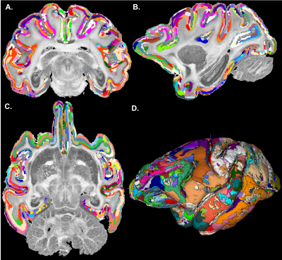 |
Automatic segmentation of cortical cytoarchitectonic domains
measured with high-resolution MAP-MRI
Kristofor Pas1,
Kadharbatcha Saleem1,
Peter J Basser1,
and Alexandru V Avram1,2,3
1Eunice Kennedy Shriver National Institute of Child Health and Human Development, National Institutes of Health, Bethesda, MD, United States, 2Center for Neuroscience and Regenerative Medicine, Bethesda, MD, United States, 3Henry M. Jackson Foundation for the Advancement of Military Medicine Inc., Bethesda, MD, United States Keywords: Microstructure, Gray Matter, MAP-MRI, cortical parcellation, cortical layers We investigate the feasibility of using advanced diffusion MRI, specifically MAP-MRI, to automatically segment microstructural domains based on subject-specific intrinsic cortical cytoarchitectonic features. Our preliminary results suggest that MAP-derived cytoarchitectonic domains delineate boundaries between cortical areas and layers in good agreement with histology. Segmentation methods that leverage the sensitivity of high-resolution MAP parameters to cortical cytoarchitecture have the potential to augment and refine current techniques for automatic cortical parcellation that rely on atlas registration. |
| 13:30 |
0962.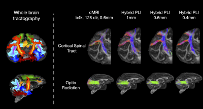 |
Tackling the gyral bias and bottleneck problems with hybrid
diffusion-microscopy tractography in the BigMac dataset
Silei Zhu1,
Istvan N. Huszar1,
Nicole Eichert1,
Michiel Cottaar1,
Greg Daubney2,
Alexandre Khrapitchev3,
Rogier B. Mars1,4,
Jeroen Mollink1,
Connor Scott5,
Adele Smart1,5,
Jerome Sallet2,
Saad Jbabdi1,
Karla Miller*1,
and Amy Howard*1
1Wellcome Centre for Integrative Neuroimaging, FMRIB, Nuffield Department of Clinical Neurosciences, University of Oxford, Oxford, United Kingdom, 2Wellcome Centre for Integrative Neuroimaging, Experimental Psychology, University of Oxford, Oxford, United Kingdom, 3Department of Oncology, University of Oxford, Oxford, United Kingdom, 4Donders Institute for Brain, Cognition and Behaviour, Radboud University Nijmegen, Nijmegen, Netherlands, 5Nuffield Department of Clinical Neurosciences, University of Oxford, Oxford, United Kingdom Keywords: Tractography & Fibre Modelling, Multimodal, diffusion MRI, microscopy We present data fusion combining precisely co-registered dMRI and microscopy to reconstruct 3D fibre orientation with micron-scale spatial resolution. We then perform hybrid dMRI-microscopy tractography to investigate two known challenges in tractography: the gyral bias and bottleneck problems. Our results using hybrid tractography suggest that the gyral bias can be overcome by increased spatial resolution, and that microstructure-informed fibre orientations can overcome the bottleneck problem, irrespective of spatial resolution. These observations can be used to inform tractography analyses in vivo. This approach builds on the complementary strengths of microscopy (high spatial resolution) and dMRI (3D whole-brain coverage). |
| 13:30 |
0963.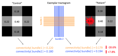 |
A multi-compartment model for pathological connectomes
Sara Bosticardo1,2,
Matteo Battocchio1,3,
Simona Schiavi4,
Cristina Granziera2,5,6,
and Alessandro Daducci1
1Diffusion Imaging and Connectivity Estimation (DICE) Lab, Department of Computer Science, University of Verona, Verona, Italy, 2Translational Imaging in Neurology (ThINK), Department of Biomedical Engineering, University of Basel, Basel, Switzerland, 3Sherbrooke Connectivity Imaging Laboratory (SCIL), Département d’Informatique, Université de Sherbrooke, Sherbrooke, QC, Canada, 4Department of Neuroscience, Rehabilitation, Ophthalmology, Genetics, Maternal and Child Health (DINOGMI), University of Genoa, Genoa, Italy, 5Research Center for Clinical Neuroimmunology and Neuroscience Basel (RC2NB), University of Basel, Basel, Switzerland, 6Department of Neurology, MS Center, University Hospital Basel, Basel, Switzerland Keywords: Diffusion/other diffusion imaging techniques, Brain Connectivity The white matter is the complex system of neuronal fibers in the brain. Any disruption to this circuitry may lead to a wide range of neurological diseases and, thus, it is fundamental to be able of detecting pathological conditions at early stages. State-of-the-art methods for studying brain connectivity assume constant properties along the fibers, but this assumption is not valid in pathological conditions that locally affect the tissue, e.g. multiple sclerosis. Here, we present a model for “multi-compartment connectomes” to explicitly consider the presence of focal lesions during the estimation of connectivity and show its effectiveness using realistic numerical simulations. |
| 13:30 |
0964.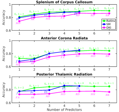 |
Characterization of normal appearing white matter in
Multiple Sclerosis with diffusion MRI using signal
representations and model parameters
Ying Liao1,
Santiago Coelho1,
Jenny Chen1,
Dmitry S. Novikov1,
and Els Fieremans1
1Radiology, NYU School of Medicine, NEW YORK, NY, United States Keywords: Diffusion/other diffusion imaging techniques, Diffusion/other diffusion imaging techniques Multiple sclerosis (MS) is a neurodegenerative and inflammatory disease characterized by focal lesions and damages to normal appearing white matter (NAWM). Diffusion MRI (dMRI) is known for its sensitivity to the microstructural changes in white matter. In this work, we study the sensitivity of dMRI metrics to MS pathology in NAWM by distinguishing MS patients from healthy controls. We found that beyond-spherical-mean rotational invariants of high-b shells contribute mostly to the classification, indicating nontrivial information content of high-b diffusion signal beyond DTI. |
| 13:30 |
0965.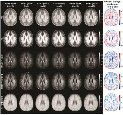 |
Age-related regional patterns in cerebral microstructure
revealed using mean apparent propagator (MAP) MRI
Mustapha Bouhrara1,
Alexandru V Avram2,
Matthew Kiely1,
Aparna Trivedi1,
and Dan Benjamini1
1National Institute on Aging, Baltimore, MD, United States, 2National Institute of Child Health and Human Development, Bethesda, MD, United States Keywords: Diffusion/other diffusion imaging techniques, Aging The relationship between brain microstructure and aging has been the subject of intense study. We used the mean apparent propagator (MAP) MRI framework, which is suitable to characterize diffusion in complex microstructure, to investigate age-related differences in a cohort of cognitively unimpaired participants, spanning a wide age range. In white matter, we established an opposing pattern of higher non-Gaussianity (NG) alongside lower propagator anisotropy (PA) among older adults. In gray matter, these two indices were consistent with one another, and exhibited regional pattern heterogeneity compared to other microstructural parameters. These results suggest that MAP-MRI provides otherwise inaccessible regional microstructural information. |
| 13:30 |
0966.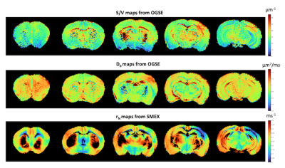 |
Permeability mapping in stroke via diffusion Standard Model
with Exchange and S/V measurements from high-frequency OGSE
Noam Shemesh1,
Rita Alves2,
Jonas Olesen3,4,
Andrada Ianus1,
and Sune Nørhøj Jespersen3,4
1Champalimaud Research, Champalimaud Foundation, Lisbon, Portugal, 2Champalimaud Foundation, Lisbon, Portugal, 3Center of Functionally Integrative Neuroscience (CFIN) and MINDLab, Department of Clinical Medicine, Aarhus University, Aarhus, Denmark, 4Department of Physics and Astronomy, Aarhus University, Aarhus, Denmark Keywords: Contrast Mechanisms, Microstructure Membrane permeability plays an important role in numerous physiological processes ranging from cell volume regulation to neural activity. Until now, diffusion MRI measurements mainly provided information on exchange rates; however, to map permeability, information on the surface-to-volume (S/V) ratio is needed. Here, we apply ultrahigh frequency OGSE to infer on S/V from the associated power law dependence (1/ω1/2,) and the Standard Model with Exchange (SMEX) model for extracting the neurite -> extracellular space exchange rates in stroked rat brains. Combined, we present quantitative measurements of permeability which show plausible values and striking contrast in stroke. |
| 13:30 |
0967.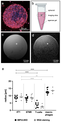 |
Profiling specific cell populations within the inflammatory
tumor microenvironment by oscillating-gradient
diffusion-weighted MRI
Emily Hoffmann1,
Mirjam Gerwing1,
Stephan Niland2,
Rolf Niehoff1,
Max Masthoff1,
Enrica Wilken1,
Philipp Berger3,
Thomas Vogl3,
Johannes A. Eble2,
Bastian Maus1,
Anne Helfen1,
Moritz Wildgruber1,4,
and Cornelius Faber1
1Clinic of Radiology, University of Münster, Münster, Germany, 2Institute of Physiological Chemistry and Pathobiochemistry, University of Münster, Münster, Germany, 3Institute of Immunology, University of Münster, Münster, Germany, 4Department of Radiology, LMU Munich, Munich, Germany Keywords: Cancer, Tumor, T-cells, macrophages, immunotherapy Immune cells are major players of the tumor microenvironment (TME), having profound effects on tumor development and metastatic progression. We present oscillating-gradient diffusion-weighted MRI (OGSE-DWI) as non-invasive imaging approach to monitor the intratumoral immune cell infiltrate, relying on size differences between cancer cells, T-cells and macrophages. By applying the Imaging Microstructural Parameters Using Limited Spectrally Edited Diffusion (IMPULSED) model to sine-shaped OGSE-DWI, changes within the TME and its specific immune cell composition were monitored and compared in syngeneic murine breast cancer models with different degrees of malignancy during tumor progression, clodronate liposome-mediated depletion of macrophages and immune checkpoint inhibitor treatment. |
| 13:30 |
0968.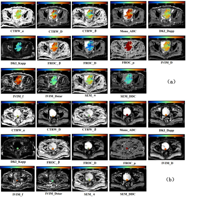 |
Assessing Muscle Invasiveness In Bladder Cancer Based On
Multiple Diffusion Models
Junting Guo1,
Lu Zhang1,
Shuo Li1,
Ding Li1,
Zhichang Fan1,
Meining Chen2,
Guoqiang Yang3,
Yan Li3,
Bin Wang3,
Le Wang3,
and Xiaochun Wang3
1College of Medical Imaging, Shanxi Medical University, Taiyuan, China, 2MR Scientific Marketing, Siemens Healthcare, Shanghai, China, 3Department of Radiology, The First Hospital of Shanxi Medical University, Taiyuan, China Keywords: Diffusion/other diffusion imaging techniques, Cancer Accurate assessment of the presence or absence of muscle invasiveness in bladder cancer is essential for selecting the best treatment options. In this study, we used 6 diffusion models including mono-exponential model (Mono), continuous-time random-walk (CTRW), incoherent motion within the voxel (IVIM), stretched exponential model (SEM), diffusion kurtosis imaging (DKI) and fractional-order calculus (FROC) model to assess muscle invasiveness in bladder cancer. The study showed that Mono,CTRW,IVIM,SEM and DKI could provide biomarkers for muscle invasion and distinguish non-muscle-invasive and muscle-invasive bladder cancer, and the combination of CTRW and FROC could further improve the classification accuracy. |
| 13:30 |
0969.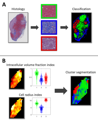 |
Cluster analysis of VERDICT MRI for identification of tumor
subregions with distinct histological features
Lukas Lundholm1,
Mikael Montelius1,
Oscar Jalnefjord1,2,
Eva Forssell-Aronsson1,2,
and Maria Ljungberg1,2
1Department of Medical Radiation Sciences, Institute of Clinical Sciences, Sahlgrenska Academy, University of Gothenburg, Gothenburg, Sweden, 2Department of Medical Physics and Biomedical Engineering, Sahlgrenska University Hospital, Gothenburg, Sweden Keywords: Diffusion/other diffusion imaging techniques, Cancer, VERDICT, preclinical, histology Characterization of tumor tissue may aid in grading and assessment of cancer treatment. Time-dependent diffusion MRI can provide non-invasive estimates of parameters relating to tissue microstructure in vivo. In this study, a cluster analysis was performed to group parameters estimated by the time-dependent diffusion MRI model VERDICT. Parameter cluster maps were compared with maps derived from classification of histological images. The results showed good agreement between VERDICT cluster maps and histology maps, indicating that the method shows potential in identifying tumor subregions of distinct morphological features. |
| 13:30 |
0970.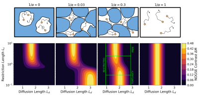 |
Selective filters of translational molecular diffusion
dynamics in white matter microstructures with preclinical
and clinical MRI
Ezequiel L. Saidman1,
Analia Zwick 1,2,3,
Stefano Tambalo 4,
Thorsten Feiweier5,
Jorge Jovicich4,
and Gonzalo A. Alvarez1,2,3
1Instituto Balseiro, CNEA, Universidad Nacional de Cuyo, S. C. de Bariloche, Argentina, 2Centro Atómico Bariloche, CONICET, CNEA, S. C. de Bariloche, Argentina, 3Instituto de Nanociencia y Nanotecnologia, CNEA, CONICET, S. C. de Bariloche, Argentina, 4Center for Mind/Brain Sciences - CIMeC, University of Trento, Rovereto, Italy, 5Siemens Healthcare GmbH, Erlangen, Germany Keywords: Diffusion/other diffusion imaging techniques, Microstructure Magnetic resonance imaging is one of the most widely used methods for non-invasive medical imaging. Gradient modulation sequences can selectively extract quantitative information at micrometer and sub-micrometer scales that might be relevant for clinical applications. We approach this topic using white matter phantoms by observing the contrast generated by Non-uniform Oscillating Gradient Spin Echo (NOGSE) sequences to selectively filter the signal of molecules confined in compartments of specific sizes. The NOGSE contrast is determined by subtracting different OGSE acquisitions on a clinical scanner. Our results demonstrate a microscopic tortuosity mechanism that provides microstructural information at short time scales. |
| 13:30 |
0971.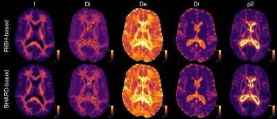 |
Rotation-invariant microstructure mapping revisited
Daan Christiaens1,2,
J-Donald Tournier3,
Stefan Sunaert2,4,
and Frederik Maes1,2
1Dept. of Electrical Engineering, ESAT/PSI, KU Leuven, Leuven, Belgium, 2Medical Imaging Research Center, UZ Leuven, Leuven, Belgium, 3Centre for the Developing Brain, School of Biomedical Engineering and Imaging Sciences, King's College London, London, United Kingdom, 4Department of Imaging & Pathology, Translational MRI, KU Leuven, Leuven, Belgium Keywords: Microstructure, Diffusion/other diffusion imaging techniques Microstructure imaging with diffusion MRI relies on non-linear fitting of a biophysical tissue model to the data, often at low signal-to-noise ratio. In this work, we derive a new rotation-invariant feature set for microstructure mapping based on a rank-1 decomposition of the multi-shell diffusion MRI signal in spherical harmonics. Simulations show that using this feature set avoids non-central-χ bias that is present at low SNR in parameter estimation based on conventional rotation-invariants. Results in human brain imaging data acquired with free waveform diffusion encoding show robust parameter estimates across white matter. |
| 13:30 |
0972.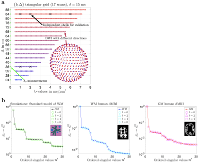 |
Zero-shell diffusion MRI: Focus on microstructure by
decoupling fiber orientations
Santiago Coelho1,
Els Fieremans1,
and Dmitry S. Novikov1
1Center for Advanced Imaging Innovation and Research (CAI2R), Department of Radiology, New York University School of Medicine, New York, NY, United States Keywords: Diffusion/other diffusion imaging techniques, Data Acquisition In brain dMRI, microstructure parameters of fiber fascicles, such as compartment fractions, exchange, diffusivities, relaxation rates, and structural disorder, are highly sought after. They can be accessed by sampling multiple diffusion weightings, b-tensor shapes, diffusion and/or echo times. Yet most scan time is spent oversampling the fiber orientation distribution function – because factoring it out nominally requires rotational invariants like the spherical mean. We show how to measure multiple inequivalent combinations of $$$(b,\,\Delta)$$$, while spending single gradient directions per unique combination. We recover signal’s rotational invariants for each one and use them for biophysical modeling of white and gray matter. |
| 13:30 |
0973.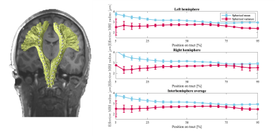 |
Axon diameter mapping is confounded by glial cells
Jelle Veraart1,
Erika P. Raven1,
Derek K. Jones2,
and Marco Palombo2,3
1Center for Biomedical Imaging, Dept. Radiology, NYU Grossman School of Medicine, New York, NY, United States, 2Cardiff University Brain Research Imaging Centre (CUBRIC), Cardiff University, Cardiff, United Kingdom, 3School of Computer Science and Informatics, Cardiff University, Cardiff, United Kingdom Keywords: Diffusion/other diffusion imaging techniques, Microstructure, Modeling, Axon Diameter Mapping The specificity of strongly diffusion-weighted MRI to intra-axonal signal contributions to neuronal processes has always been presumed. Here we demonstrate empirically that strongly diffusion-weighted MRI data is also sensitive to a distinct isotropic compartment that may bias axon diameter estimates. We hypothesize that such a compartment represents isotropically-distributed glial processes. We use in vivo human data and Monte Carlo simulations to support our hypothesis and evaluate a novel strategy to suppress signal arising from glial cell bodies and processes to promote more accurate axon diameter mapping. |
| 13:30 |
0974.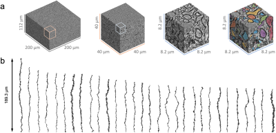 |
Diffusion simulations in human myelinated axons from 3d EM
evaluate the impact of realistic axon geometry on diameter
mapping
Hong-Hsi Lee1,2,
Qiyuan Tian1,2,
Ricardo Coronado-Leija3,
Els Fieremans3,
Dmitry S Novikov3,
and Susie Y Huang1,2
1Radiology, Athinoula A. Martinos Center for Biomedical Imaging, Massachusetts General Hospital, Charlestown, MA, United States, 2Radiology, Harvard Medical School, Boston, MA, United States, 3Center for Advanced Imaging Innovation and Research, New York University School of Medicine, New York, NY, United States Keywords: Validation, Microstructure To disentangle the effect of complex features on axon diameter mapping, we segmented myelinated axons in a human brain electron microscopy data and use these to create artificial cylinders with undulations and caliber variations that can be tuned to varying degrees. Diffusion simulations in segmented real axons and artificial axons showed that undulations and beadings lead to overestimation and underestimation of the axon diameter, respectively. |
| 00:00 |
0975.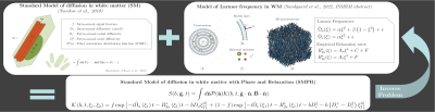 |
Incorporating susceptibility effects into the Standard Model
of diffusion in white matter
Anders Dyhr Sandgaard1,
Valerij G. Kiselev2,
Noam Shemesh3,
and Sune Nørhøj Jespersen1,4
1Center for functionally integrative neuroscience, department of clinical medicine, Aarhus University, Aarhus, Denmark, 2Division of Medical Physics, Department of Radiology, University Medical Center Freiburg, Freiburg, Germany, 3Champalimaud Research, Champalimaud Centre for the Unknown, Lisbon, Portugal, 4Department of Phsysics and Astronomy, Aarhus University, Aarhus, Denmark Keywords: Microstructure, Diffusion/other diffusion imaging techniques Estimating parameters of the Standard Model (SM) of white matter (WM) is an ill-posed problem (WM). To overcome this degeneracy, models extensions have been proposed adding echo-time dependent information. Here we investigate the feasibility of incorporating our analytical solution for the frequency shift from white matter axons accounting for magneto-structural anisotropy along with an empirical orientation dependence of relaxation into SM parameter estimation. This may also help achieving rotation-free mapping of susceptibility-related parameters using diffusion MRI instead of the impractical sample rotation in the scanner. |
| 00:00 |
0977.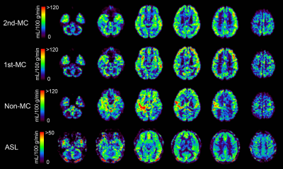 |
Motion-compensated diffusion imaging with phase-contrast
(MC-DIP) for the quantification of regional cerebral blood
flow
Naoki Ohno1,
Tosiaki Miyati1,
Genki Nambu1,
Daichi Tanaka1,
Yuki Makino1,
Noam Alperin2,
Yu Ueda3,
Marc Van Cauteren3,
Toshifumi Gabata1,
and Satoshi Kobayashi1
1Kanazawa University, Kanazawa, Japan, 2University of Miami, Miami, FL, United States, 3Philips Japan, Tokyo, Japan Keywords: Diffusion/other diffusion imaging techniques, Diffusion/other diffusion imaging techniques Diffusion imaging with phase-contrast (DIP) can quantitatively evaluate regional cerebral blood flow (rCBF), in which total CBF (tCBF) from phase-contrast magnetic resonance imaging (PC-MRI) converts the brain’s perfusion-related diffusion parameters into rCBF values. However, it is sensitive to bulk motion (i.e., brain pulsation), potentially causing an overestimation of DIP-derived rCBF values. To overcome this issue, we propose a motion-compensated DIP (MC-DIP) that incorporates motion-compensated diffusion gradients. DIP with second-order motion-compensated diffusion gradients (2nd-MC-DIP) improved the fitting accuracy of the biexponential analysis and showed the ability to quantitatively evaluate rCBF in gray and white matter. |
| 00:00 |
0978.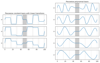 |
A piecewise-polynomial basis for generalized diffusion
encoding gradient waveforms
Emmanuel Caruyer1
1IRISA UMR 6074, Empenn ERL 1228, Univ Rennes, CNRS, Inria, Inserm, Rennes, France Keywords: Diffusion/other diffusion imaging techniques, Data Acquisition Diffusion-encoding gradient waveforms are a key parameter of experimental design in diffusion magnetic resonance imaging. Their optimization is important for e.g. maximizing the efficiency in B-tensor encoding, or improving the sensitivity and specificity to microstructure parameters of biophysical models. The main challenge in optimizing gradient waveforms and trajectories is the large dimension of the search space and the constraints associated with physical and physiological limitations. Here we propose an original piecewise polynomial representation, which natively enforces the linear constraints that arise from refocusing and regularity. We illustrate the basis on B-tensor encoding and Monte-Carlo simulation of the diffusion signal. |
 Back to Meeting Home
 Back to the Program-at-a-Glance
The International Society for Magnetic Resonance in Medicine is accredited by the Accreditation Council for Continuing Medical Education to provide continuing medical education for physicians.
 Back to Meeting Home
 Back to the Program-at-a-Glance
 Full Session Video