Power Pitch
Pitch: MSK Exotica
ISMRM & ISMRT Annual Meeting & Exhibition • 03-08 June 2023 • Toronto, ON, Canada

| 08:15 |
0084.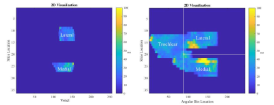 |
Repeatability of in vivo T2 Relaxation Times Regional and
Cluster Analysis in the Loaded Knee
Kaitlin G Sofko1,
Ibukunoluwa Elebute1,
Lumeng Cui2,
Marianne S Black3,
Natasha M Bzowey1,
and Emily J McWalter4
1Department of Mechanical Engineering, University of Saskatchewan, Saskatoon, SK, Canada, 2Siemens Healthcare Limited, Burnaby, BC, Canada, 3Department of Mechanical Engineering, University of Victoria, Victoria, BC, Canada, 4Department of Biomedical Engineering, University of Saskatchewan, Saskatoon, SK, Canada Keywords: Cartilage, Cartilage T2 relaxation time mapping of articular cartilage is most often assessed in relaxed knees; however, it’s primary function is resisting and distributing forces. Further, most work has focused on regional analysis rather than local changes. The aim of this work was to assess the repeatability of T2 relaxation time in the loaded knees of five healthy participants using both regional and local (cluster-based) approaches. We show that regional and local assessments are sufficiently repeatable for in vivo studies. |
| 08:15 |
0085.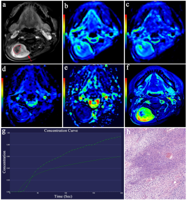 |
Differentiation of Malignant and Benign Musculoskeletal
Tumors Using IVIM-DWI Versus DCE-MRI
Miaomiao Cheng1,
Hua Zhang1,
Jixian Li1,
Zhiyan Xie1,
Jiufa Cui1,
and Xuejun Liu1
1The Affiliated Hospital of Qingdao University, Qing Dao, China Keywords: Bone, Diffusion/other diffusion imaging techniques, diagnostic value This study aimed to investigate the diagnostic value of IVIM-DWI and DCE-MRI in differentiating benign and malignant musculoskeletal tumors. IVIM parameters (FP, D, and DP) were statistically compared with ADC and DCE-MRI (Ktrans, Kep, Ve, and iAUC) within two groups. D, Ktrans and Kep have good diagnostic performance in identification of benign and malignant musculoskeletal tumors, DCE-MRI and IVIM have similar diagnostic efficacy, and combining these two methods can further improve the diagnostic performance. IVIM-DWI may be an alternative to DCE-MRI in patients with contrast contraindications. |
| 08:15 |
0086.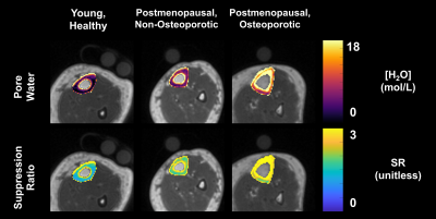 |
Automated, reference-free quantification of cortical bone
parameters detects impairments in postmenopausal
osteoporosis
Brandon Clinton Jones1,2,
Felix Werner Wehrli1,
Nada Kamona1,2,
Brian-Tinh Duc Vu1,2,
Hyunyeol Lee1,3,
Hee Kwon Song1,
Mona al Mukaddam4,
Peter J Snyder4,
Trevor Chan1,
Walter RT Witschey1,
Matthew MacLean1,
Nicholas J Josselyn1,5,
Srikant Kamesh Iyer1,
and Chamith Sudesh Rajapakse1
1Radiology, University of Pennsylvania, Philadelphia, PA, United States, 2Bioengineering, University of Pennsylvania, Philadelphia, PA, United States, 3School of Electronics, Kyungpook National University, Daegu, Korea, Republic of, 4Endocrinology, University of Pennsylvania, Philadelphia, PA, United States, 5Data Science, Worcester Polytechnic Institute, Worcester, MA, United States Keywords: Bone, Aging, Osteoporosis, Porosity, ultrashort echo time While ultrashort echo time (UTE) measures of pore water have shown promise in assessing cortical bone porosity, most are hindered by the need for complicated processing and reference samples. The suppression ratio (SR) is a marker of porosity which is simply calculated as the voxel-wise ratio of two UTE magnitude images, one without and with long-T2 suppression. Automated cortical bone segmentation via deep learning showed elevated SR in postmenopausal women with osteoporosis (P=0.001) and was strongly associated with pore water density (R=0.93) and with pQCT BMD (R=-0.88). Results suggest that SR can detect elevated porosity in postmenopausal osteoporosis. |
| 08:15 |
0087.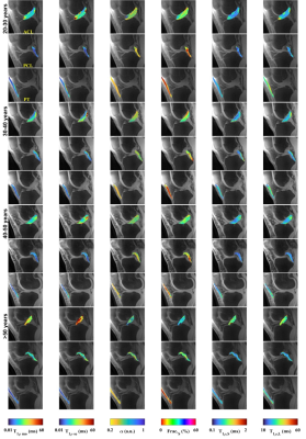 |
Age and gender-dependence of T1ρ-prepared PETRA sequence in
knee ligaments and tendon
Hector Lise de Moura1,
Richard Kijowski1,
Azadeh Sharafi2,
Marcelo Zibetti1,
and Ravinder Regatte1
1Center for Biomedical Imaging, NYU Langone Health, New York City, NY, United States, 2Medical College of Wisconsin, Wauwatosa, WI, United States Keywords: Tendon/Ligament, Osteoarthritis This study was performed to investigate gender-related and age-related differences in ultra-short echo-time (UTE) T1ρ parameters in the anterior cruciate ligament (ACL), posterior cruciate ligament (PCL), and patellar tendon (PT). A pointwise encoding time reduction with radial acquisition (PETRA) sequence was used to measure single-component and multi-component PETRA-T1ρ parameters in the ACL, PCL, and PT at 3.0T in 18 healthy volunteers. There were significant correlations between age and single-component and multi-component PETRA-T1p parameters in the ACL and PCL but not the PT. There were no significant differences in PETRA-T1ρ parameters in the ACL, PCL, or PT between males and females. |
| 08:15 |
0088.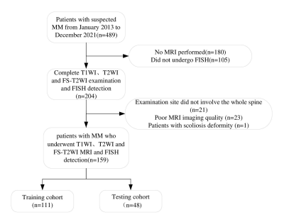 |
Radiomic nomograms from pre-treatment magnetic resonance
imaging-based may predict high-risk cytogenetic status in
multiple myeloma
Liu Suwei1
1Second Hospital of Lanzhou University, Lanzhou, China Keywords: Skeletal, Skeletal Our study aimed to develop and validate one or more clinically relevant radiomic nomograms based on multisequence MRI radiomic features to predict the pre-treatment HRC status of patients with MM. We developed radiomic nomograms for 14 models at a single center using clinically obtained whole-spine MRI images of 159 patients with MM. This study revealed that radiomic features of pre-treatment MRI images are associated with HRC status in patients with MM. Among the proposed models, the nomograms of the FT2, FT2+1, and FT2+2+1 models were identified as outstanding at distinguishing patients with HRC from those with non-HRC status. |
| 08:15 |
0089.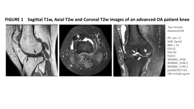 |
A linear model analysis of pain sensitisation in knee
osteoarthritis assessed by MRI in relation to the blood
biomarker thrombospondin-4
Franklyn A Howe1,
Soraya Khoushesh2,
Anna Blundell2,
Christine Heron3,
Vivienne Ejindu3,
Abiola Harrison4,
and Nidhi Sofat2,5
1Molecular and Clinical Sciences, St George's, University of London, London, United Kingdom, 2Infection & Immunity, St George's, University of London, London, United Kingdom, 3Radiology, St George's University Hospital Foundation Trust, London, United Kingdom, 4Infection & Immunity, St George's University Hospital Foundation Trust, London, United Kingdom, 5Rheumatology, St George's University Hospital Foundation Trust, London, United Kingdom Keywords: Osteoarthritis, Degenerative, Pain We investigated how pain in knee osteoarthritis (OA) relates to physical damage, clinical parameters, pain sensitisation and serum levels of thrombosponin-4 (TSP4). Across all patients (mild and advanced OA), the number of osteophytes (nOst) and TSP4 levels are significant factors in determining pain levels, with BMI and HADS the strongest factors. Grouping the patients according to low and high TSP4 levels, suggests a TSP4 phenotype that relates to central sensitisation. For high TSP4, pain pressure thresholds are a significant factor in reported knee pain in addition to HADS, BMI and physical damage. |
| 08:15 |
0090.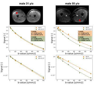 |
Microcirculation in aging skeletal muscle assessed with
flow-compensated Intravoxel Incoherent Motion (IVIM)
Valentina Mazzoli1,
Marco Barbieri1,
Yael Vainberg1,
Michelle Li 1,
Matthew Middione1,
Daniel B Ennis1,
Feliks Kogan1,
and Garry E Gold1
1Department of Radiology, Stanford University, Stanford, CA, United States Keywords: Muscle, Diffusion/other diffusion imaging techniques Assessment of microcirculation is extremely important in skeletal muscle aging reasearch. While IVIM can provide extremely valuable information on perfusion, this is usually dominated by flow in larger vessel, resulting in limited sensitivity to microcirculation. In this study we propose the use of flow and non-flow-compensated diffusion encoding waveforms for IVIM of skeletal muscle. Using this approach we show diffrences in IVIM parameters between younger and older adults, and show correlation between the difference in IVIM parameters and age |
| 08:15 |
0091.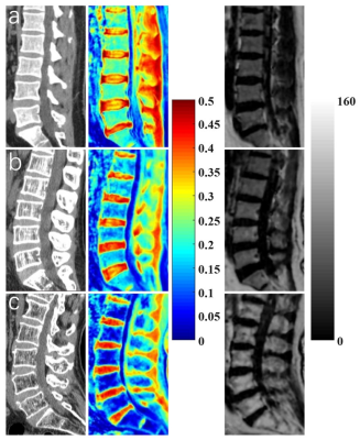 |
Assessment of Osteoporosis in Lumbar Spine Using Ultrashort
Echo Time Magnetization Transfer (UTE-MT) Imaging
Jin Liu1 and
Ya-Jun Ma1
1Department of Radiology, University of California San Diego, San Diego, CA, United States Keywords: Bone, Skeletal The bone collagen matrix makes a crucial contribution to the mechanical properties of bone including elasticity and tensile strength. Its changes can be accessed by the ultrashort echo time magnetization transfer (UTE-MT) technique. This study aims to investigate the feasibility of the UTE-MT ratio (UTE-MTR) in the assessment of lumbar osteoporosis (OP). Our results demonstrated that the UTE-MTR is highly correlated with bone mineral density and Fracture Risk Assessment Tool scores and has a high ability to distinguish people with different bone masses, which indicates that the UTE-MTR has great potential in the diagnosis of the patient with OP. |
| 08:15 |
0092.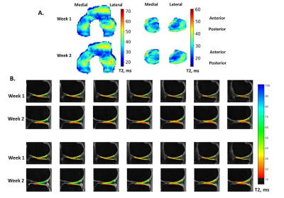 |
Repeatability and Sensitivity of qDESS T2 Mapping of
Cartilage
Ashley A. Williams1,
Jessica L Asay2,
Daniella Asare1,
Arjun D. Desai2,
Gordhan B. Mahtani1,
Jade He1,
Adam L. C. Wadsworth1,
Sachi Bansal1,
Garry E. Gold2,
Brian Hargreaves2,
Akshay Chaudhari2,
and Constance R. Chu1
1Orthopaedic Surgery, Stanford University, Stanford, CA, United States, 2Department of Radiology, Stanford University, Stanford, CA, United States Keywords: Cartilage, Relaxometry, Repeatability, T2, qDESS Intra- and inter-day repeatabilities of qDESS T2 in knee cartilage, assessed by fully-automatic segmentation using a deep-learning, open-source framework for musculoskeletal MRI analysis (DOSMA) and also by manual segmentation of tread mark regions of known tibiofemoral contact areas, were assessed and compared in 10 uninjured participants. qDESS T2 RMSA-CVs were less than 6% for all ROIs examined and showed good to excellent ICCs for the majority of ROIs assessed. A preliminary sensitivity analysis found that both segmentation schemes detected significant T2 changes over time in lateral tibial cartilage while only DOSMA segmentation detected T2 change to medial tibial cartilage. |
| 08:15 |
0093.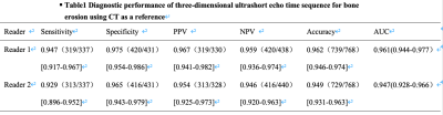 |
Assessment of the sacroiliac joint in patients with
ankylosing spondylitis by three-dimensional ultrashort echo
time MRI
Cui Ren1,
Qing Li2,
Qiao Zhu1,
Sommer Stefan3,4,5,
and Huishu Yuan1
1Peking University Third Hospital, Beijing, China, 2MR Collaborations, Siemens Healthineers Ltd, Shanghai, China, 3Siemens Healthineers International AG, Zurich, Switzerland, 4Swiss Center for Musculoskeletal Imaging (SCMI), Zurich, Switzerland, 5Advanced Clinical Imaging Technology (ACIT), Zurich, Switzerland Keywords: Joints, Cartilage, sacroiliac joint To assess the diagnostic performance of three-dimensional ultrashort echo time sequence (3D-UTE) in bone erosion detection of the sacroiliac joint (SIJ) in patients with ankylosing spondylitis (AS) and to test whether SIJ cartilage T2 * values might help in identification of patients with AS. |
| 08:15 |
0094.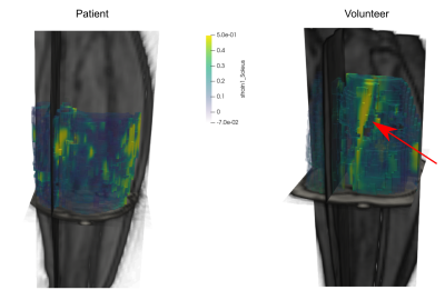 |
Contraction characteristics of the triceps surae in muscular
diseases assessed by 3D dynamic imaging with synchronized
electrical stimulation
Francesco Santini1,2,
Xeni Deligianni1,2,
Matteo Paoletti3,
Sabrina Ravaglia3,
Arianna Faggioli3,
Ning Jin4,
and Anna Pichiecchio3,5
1Basel Muscle MRI, Department of Biomedical Engineering, University of Basel, Basel, Switzerland, 2Research Coordination Team, Department of Radiology, University Hospital Basel, Basel, Switzerland, 3Advanced Imaging and Radiomics Center, IRCCS Mondino Foundation, Pavia, Italy, 4Cardiovascular MR R&D, Siemens Medical Solutions USA, Inc., Cleveland, OH, United States, 5Department of Brain and Behavioural Sciences, University of Pavia, Pavia, Italy Keywords: Muscle, Rare disease, Dynamic MRI, Muscle Function Accelerated three-dimensional. three-directional dynamic acquisition of muscle contraction during electrical muscle stimulation was applied on a cohort of 10 patients with metabolic diseases affecting the muscles and 14 healthy controls. Maximum strain and contraction rates were calculated on the three muscles of the triceps surae. The strain and the buildup rate (rate to reach maximum strain) showed a clear trend for the differentiation of patients and volunteers, especially in the Soleus muscle. In conclusion, dynamic muscle MRI during electrical stimulation is a promising marker for muscle health in muscular diseases. |
| 08:15 |
0095.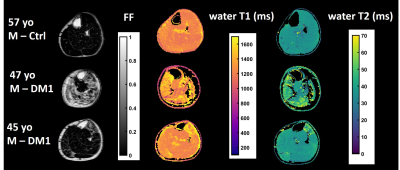 |
Longitudinal evaluation of the muscles of patients with
Myotonic Dystrophy type 1 treated by metformin, using
quantitative 1H and 23Na MRI
Benjamin Marty1,
Pierre-Yves Baudin1,
Aurélie Canal2,
Jean-Yves Hogrel2,
Melinda Gyenge3,
Nuria Jebrouni3,
Teresa Gerhalter4,
Armin M Nagel4,
Harmen Reyngoudt1,
and Guillaume Bassez3
1NMR Laboratory, Neuromuscular Investigation Center, Institute of Myology, Paris, France, 2Neuromuscular Physiology and Evaluation Laboratory, Neuromuscular Investigation Center, Institute of Myology, Paris, France, 3Institute of Myology, Paris, France, 4Institute of Radiology, University Hospital Erlangen, FAU, Erlangen, Germany Keywords: Muscle, Tissue Characterization Myotonic dystrophy type 1 (DM1) is a neuromuscular disorder resulting in progressive muscle wasting and dysfunction. We aimed at determining the relationship between several 1H and 23Na MRI indices of disease severity and disease activity in the muscles of DM1 patients, and evaluating their response to a 12-month metformin treatment. We showed that these indices could differentiate DM1 patients from healthy controls. Variables related to disease severity correlated with functional tests and indices related to disease activity were increased and mutually correlated. Over the 12-months treatment interval, MRI variables were more sensitive than functional outcomes to detect disease progression. |
| 08:15 |
0096.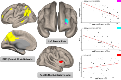 |
Fibromyalgia associates with pain-promoting and inhibitory
functional connectivity of the default mode network in
psoriatic arthritis.
Kristian Stefanov1,
Flavia Sunzini1,
Salim Al-Wasity1,
Steven Harte2,
Richard Harris3,
Daniel J. Clauw2,
Gordon Waiter4,
Jonathan Cavanagh1,
and Neil Basu1
1School of Infection & Immunity, University of Glasgow, Glasgow, United Kingdom, 2Chronic Pain and Fatigue Research Center, University of Michigan, Ann Arbor, MI, United States, 3University of Michigan, Ann Arbor, MI, United States, 4Aberdeen Biomedical Imaging Centre, University of Aberdeen, Aberdeen, United Kingdom Keywords: Rheumatoid Arthritis, Brain Connectivity, Brain, fMRI (Resting State), Inflammation, Multimodal Patients with the musculoskeletal disorder psoriatic arthritis improve their inflammation with current treatments but still experience pain. Previous neuroimaging findings have identified functional connectivity of the resting-state default mode network and we explored how such features associate with nociplastic pain using both agonistic and selective approaches. We observed increased connectivity between the default mode network and regions of the brain related to both increased pain intensity and decreased pain inhibition. The implications of such findings shift the focus onto targeting central pain pathways in people with psoriatic arthritis. |
| 08:15 |
0097.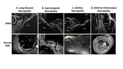 |
Sensitivity and Reproducibility of MRI Detection of
Hourglass-Like Constrictions in Parsonage-Turner Syndrome
Clare Nimura1,
Darryl Sneag1,
Philip Colucci1,
Casey Urban2,
Tim Li3,
Emily Pedrick1,
Joseph Feinberg4,
Carlo Milani4,
and Ek Tsoon Tan1
1Department of Radiology and Imaging, Hospital for Special Surgery, New York, NY, United States, 2Hand and Upper Extremity Service, Hospital for Special Surgery, New York, NY, United States, 3Weill Cornell Medical College, New York, NY, United States, 4Department of Physiatry, Hospital for Special Surgery, New York, NY, United States Keywords: Neurography, Nerves, Parsonage-Turner syndrome; electromyography A retrospective analysis of 123 patients diagnosed with Parsonage-Turner Syndrome (PTS; neuralgic amyotrophy) found that magnetic resonance neurography (MRN)-based detection of hourglass-like constrictions (HGCs) in affected nerves was 91.2-92.0% sensitive to electromyography-confirmed PTS. Post-hoc inter-rater reliability analysis revealed an inter-reliability of 91.3-94.3% for detection of HGCs. This retrospective study confirmed that MRN detection of HGCs is sensitive and reliable for diagnosing PTS and may be used as an objective diagnostic tool for the syndrome. |
| 08:15 |
0098.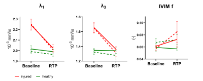 |
IVIM-corrected DTI in acute hamstring injury in a clinically
feasible acquisition time: a high-b DTI and multiband
acceleration approach
Susanne S. Rauh1,
Joep Suskens2,
Jithsa R. Monte3,
Frank Smithuis3,
Oliver J. Gurney-Champion3,
Johannes L. Tol2,
Mario Maas3,
Aart J. Nederveen3,
Gustav J. Strijkers1,
and Melissa T. Hooijmans3
1Department of Biomedical Engineering and Physics, Amsterdam Movement Sciences, Amsterdam UMC, location AMC, Amsterdam, Netherlands, 2Department of Orthopedic Surgery, Amsterdam UMC, location AMC, Amsterdam, Netherlands, 3Department of Radiology and Nuclear Medicine, Amsterdam UMC, location AMC, Amsterdam, Netherlands Keywords: Muscle, Diffusion Tensor Imaging Diffusion tensor imaging (DTI) with correction for intravoxel incoherent motion (IVIM) is a potential biomarker to assess hamstring injury recovery and predict return-to-play time. However, the long acquisition times hinder the use in clinical practice. By accelerating in the b-value space and using multiband acceleration the scan time can be reduced to clinically acceptable levels. In this work we showed that those methods preserve the sensitivity to hamstring injuries and that high-b DTI in combination with multiband factor 2 can reduce the scan time to 3:40min. |
| 08:15 |
0099.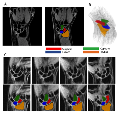 |
Development and Analysis of MRI-Derived Carpal Motion Data
Elements
Kevin Koch1,
Andrew S Nencka1,
Mohammad Zarenia2,
and Rajeev Mannem1
1Radiology, Medical College of Wisconsin, Milwaukee, WI, United States, 2Radiation Oncology, Medical College of Wisconsin, Milwaukee, WI, United States Keywords: Joints, Joints We present the construction and analysis of dynamic carpal data elements derived from 4D-MRI of the moving wrist. Across a clinically asymptomatic cohort of 31 subjects, dynamic data elements were constructed from capitate-normalized profiles of the scaphoid and lunate bones in a subject-specific radius-based coordinate system. Along with computing stability estimates of these measures, they are utilized to perform preliminary logistic regression modeling of radiologically-identified asymptomatic abnormalities within the study cohort. The results of this analysis suggests that the derived data elements have promising capabilities for characterizing carpal pathology and abnormalities. |
| 08:15 |
0100.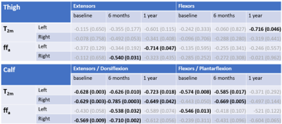 |
Lower limb muscle-water T2 as a marker of disease severity
and progression in amyotrophic lateral sclerosis
Nick Zafeiropoulos1,
Uros Klickovic1,
Luca Zampedri1,
Stephen Wastling1,
Jasper Morrow1,
Tarek Yousry1,
Michael Hanna1,
Linda Greensmith1,
Pietro Fratta1,
and John Thornton1
1Queen Square Institute of Neurology, University College London, London, United Kingdom Keywords: Muscle, Quantitative Imaging Using optimised maximum-likelihood-estimation extended-phase-graph-model relaxometry in ALS patients, elevated muscle water T2 (T2m) were revealed for all calf muscles and most thigh muscles vs. controls, with evidence of sparing of particular muscles. Similar but les consistent increases were seen in the associated fat fraction (ffa, relaxometry estimated). T2m correlated significantly with both functional rating scores and myometry, most consistently at the calf level. Associated ffa changes and correlations were less significant. |
08:15 |
0101.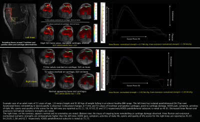 |
Towards the understanding of the the role of Functional
strength markers in Cartilage-Bone Cross Talk: a PET/MRI
study in isolated PFJOA patients
Rupsa Bhattacharjee1,
Eric C. Hammond2,
Koren E. Roach1,
Emma Bahroos1,
Richard B. Souza1,2,
Sharmila Majumdar1,
and Valentina Pedoia1
1Department of Radiology and Biomedical Imaging, University of California, San Francisco (UCSF), San Francisco, CA, United States, 2Department of Physical Therapy and Rehabilitation Science, University of California, San Francisco (UCSF), San Francisco, CA, United States Keywords: Bone, PET/MR, SUV, T1rho, flexion strength, extension strength, patellofemoral OA, Cartilage-Bone-CrossTalk In this exploratory study, we investigated standardized-uptake-values(SUV) from simultaneous bilateral PET-MRI, and individual joint loading in knees cross-sectionally in patients with isolated PFJOA. Additionally, to discover regions of colocalized changes in bone-cartilage interactions, we explored SUV interactions with cartilage biomarker T1rho relaxation values from high-resolution bilateral axial MAPSS. Strong negative correlations were between knee normalized isometric strength vs. maximum SUV in the lateral patella and medial trochlea, suggest higher degree of ongoing bone-remodelling combined with decreased knee flexion and extention strength. The existance of parallel higher trochlear bone remodelling and cartilage loss (disease progression) or vice-versa was also noted. |
| 08:15 |
0102.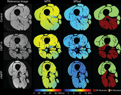 |
Muscle magnetization transfer ratio is promising biomarker
of hereditary neuropathy with liability to pressure palsy
Alison R Roth1,
Ashley J Minks1,
Yongsheng Chen2,
Jun Li3,
and Richard D Dortch1
1Barrow Neuroimaging Innovation Center, Barrow Neurological Institute, Phoenix, AZ, United States, 2Department of Neurology, Wayne State University School of Medicine, Detroit, MI, United States, 3Department of Neurology, Houston Methodist Hospital, Houston, TX, United States Keywords: Muscle, CEST & MT Magnetization transfer ratio (MTR) and volume of thigh muscles were investigated as imaging biomarkers in Charcot-Marie-Tooth type 1A (CMT1A) and hereditary neuropathy with liability to pressure palsy (HNPP) patients. MTR was significantly different between HNPP and healthy control subjects; muscle volume was significantly different between CMT1A patients and healthy control subjects and HNPP patients. MTR and volume were found to be repeatable and reliable. |
| 08:15 |
0103.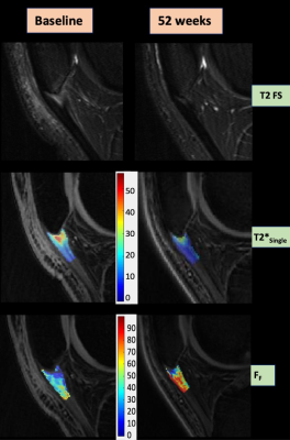 |
Association Between Ultrashort Echo-Time T2* Parameters and
Clinical Outcome in an RCT of Patellar Tendinopathy
Rianne A van der Heijden1,
Robert Moskwa2,
Fang Liu3,
John Wilson4,
Bryan Heiderscheit4,
Scott J Hetzel5,
Zachary E Stewart3,
Kenneth Lee1,
and Richard Kijowski6
1Department of Radiology, University of Wisconsin-Madison, Madison, WI, United States, 2Department of Medical Physics, University of Wisconsin-Madison, Madison, WI, United States, 3Massachusetts General Hospital/Harvard Medical School, Boston, MA, United States, 4Department of Orthopedics and Rehabilitation, University of Wisconsin-Madison, Madison, WI, United States, 5Department of Biostatistics and Medical Informatics, University of Wisconsin-Madison, Madison, WI, United States, 6Department of Radiology, New York University Grossman School of Medicine, New York, NY, United States Keywords: Tendon/Ligament, Quantitative Imaging, UTE This study was performed to investigate the association between bi-component UTE-T2* parameter of the patellar tendon and clinical pain scores over time in a randomized control trial for patellar tendinopathy (PT). The fraction of the fast-relaxing macromolecular bound water (FF) of the proximal patellar tendon and VAS pain scores were assessed at 0, 16 and 52-weeks in 29 patients with PT randomized into three treatment groups. All treatment groups showed a significant improvement (p<0.05) in pain and a significant increase (p<0.05) in FF over time, with a significant inverse correlation (p<0.05) between the increase in FF and decreased pain. |
 Back to Meeting Home
 Back to the Program-at-a-Glance
The International Society for Magnetic Resonance in Medicine is accredited by the Accreditation Council for Continuing Medical Education to provide continuing medical education for physicians.
 Back to Meeting Home
 Back to the Program-at-a-Glance
 Full Session Video