Power Pitch
Pitch: Advances in Brain Connectivity
ISMRM & ISMRT Annual Meeting & Exhibition • 03-08 June 2023 • Toronto, ON, Canada

Power Pitch
Pitch: Advances in Brain Connectivity
| 13:45 |
0219.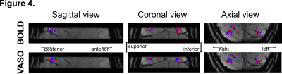 |
Mapping curvature responses in human V4 using VASO at 3T
Elisa Zamboni1,2,
Isaac Watson2,3,
Sharyfah Alasiri2,4,
Elizabeth Fear2,5,
Elia Formisano6,7,
Rainer Goebel7,8,
Rüdiger Stirnberg9,
Laurentius Huber6,7,
Aneurin Kennerley2,10,
and Antony Morland1,2,11
1Psychology, University of York, York, United Kingdom, 2York Neuroimaging Centre, University of York, York, United Kingdom, 3School of Physics, Engineering, and Technology, University of York, York, United Kingdom, 4University of York, York, United Kingdom, 5Biomolecular Sciences, University of Urbino Carlo Bo, Urbino, Italy, 6Cognitive Neuroscience, Maastricht University, Maastricht, Netherlands, 7Maastricht Brain Imaging Centre, Maastricht University, Maastricht, Netherlands, 8Cognitive Neuroscience, Maastricht Univeristy, Maastricht, Netherlands, 9German Center for Neurodegenerative Diseases (DNZE), Bonn, Germany, 10Institute of Sport, Manchester Metropolitan University, Manchester, United Kingdom, 11York Biomedical Research Institute, University of York, York, United Kingdom Keywords: Gray Matter, fMRI (task based), VASO The intermediate processing steps in human vision are not well characterised. We show however that the high specificity of VASO fMRI permits investigation functional organisation of curvature responses in human visual area V4, which is an intermediate region in the visual system. Understanding how the functional architecture and hierarchical integration of local contours (curvature) contributes to formation of shapes can inform computational models of object recognition. The emergence of inter-individual differences in these organisations can explain individual differences in healthy and impaired visual perception. |
| 13:45 |
0220.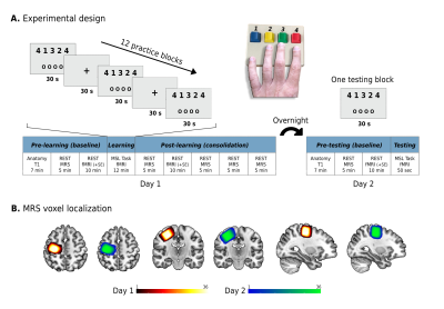 |
Excitation and inhibition in the human M1 during motor
memory consolidation and the relation to neural plasticity:
a multimodal 7T study
Tamir Eisenstein1,
Edna Furman-Haran2,
and Assaf Tal1
1Department of Chemical and Biological Physics, Weizmann Institute of Science, Rehovot, Israel, 2Life Sciences Core Facilities, Weizmann Institute of Science, Rehovot, Israel Keywords: Brain Connectivity, Spectroscopy, learning and plasticity Here, we show, using ultra-high field MRS and functional and structural MRI, how changes in glutamate and GABA in the human motor cortex following motor skill learning may play key roles in promoting motor memory consolidation and neuroplasticity. Increased glutamate after learning was associated with overnight skill performance improvements, and increased functional connectivity of M1 with the striatum, suggesting for functional plasticity. Greater reduction in GABA following learning was associated with increased grey matter volume in M1 overnight, suggesting for structural plasticity. We therefore highlight the importance of these neurochemical modifications in promoting learning and plasticity in the human brain. |
| 13:45 |
0221.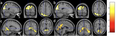 |
Dynamic ASL and Multi-echo BOLD in Hippocampus Functional
Connectivity of Aging
Zongpai Zhang1,
Shichun Chen1,
Yanchen Guo1,
Yakun Zhang1,
Lissa Riley2,
Adam K. Anderson2,
and Eve DeRosa2
1State University of New York at Binghamton, Binghamton, NY, United States, 2Cornell University, Ithaca, NY, United States Keywords: Brain Connectivity, Aging The effect sizes of aging in hippocampus (HPC) resting-state functional connectivity (rsFC) using dASL and multi-echo BOLD (ME-BOLD) imaging were evaluated in 20 subjects (10 young adults, 10 older adults). Both dASL and ME-BOLD showed reduced HPC rsFC in older adults but with larger extended region in dASL results. dASL demonstrated significantly higher effect sizes of aging in HPC rsFC compared to ME-BOLD, supporting more sensitivity of dASL in detecting the aging effect in HPC rsFC. |
| 13:45 |
0222.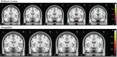 |
Multi-Echo EPI Increases Sensitivity to BOLD Activation in
the Olfactory Network Compared to Single-Echo EPI
Sichen Ludwig Zhao1,2,
Clara U Raithel2,3,
Jay A Gottfried2,3,
John A Detre2,
and M Dylan Tisdall4
1Department of Bioengineering, School of Engineering and Applied Science, University of Pennsylvania, Philadelphia, PA, United States, 2Department of Neurology, Perelman School of Medicine, University of Pennsylvania, Philadelphia, PA, United States, 3Department of Psychology, School of Arts and Science, University of Pennsylvania, Philadelphia, PA, United States, 4Department of Radiology, Perelman School of Medicine, University of Pennsylvania, Philadelphia, PA, United States Keywords: Brain Connectivity, fMRI, Multi-echo EPI The human olfactory network presents technical challenges for functional MRI due to its location in regions of high static susceptibility. We developed a novel multi-echo EPI (ME-EPI) protocol, optimized for olfactory regions, and compared this protocol to a conventional single-echo EPI (1E-EPI). We show that the optimized ME-EPI increases sensitivity to BOLD activation response for olfactory regions and reduces the numbers of subjects required to detect significant group effects. |
| 13:45 |
0223.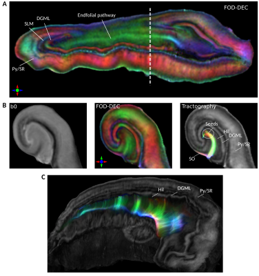 |
Investigating the endfolial pathway of the human hippocampus
using ex vivo dMRI at high spatio-angular resolution
Jonathan Scharff Nielsen1 and
Manisha Aggarwal1
1Department of Radiology and Radiological Science, Johns Hopkins University, Baltimore, MD, United States Keywords: Brain Connectivity, Tractography & Fibre Modelling, Hippocampus internal circuitry; probabilistic tractography. The endfolial pathway is a collection of fibers within the hilus of the human hippocampus that forms part of the complex intra-hippocampal circuitry. We investigate this pathway using high-field (11.7 T), high spatio-angular resolution diffusion MRI (dMRI) of 3 intact, excised hippocampi. Using probabilistic tractography and computational unfolding of the hippocampal strata, we demonstrate a unique sensitivity of dMRI to the 3D trajectory of the intra-hippocampal fibers. Keywords: Ex vivo high spatio-angular resolution diffusion MRI; hippocampus internal circuitry; probabilistic tractography. |
| 13:45 |
0224.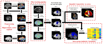 |
Hippocampal-cortical connections with high-resolution ex
vivo and in vivo diffusion MRI of human brain at 7T
Zuozhen Cao1,
Zhiyong Zhao1,
Qinfeng Zhu1,
Keqing Zhu2,
Jing Zhang3,
and Dan Wu1
1Department of Biomedical Engineering, Zhejiang University, HangZhou, China, 2China Brain Bank and Department of Neurology in Second Affiliated Hospital, Zhejiang University, HangZhou, China, 3Department of Pathology, The First Affiliated Hospital and School of Medicine, Zhejiang University, HangZhou, China Keywords: Brain Connectivity, Diffusion/other diffusion imaging techniques, Ex-vivo, hippocampus, structure connectivity Hippocampus is a critical brain structure associated with many brain functions. The connectivity between hippocampal sub-regions and cerebral cortex hasn’t been fully characterized, and spatial resolution is the key to resolve such connectivity. We utilized 3D high-resolution ex vivo diffusion MRI (dMRI) at 7T to investigate the structure connectivity between hippocampal sub-regions and cortex and compared the results with in vivo data. We found that different sub-regions demonstrated unique fiber projections to cortex, and the high-resolution ex-vivo dMRI resulted in more connections with temporal-occipital lobe and less connections to central gyrus and frontal lobe, compared to lower-resolution in vivo dMRI. |
| 13:45 |
0225.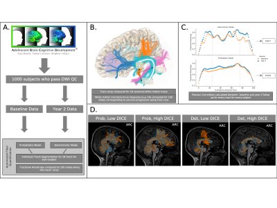 |
Optimizing Reproducibility of White Matter Tractography in
Longitudinal Imaging of a Large Sample of Developing
Adolescents
Pierre Nedelec1,
Samuel Lashof-Regas1,
Andreas Rauschecker1,
and Leo Sugrue1
1Radiology & Biomedical Imaging, University of California, San Francisco, San Francisco, CA, United States Keywords: Brain Connectivity, Adolescents The best method for automatically producing replicable white matter (WM) tracts from diffusion imaging is not known. We use Automated Fiber Quantification (AFQ) to compare probabilistic and deterministic models’ performance generating 18 WM tracts for each of 1000 adolescents at baseline and 2-year follow up. We gauged performance on the correlation between fractional anisotropy (FA) values and degree of volumetric overlap (DICE score). Based on these measures we found that a probabilistic approach was able to more reliably generate tract profiles over time within individual subjects. |
| 13:45 |
0226.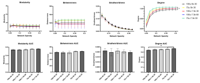 |
The Influence of b value and Resolution on MR Tractography
and Connectome Construction in Adult Mouse Brain at 16.4
Tesla
Majd Alkhalily1,
Laura Currey2,
Michael Piper2,
and Nyoman D Kurniawan1
1Centre for Advanced Imaging, The University of Queensland, Brisbane, Australia, 2The School of Biomedical Sciences, The University of Queensland, Brisbane, Australia Keywords: Brain Connectivity, Diffusion/other diffusion imaging techniques, Tractography Several pipelines are available to quantify mouse brain connectome using diffusion-tractography. However, data acquisition and processing methods used a wide range of parameters. In this study, ex-vivo adult mouse brains were scanned using a range of b values, and spatial and angular resolutions at 16.4 T. iFOD2 were used for fibertracking to generate the connectomes. Network analysis revealed increased nodal degree and reduced connectivity at 75 μm and this change increases at higher b value. Comparison with Allen mouse brain atlas showed consistent connectome profiles were achieved by acquiring diffusion with b=5000 s/mm2 at 100 μm resolution and 30 directions. |
13:45 |
0227.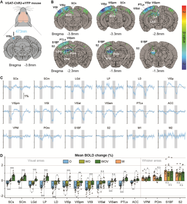 |
Separation of cortical and non-cortical networks to
visual-somatosensory interaction by fMRI with optogenetic
silencing
Thi Ngoc Anh Dinh1 and
Seong-Gi Kim1
1Department of Biomedical Engineering, Sungkyunkwan University (SKKU), Suwon 16419, South Korea, Center for Neuroscience Imaging Research, Institute for Basic Science (IBS), Suwon 16419, South Korea, Suwon, Korea, Republic of Keywords: Brain Connectivity, fMRI (task based) Multisensory integration is necessary for the animal to survive in the real world. Multiple studies with conventional methods established the multisensory integration process in multiple brain areas. This study aimed to investigate interactions between visual and somatosensory networks in a whole brain scale using 15.2T BOLD fMRI with the assistance of cortical silencing by optogenetic stimulation.Keywords: ultrahigh field fMRI, optogenetics, multimodal integration. |
| 13:45 |
0228.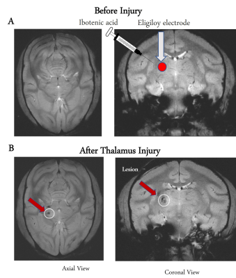 |
Reorganization of functional networks in squirrel monkey
brain after thalamic injury and their relations to
task-specific behaviors
Anirban Sengupta1,
Daniela Hernandez Duque1,
Arabinda Mishra1,
Jamie L Reed1,
Pai-Feng Yang1,
Feng Wang1,
Zhangyan Yang1,
Li Min Chen2,
and John C Gore1
1Vanderbilt University Medical Center, Nashville, TN, United States, 2Vanderbilt University Medical Center, NASHVILLE, TN, United States Keywords: Brain Connectivity, Brain, Thalamus lesion Resting state fMRI was used to assess whole brain functional networks before and after a targeted, unilateral lesion in the hand and arm representation of the ventroposterior lateral nucleus of the thalamus of squirrel monkeys. Using Independent Component Analysis, the overall whole brain resting state functional connectivity (rsFC) dropped after injury but slowly recovered. The trajectory of network changes corresponded well with quantitative metrics of animal performance on a behavioral task using the affected hand. Some functional networks showed reductions in connectivity post injury followed by recoveries while others reflected compensatory increases that began in the first week after injury. |
|
13:45 |
0229.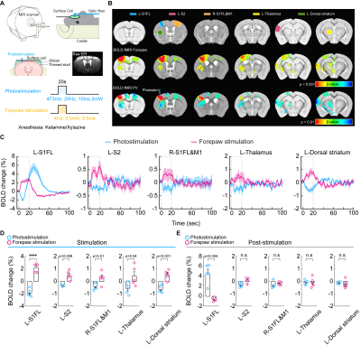 |
Biphasic fMRI responses to optogenetic stimulation of
parvalbumin interneurons
Tan Thanh Vo1,2,3,
Geun Ho Im3,
Patrick J Drew4,
and Seong-Gi Kim1,2,3
1Department of Biomedical Engineering, Sungkyunkwan University, Suwon 16419, Republic of Korea, Suwon, Korea, Republic of, 2Department of Intelligent Precision Healthcare Convergence, Sungkyunkwan University, Suwon 16419, Korea, Suwon, Korea, Republic of, 3Center for Neuroscience Imaging Research (CNIR), Institute for Basic Science (IBS), Suwon 16419, Republic of Korea, Suwon, Korea, Republic of, 4Center for Neural Engineering, Departments of Engineering Science and Mechanics, Neurosurgery, Biology, and Biomedical Engineering, The Pennsylvania State University, University Park, PA 16802, USA, University Park, PA, United States Keywords: Brain Connectivity, fMRI (task based) Parvalbumin (PV) neurons are the largest population of cortical interneurons, thus their role in neurovascular coupling is highly important in interpreting fMRI data, especially optogenetic fMRI. However, PV-based hemodynamic response is not well-understood. Here, we observed the biphasic BOLD response with initial vasoconstriction and follow-up ultraslow vasodilation at the stimulation site by PV photostimulation, while negative BOLD was observed at the downstream output. CBV-fMRI revealed that vasoconstriction localizes at middle to deeper layers matching with the distribution of glutamatergic neurons, while vasodilation propagated from superficial layers driven by neuropeptide substance P signaling. |
| 13:45 |
0230.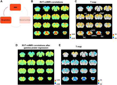 |
Neural underpinning of a respiration-related resting-state
fMRI network
Nanyin Zhang1 and
Wenyu Tu1
1The Pennsylvania State University, University Park, PA, United States Keywords: Brain Connectivity, Animals, Respiration, Physiological impacts Respiration can induce non-neural artifacts in the resting-state fMRI (rsfMRI) signal. In the meantime, as a crucial physiologic process, respiration that can directly drive neural activity change, and may thereby modulate the rsfMRI signal. Nonetheless, this potential neural component in the respiration-fMRI relationship remains elusive. To elucidate this issue, we developed a platform to achieve a concurrent measure of electrophysiology, respiration, and whole brain rsfMRI signals, and identified a respiration-associated network that was underpinned by neural activity, which represents a novel component in the respiration-rsfMRI relationship that is distinct from respiration-related rsfMRI artifacts. |
|
13:45 |
0231.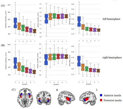 |
Mapping functional connectivity of insular subdivisions
using connectivity-based parcellation in adolescents with
major depressive disorder
Zilin Zhou1,
Lingxiao Cao1,
Yingxue Gao1,
Ruohan Feng2,
Yang Li3,
Weijie Bao1,
Kaili Liang1,
Lihua Zhuo2,
Guoping Huang3,
and Xiaoqi Huang1,4
1Huaxi MR Research Center (HMRRC), Functional and Molecular Imaging Key Laboratory of Sichuan Province, Department of Radiology, West China Hospital of Sichuan University, Chengdu, China, Chengdu, China, 2Department of Radiology, the Third Hospital of Mianyang, Sichuan Mental Health Center, Mianyang, China, Mianyang, China, 3Department of Psychiatry, the Third Hospital of Mianyang, Sichuan Mental Health Center, Mianyang, China, Mianyang, China, 4Psychoradiology Research Unit of the Chinese Academy of Medical Science , West China Hospital of Sichuan University, Chengdu, Sichuan, China, Chengdu, China Keywords: Psychiatric Disorders, Adolescents, major depressive disorder Using a data-driven connectivity-based parcellation technique, we identified anterior and posterior functional subdivisions of insula per hemisphere in adolescent population based on their similar functional connectivity profiles. Then, we investigated the distinct functional connectivity of each insular subregion between depressive adolescents and typical developmental controls. The significant unbalanced connectivity of insular subregions with left superior frontal gyrus (SFG) between two groups was revealed, which associated with interpersonal relation, emotional expressiveness, and cognition impairments in depressive adolescents. Our findings indicate that abnormalities in functional architectures of insular subdivisions may be the potential neuroimaging mechanism underlie the specific manifestations in adolescent depression. |
| 13:45 |
0232.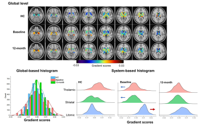 |
Distinct subcortical and cortical functional gradient
dysfunction in schizophrenia and the treatment effects after
antipsychotics
Chengmin Yang1,
Wenjing Zhang1,
Jiajun Liu2,
Zhipeng Yang2,
and Su Lui1
1Huaxi MR Research Center (HMRRC), Functional and molecular imaging Key Laboratory of Sichuan Province, Department of Radiology, West China Hospital, Sichuan University, Chengdu, China., Chengdu, China, 2College of Electronic Engineering, Chengdu University of Information Technology, Chengdu, P.R. China., Chengdu, China Keywords: Psychiatric Disorders, Gradients By characterizing the connectome gradient changes of subcortex and cortex in drug-naive first-episode schizophrenia and the treatment effect after antipsychotics, we found that the distinct fundamental functional segregation of subcortex and functional integration in cortex in patients at baseline when compared to healthy controls, and the longitudinal analyses indicated that the treatment would normalize the altered gradients. The improved subcortical gradient changes were associated with significant improvement of symptoms. This study provided the new perspective on the abnormal subcortical and cortical hierarchy organization in schizophrenia and its longitudinal subcortical gradient changes could be sensitive to reflect the antipsychotic treatment effect. |
| 13:45 |
0233.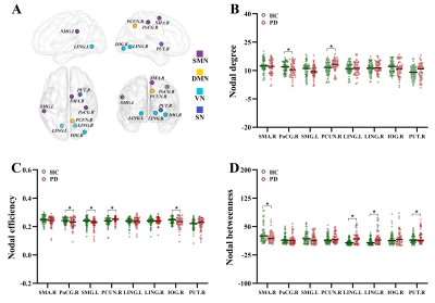 |
Topological Disruption of High-Order Functional Networks in
Cognitively Preserved Parkinson’s Disease
Song'an Shang1,
Weiqiang Dou2,
and Ye Jing1
1Clinical Medical College, Yangzhou University, Yangzhou, China, 2GE Healthcare, MR Research China, Beijing, China Keywords: Brain Connectivity, Parkinson's Disease Parkinson’s disease (PD) is not solely a disruption of motor-related networks but a consequence of impaired high-level cognitive processes, indicating that high-order information exchange in cognitively preserved patients with PD could be sensitively revealed in the high-order-functional connection (HOFC) networks. We thus attempted to characterize the topological alterations and classification performance of HOFC networks in normally cognitive patients with PD. Our findings identified the disrupted topology of functional interactions at high-level with extensive alterations of topological properties and improved differentiating ability in patients with PD prior to clinical symptoms of cognitive impairment, providing complementary insights into complex neurodegeneration in PD. |
| 13:45 |
0234.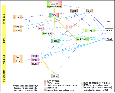 |
Functional connectivity changes of brainstem nuclei in
prodromal Parkinson’s disease: A 7 Tesla resting-state study
Kavita Singh1,
Maria Guadalupe Garcia Gomar1,2,
Ambra Stefani3,
Aleks Videnovic3,
and Marta Bianciardi1,4
1Brainstem Imaging Laboratory, Department of Radiology, Athinoula A. Martinos Center for Biomedical Imaging, Massachusetts General Hospital and Harvard Medical School, Boston, MA, United States, 2Escuela Nacional de Estudios Superiores Unidad Juriquilla, Universidad Nacional Autónoma de México, Querétaro, Mexico, 3Department of Neurology, Massachusetts General Hospital and Harvard Medical School, Boston, MA, United States, 4Division of Sleep Medicine, Harvard University,, Boston, MA, United States Keywords: Parkinson's Disease, fMRI (resting state), 7 Tesla MRI, REM sleep behavior disorder, prodromal PD, alpha synucleopathies, brainstem Based on animal and human lesion studies, isolated rapid-eye-movement (REM) sleep behavior disorder (iRBD) is due to changes in arousal/motor brainstem nuclei structure and function and is recognized as the hallmark of Parkinson disease (PD). Resting-state fMRI studies in iRBD in humans mostly studied changes in nigrostriatal/nigrocortical connectivity pathway but failed to assess the brainstem circuits involved. To fill this gap, we investigated the functional connectivity of 20 brainstem nuclei relevant for iRBD using high-sensitivity and high spatial-resolution 7 Tesla resting-state fMRI, as well as a recently developed in-vivo probabilistic atlas of brainstem nuclei. |
| 13:45 |
0235.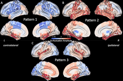 |
Dynamic co-fluctuation patterns of fMRI brain activity in
temporal lobe epilepsy
Lucas E Sainburg1,
Baxter P Rogers2,
Catie Chang3,
Dario J Englot4,
and Victoria L Morgan2
1Biomedical Engineering, Vanderbilt University, Nashville, TN, United States, 2Radiology and Radiological Sciences, Vanderbilt University Medical Center, Nashville, TN, United States, 3Electrical and Computer Engineering, Vanderbilt University, Nashville, TN, United States, 4Neurological Surgery, Vanderbilt University Medical Center, Nashville, TN, United States Keywords: Brain Connectivity, Epilepsy Temporal lobe epilepsy (TLE) presents with transient epileptic activity at rest, typically originating from the hippocampus. Here we measure dynamic functional connectivity (FC) at single functional MRI timepoints with edge timeseries to detect recurring hippocampal co-fluctuation patterns and their alterations in TLE. Three anterior hippocampus co-fluctuation patterns were detected in healthy controls. One pattern was altered in TLE, while another pattern was more frequent in TLE than controls. These changes reflect alterations in dynamic spatial patterns of FC from the epileptic focus in TLE. |
| 13:45 |
0236.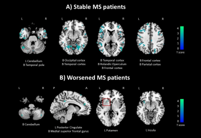 |
Longitudinal Changes of Functional Connectivity Dynamism Are
Relevant for Disability Worsening in Multiple Sclerosis: A
2.5-Year Study
Paola Valsasina1,
Giulia d'Amore1,2,
Paolo Preziosa1,2,3,
Monica Margoni1,2,
Massimo Filippi1,2,3,4,5,
and Maria Assunta Rocca1,2,3
1Neuroimaging Research Unit, Division of Neuroscience, IRCCS San Raffaele Scientific Institute, Milan, Italy, 2Neurology Unit, IRCCS San Raffaele Scientific Institute, Milan, Italy, 3Vita-Salute San Raffaele University, Milan, Italy, 4Neurorehabilitation Unit, IRCCS San Raffaele Scientific Institute, Milan, Italy, 5Neurophysiology Service, IRCCS San Raffaele Scientific Institute, Milan, Italy Keywords: Brain Connectivity, Multiple Sclerosis Here, we investigated changes in time-varying functional connectivity over 2.5 years of follow-up in 129 multiple sclerosis patients and their association with disability progression. At follow-up, 25/129 (19.3%) patients worsened clinically. At baseline, multiple sclerosis patients showed reduced time-varying functional connectivity vs controls in orbitofrontal, cerebellar, precuneal and thalamic regions. At 2.5-year follow-up, patients exhibited widespread reduction of time-varying functional connectivity over time. Such a pattern was confirmed when looking at clinically stable patients. Conversely, clinically worsened patients presented peculiar reductions of time-varying functional connectivity in default-mode network areas and in basal ganglia, this latter significant at time-by-group interaction analysis. |
| 13:45 |
0237.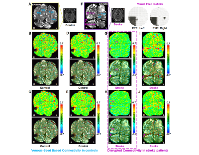 |
Disrupted vein-dominated connectivity and increased CSF
involvement in ischemic stroke human brain
Jianyu Yuan1,
Chaogang Tang2,
Lei Zhang2,
and Yi He1
1Guangdong Provincial Key Laboratory of Biomedical Imaging and Guangdong Provincial Engineering Research Center of Molecular Imaging, The Fifth Affiliated Hospital, Sun Yat-sen University, Zhuhai, China, 2Department of Cerebrovascular Disease, The Fifth Affiliated Hospital, Sun Yat-sen University, Zhuhai, China Keywords: Brain Connectivity, fMRI (resting state) High-resolution resting-state (rs) fMRI enables the functional mapping of vein-dominated connectivity, correlated with neuronal calcium signals. Here, we performed high-resolution rs-fMRI to examine 30 ischemic stroke patients with binocular isotropic hemianopia and 10 healthy controls. The results of seed-based and independent component analysis (ICA) demonstrated the reduced vein-dominated correlation patterns. Interestingly, ICA also found increased correlations between cerebrospinal fluid (CSF) and ischemic lesions in stroke patients, showing ultra-slow oscillation frequencies up to 0.04 Hz. Our findings suggest that ischemic lesions are associated with CSF, prompting disruption of vein-dominated connectivity. |
| 13:45 |
0238.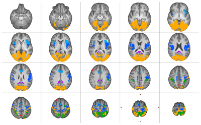 |
A comparison of multi-echo and single-echo fMRI resting
state mapping in neurosurgical patients
Ahmed Radwan1,
Louise Emsell1,
Ronald Peeters2,
Tom Theys3,
Patrick Dupont4,
and Stefan Sunaert1
1Department of Imaging and pathology, Translational MRI, KU Leuven, Leuven, Belgium, 2Department of Radiology, University Hospitals Leuven, Leuven, Belgium, 3Department of Neurosciences, Research Group Experimental Neurosurgery and Neuroanatomy, KU Leuven, Leuven, Belgium, 4Department of Neurosciences, Laboratory for Cognitive Neurology, KU Leuven, Leuven, Belgium Keywords: Brain Connectivity, fMRI (resting state), multi-echo fMRI, Neurosurgery We compared multi-echo (mTE) resting-state (rs) fMRI to single-echo (sTE) fMRI for seed-based mapping of seven canonical resting state networks (RSN) in 69 pre-surgical patients. Acquisition parameters were identical except for TE and statistical analyses were constrained by group-specific RSN masks. mTE-rsfMRI showed significantly higher functional-connectivity (fc) for all RSNs, while sTE showed comparatively fewer RSNs with areas of significantly higher fc (PFWE<0.05). Our results suggest that mTE-rsfMRI may be more sensitive than sTE-rsfMRI for mapping of the sensory-motor, default mode, salience, frontoparietal, dorsal attention, language and visual networks in a neurosurgical setting. |
 Back to Meeting Home
 Back to the Program-at-a-Glance
The International Society for Magnetic Resonance in Medicine is accredited by the Accreditation Council for Continuing Medical Education to provide continuing medical education for physicians.
 Back to Meeting Home
 Back to the Program-at-a-Glance
 Full Session Video