Power Pitch
Pitch: Neurodegeneration in Human & Animal Models
ISMRM & ISMRT Annual Meeting & Exhibition • 03-08 June 2023 • Toronto, ON, Canada

Power Pitch
Pitch: Neurodegeneration in Human & Animal Models
| 13:30 |
0574.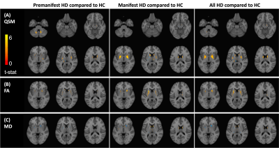 |
7T MRI Reveals Abnormal Iron Deposition and Microstructure
in Premanifest and Manifest Huntington’s Disease
Jingwen Yao1,
Melanie A. Morrison1,2,
Angela Jakary1,
Sivakami Avadiappan1,
Julia Glueck3,
Theresa Driscoll3,
Michael Geschwind3,
Alexandra Nelson3,
Duan Xu1,2,
Christopher P. Hess1,3,
and Janine M. Lupo1,2
1Department of Radiology and Biomedical Imaging, University of California, San Francisco, San Francisco, CA, United States, 2UCSF/UCB Graduate Program in Bioengineering, University of California, San Francisco, San Francisco, CA, United States, 3Department of Neurology, University of California, San Francisco, San Francisco, CA, United States Keywords: Neurodegeneration, Neurodegeneration, Huntington's disease, Movement disorder We used QSM and DTI at 7T to investigate iron dysregulation and microstructure disruption in subcortical regions in Huntington’s disease (HD). We observed significant volume loss and increased iron deposition in the striatum and globus pallidus, and increased FA in the striatum. The deep cerebellar nuclei (dentate nuclei) showed a unique transient increase in volume and susceptibility in premanifest patients, implicating it as a new marker of HD disease progression that is sensitive to the pre-symptomatic window of HD. We also found varying relationships between imaging features in different brain regions, warranting further analyses of subregional changes. |
| 13:30 |
0575.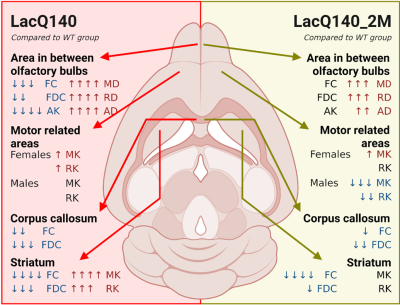 |
Diffusion MRI reveals rescue of structural changes in a
mouse model of Huntington’s disease by mutant Huntingtin
lowering
Joëlle van Rijswijk1,2,
Nicholas Vidas-Guscic1,2,
Johan Van Audekerke1,2,
Tamara Vasilkovska1,2,
Dorian Pustina3,
Haiying Tang3,
Roger Cachope3,
Deanna M. Marchionini3,
Ignacio Munoz-Sanjuan3,
Annemie Van der Linden1,2,
Mohit H Adhikari1,2,
and Marleen Verhoye1,2
1Bio-Imaging Lab, Department of Biomedical Sciences, University of Antwerp, Antwerp, Belgium, 2µNEURO Research Centre of Excellence, University of Antwerp, Antwerp, Belgium, 3CHDI Management/CHDI Foundation, Princeton, NJ, United States Keywords: Neurodegeneration, Diffusion/other diffusion imaging techniques, Huntington's Disease Huntington’s disease (HD) is a neurodegenerative disorder for which no cure is available. There is an urgent need to find early biomarkers which are sensitive enough to determine and follow up the efficacy of novel therapies. In this study, we present the outcome of a treatment study in the LacQ140 HD mouse model using diffusion tensor/kurtosis imaging (DTI/DKI) and fixel-based analysis (FBA). We observed that early, moderate lowering of mutant Huntingtin (mHtt) rescues structural alterations measured in this mouse model with diffusion MRI in the olfactory bulb and striatum, which are prominent regions affected in HD. |
| 13:30 |
0576.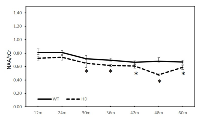 |
A longitudinal study of cerebral metabolite alteration in
the developmental monkey brain with Huntington's disease
Chunxia Li1 and
Xiaodong Zhang1
1Emory National Primate Research Center, Emory University, Atlanta, GA, United States Keywords: Neurodegeneration, Spectroscopy, Huntington's disease Previous studies have demonstrated in vivo MRS could be an effective approach to assess the metabolite changes in Huntington Disease (HD) patients and models. However, its sensitivity to detect the metabolite abnormality is still in dispute. The metabolic changes in the striatum of transgenic monkeys of HD were investigated with MRS from 12 to 60 months of age here. A progressive and significant reduction of NAA/tCr and NAA/tCho in striatum was observed at 30 and 36 months and old, respectively, suggesting the sensitivity of the in vivo MRS to assess the neurochemical alteration in the evolution of the disease. |
| 13:30 |
0577.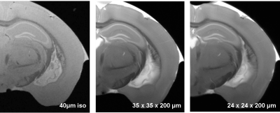 |
Ultra-high spatial resolution for ex-vivo structural brain
MRI
Alireza Abaei1,
Stefano Antonucci2,
Jelena Scekic-Zahirovic2,
Florian olde Heuvel2,
Francesco Roselli2,
and Volker Rasche1
1Core Facility Small Animal Imaging (CF-SANI), University of Ulm, Ulm, Germany, 2German Center for Neurodegenerative Diseases (DZNE), Ulm, Germany Keywords: Neurodegeneration, Preclinical, ultra-high resolution neuro neuroimaging This study aims at demonstrating the feasibility of ex vivo MRI for structural imaging of brain. High spatial resolution in the range of 15-20 µm is obtained by an optimized FLASH sequence together with a cryogenically cooled RF coil. A 15µm-resolution was obtained for the brain, revealing cortical grey matter lamination, white matter and vascular architecture. As proof of concept, we showed in an Amyotrophic Lateral Sclerosis ( FUSΔNLS) mouse the pattern of atrophy as proxy of vulnerability using high degree of anatomical granularity. Overall, we demonstrate an ex-vivo MRI strategy with histology-grade resolution, comprehensive and non-destructive brain sampling. |
13:30 |
0578.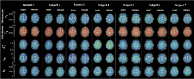 |
A Method for Detection of Subtle Blood-Brain Barrier
Disruption using Non-Contrast MR Fingerprinting
Emma L Thomson1,2,
Elizabeth Powell1,
Claudia A M Gandini Wheeler-Kingshott3,4,5,
and Geoff J M Parker1,6,7
1Centre for Medical Image Computing, Department of Medical Physics and Biomedical Engineering, University College London, London, United Kingdom, 2UCL Queen Square Institute of Neurology, Faculty of Brain Sciences, London, United Kingdom, 3NMR Research Unit, Queen Square MS Centre, Department of Neuroinflammation, London, United Kingdom, 4Department of Brain & Behavioural Sciences, University of Pavia, Pavia, Italy, 5Brain Connectivity Centre Research Department, IRCCS Mondino Foundation, Pavia, Italy, 6NMR Research Unit, Queen Square MS Centre, Department of Neuroinflammation, UCL Queen Square Institute of Neurology, Faculty of Brain Sciences, London, United Kingdom, 7Bioxydyn Limited, Manchester, United Kingdom Keywords: Neurodegeneration, Blood vessels Using magnetic resonance fingerprinting (MRF), we propose a method for regional quantification of blood volume (νb) and BBB water exchange (by quantifying capillary water residence time, τb), as a metric of BBB function/dysfunction. A single axial slice was acquired for seven healthy volunteers using an MRF SPGR sequence. Matching to a precomputed dictionary was performed to simultaneously quantify intra and extravascular T1, B1+, νb, and τb. Initial findings in volunteers show that it is possible to quantify these five parameters simultaneously. Our results show promise for patient studies of blood-brain barrier disruption. |
| 13:30 |
0579.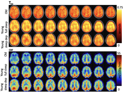 |
Investigating application of multi-echo, multi-delay
arterial spin labeling (ASL) to study endothelial exchange
of water
Swati Rane Levendovszky1,
Lena Vaclavu2,
Jaqueline Flores1,
Elaine Peskind3,
Jeffrey Iliff3,
and Matthias J.P. van Osch2
1Radiology, University of Washington School of Medicine, Seattle, WA, United States, 2C.J. Gorter MRI Center, Radiology, Leiden University Medical Center, Leiden, Netherlands, 3Mental Illness Research, Education and Clinical Center, VA Puget Sound, Seattle, WA, United States Keywords: Neurodegeneration, Perfusion, sleep, aging We applied multi-echo Hadamard encoded ASL imaging to study blood flow (CBF) and trans-endothelial exchange of water (Tex) as markers of vascular function to study two common risk factors of dementia; aging and sleep deprivation. We found that older adults had significantly lower CBF, and shorter Tex compared to young adults. In the same young adults, we compared CBF and Tex after a normal night of sleep and after sleep deprivation. Both CBF and Tex were insignificantly lower when measured after sleep deprivation compared to measurements after normal sleep. |
| 13:30 |
0580.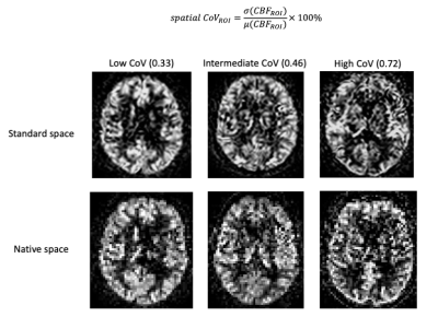 |
ASL spatial coefficient of variance predicts increased white
matter hyperintensities volume over time in cognitively
unimpaired subjects
Beatriz Padrela1,
Lyduine E Collij1,
Luigi Lorenzini1,
Silvia Ingala1,
Carole Sudre1,
Pieter Jelle Visser2,
Anouk den Braber2,
Frederik Barkhof1,3,
Jan Petr4,
and Henk J.M.M. Mutsaerts1
1Radiology and Nuclear Medicine, Amsterdam UMC, Amsterdam, Netherlands, 2Department of Neurology, Neuroscience Campus Amsterdam, VU University Medical Center, Location VUmc, Alzheimer Center, Amsterdam, Netherlands, 3University College London, Centre for Medical Image Computing (CMIC), London, United Kingdom, 4Institute of Radiopharmaceutical Cancer Research, Helmholtz-Zentrum Dresden-Rossendorf, Dresden, Germany Keywords: Neurodegeneration, Perfusion, Cerebral blood flow, Dementia Arterial transit artifacts (ATAs) in arterial spin labeling (ASL) images are common in populations with prolonged arterial transit time (ATT) and may be associated with vascular insufficiency. The spatial coefficient of variance (sCoV) of ASL images can quantify the presence of these artifacts. Vascular insufficiency could contribute to the development of white matter hyperintensities (WMH), a common marker of cerebral small vessel disease. We demonstrated that baseline sCoV of CBF is associated with WMH at baseline and predicts WMH volume change, in a cognitively unimpaired population of 88 subjects. |
| 13:30 |
0581.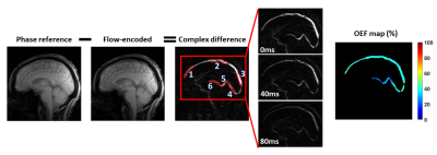 |
Region-specific characteristics of brain oxygen extraction
fraction: relationships to aging and white matter
hyperintensities
Jie Song1,
Wen Shi2,
Kaisha Hazel3,
George Pottanat3,
Ebony Jones3,
Cuimei Xu3,
Doris Lin3,
Sevil Yasar4,
Marilyn Albert5,
Hanzhang Lu3,
and Dengrong Jiang3
1Department of Biomedical Engineering, Johns Hopkins University School of Engineering, Baltimore, MD, United States, 2Department of Biomedical Engineering, Johns Hopkins University School of Medicine, Baltimore, MD, United States, 3Department of Radiology, Johns Hopkins University School of Medicine, Baltimore, MD, United States, 4Department of Medicine, Johns Hopkins School of Medicine, Baltimore, MD, United States, 5Department of Neurology, Johns Hopkins University School of Medicine, Baltimore, MD, United States Keywords: Neurodegeneration, Aging Aging is a multifaceted process involving both structural and metabolic alterations. Cerebral oxygen-extraction-fraction (OEF) is an important physiological parameter indexing the brain’s oxygen metabolism. In this work, we assessed regional OEF in young and older adults, and investigated its associations with aging and white-matter-hyperintensities. We observed significant age-related increase in cortical OEF but not in subcortical OEF, suggesting that aging may have different effects on tissue metabolism in cortical and subcortical regions. Furthermore, we found a significant inverse correlation of WMH with OEF in internal-cerebral-veins, implying that OEF of subcortical structures may be useful in predicting WMH. |
| 13:30 |
0582.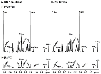 |
Chronic stress impacts energy metabolism and triggers motor
neuropathy in an Optineurin knockout mouse model
Bedaballi Dey1,2,
Dipak Roy1,
Sumana Chakravarty2,3,
Ghanshyman Swarup1,
Arvind Kumar1,2,
and Anant Bahadur Patel1,2
1CSIR-Centre for Cellular & Molecular Biology (CCMB), Hyderabad, India, 2Academy of Scientific and Innovative Research (AcSIR), Ghaziabad, India, 3CSIR-Indian Institute of Chemical Technology (IICT), Hyderabad, India Keywords: Neurodegeneration, Spectroscopy, Amyotrophic Lateral Sclerosis Amyotrophic Lateral Sclerosis (ALS) is the most common adult-onset progressive motor neurodegenerative disease. To study whether in vivo loss of the multifunctional adaptor protein Optineurin (OPTN) triggers ALS, a whole-body optineurin knock-out (Optn KO) mice was generated. In the absence of a clinical phenotype, we investigated the role of chronic stress as a potential ‘determinant’ of motor phenotype in Optn deficient background. Progressive motor impairment was observed after 30 days from chronic variable mild stress (CVMS) exposure in KO stressed mice. This occurs as a result of neurodegeneration and reactive gliosis associated with neuronal glucose hypometabolism and astroglial hypermetabolism. |
| 13:30 |
0583.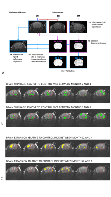 |
MRI detects macro- and microstructural changes to normal
brain tissue following hemi-brain radiotherapy
Ben R Dickie1,
Duncan Forster1,
Abigail Bryce-Atkinson2,3,
Izabelle Lövgren2,3,
Azadeh Abravan4,
Marcel van Herk2,3,
and Kaye Williams5
1Division of Informatics, Imaging and Data Sciences, Faculty of Biology, Medicine and Health, University of Manchester, UK, Manchester, United Kingdom, 2Division of Cancer Sciences, School of Health Sciences, Faculty of Biology, Medicine and Health, University of Manchester, UK, Manchester, United Kingdom, 3Department of Radiotherapy Related Research, The Christie NHS Foundation Trust, Manchester, United Kingdom, Manchester, United Kingdom, 4Division of Cancer Sciences, School of Health Sciences, Faculty of Biology, Medicine and Health, University of Manchester, UK, Manchester, UT, United Kingdom, 5Division of Pharmacy and Optometry, School of Health Sciences, Faculty of Biology, Medicine and Health, University of Manchester, UK, Manchester, United Kingdom Keywords: Neurodegeneration, Radiotherapy Approximately 50-90% of patients that survive treatment for brain tumours experience dementia-like cognitive impairments. Here we use MRI and behavioural testing to assess longitudinal changes to brain macro- and microstructure and cognitive dysfunction following hemi-brain radiotherapy. We show shrinkage of cortical and hippocampal regions in the irradiated hemisphere, and expansion of cortical tissue in the non-irradiated hemisphere relative to non-irradiated control mice. Brain microstructure was also changed including increases in gray matter diffusivity, and decreases in white matter fractional anisotropy. Changes on MRI were accompanied by early and persistent deficits in novel object recognition. |
| 13:30 |
0584.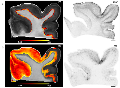 |
Quantitative correlations of propagator metrics with
phosphorylated tau and astrogliosis in chronic traumatic
encephalopathy
Mihika Gangolli1,2,3,
Sinisa Pajevic4,5,
Elizabeth B. Hutchinson5,6,
Joong Hee Kim1,2,7,
Dan Benjamini1,8,
and Peter J. Basser1,5
1Center for Neuroscience and Regenerative Medicine, Bethesda, MD, United States, 2The Henry M. Jackson Foundation for the Advancement of Military Medicine, Inc., Bethesda, MD, United States, 3Radiology and Imaging Sciences, Clinical Center, National Institutes of Health, Bethesda, MD, United States, 4Section on Critical Brain Dynamics, National Institute of Mental Health, National Institutes of Health, Bethesda, MD, United States, 5Section on Quantitative Imaging and Tissue Sciences, Eunice Kennedy Shriver National Institute of Child Health and Human Development, National Institutes of Health, Bethesda, MD, United States, 6Biomedical Engineering, University of Arizona, Tucson, AZ, United States, 7Laboratory of Functional and Molecular Imaging, National Institute of Neurological Disorders and Stroke, National Institutes of Health, Bethesda, MD, United States, 8Multiscale Imaging and Integrative Biophysics Unit, National Institute on Aging, National Institutes of Health, Bethesda, MD, United States Keywords: Neurodegeneration, Diffusion/other diffusion imaging techniques Propagator metrics computed from high spatial resolution diffusion MRI data are correlated with histopathological assessments of phosphorylated tau (p-tau) and astrogliosis in tissue specimen with a diagnosis of Stage III/IV chronic traumatic encephalopathy (CTE). Region of interest based analysis showed significant correlations of p-tau with non-Gaussianity (NG) in deep cortical gray matter and of propagator anisotropy (PA) with astrogliosis in superficial cortical white matter. An unsupervised clustering approach with PA and NG as inputs is then used to segment MR data of tissue specimen into clusters to determine whether propagator metrics can be utilized to detect underlying pathology. |
| 13:30 |
0585.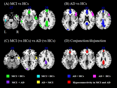 |
Shared and differed functional connectivity abnormalities
for default mode network in mild cognitive impairment and
Alzheimer’s disease
Yaxuan Wang1,
Fenghua Long1,
Qiyong Gong1,
Su Lui1,
and Fei Li1
1Huaxi MR Research Center (HMRRC), Department of Radiology, West China Hospital of Sichuan University, Chengdu 610041, Sichuan, P.R. China., Chengdu, China Keywords: Neurodegeneration, Brain Connectivity We performed a meta-analysis to investigate shared and different alterations of resting-state functional connectivity (rsFC) for default mode network (DMN) in mild cognitive impairment (MCI) and Alzheimer’s disease (AD). We found both patients with MCI and AD showed hypoconnectivity within DMN in bilateral medial prefrontal cortex (mPFC)/anterior cingulate cortex (ACC) and precuneus/posterior cingulate gyrus (PCC), indicating possibly shared neuropathologic mechanism underlying common cognitive dysfunction. Moreover, we identified distinct alterations of rsFC for DMN in some brain regions, which suggested potential imaging markers to distinguish the two diseases. |
| 13:30 |
0586.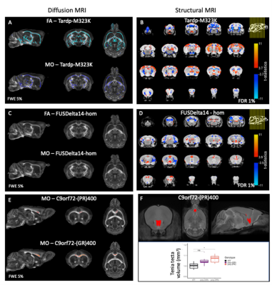 |
Distinct patterns of microstructural changes in mouse models
of ALS/FTD with mutations in Tardbp, Fus and C9orf72
Aurea Martins Bach1,
Cristiana Tisca1,
Mohamed Tachrount1,
Carmelo Milioto2,3,
Mireia Carcolé2,3,
Shoshana Spring4,
Remya R. Nair5,6,
Thomas J. Cunningham6,7,
Elizabeth M. C. Fisher8,
Adrian M. Isaacs2,3,
Brian J. Nieman4,
Jason Lerch1,
and Karla Miller1
1Wellcome Centre for Integrative Neuroimaging, FMRIB, Nuffield Department of Clinical Neurosciences, University of Oxford, Oxford, United Kingdom, 2UK Dementia Research Institute at UCL, Faculty of Brain Sciences, University College London, London, United Kingdom, 3Department of Neurodegenerative Disease, UCL Queen Square Institute of Neurology, University College London, London, United Kingdom, 4Mouse Imaging Centre, The Hospital for Sick Children, Toronto, ON, Canada, 5Department of Physiology, Anatomy and Genetics, University of Oxford, Oxford, United Kingdom, 6Mammalian Genetics Unit, MRC Harwell Institute, Oxford, United Kingdom, 7MRC Prion Unit and Institute of Prion diseases, University College London, London, United Kingdom, 8Department of Neuromuscular Diseases, UCL Queen Square Institute of Neurology, University College London, London, United Kingdom Keywords: Neurodegeneration, Preclinical Amyotrophic lateral sclerosis (ALS) and Frontotemporal dementia (FTD) form a disease spectrum with shared clinical, pathological and genetic features. The most common mutations in ALS/FTD are in the genes C9Orf72, TARDBP and FUS. We used post-mortem diffusion kurtosis imaging to assess microstructure imaging phenotypes in mouse models of ALS/FTD with mutations in these genes. While mice with mutation in Tardbp presented reduced FA and MO in various white matter tracts, no difference could be detected in mice with mutation in Fus, and increased MO in a portion of the corpus callosum was observed in mice with mutation in C9orf72. |
| 13:30 |
0587.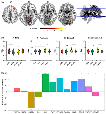 |
The interaction between first-episode schizophrenia and age
based on gray matter volume and its molecular analysis: A
multimodal MRI study
Jingli Chen1,2,
Yarui Wei1,2,
Kangkang Xue1,2,
and Jingliang Cheng1,2
1Department of Magnetic Resonance Imaging, The First Affiliated Hospital of Zhengzhou University, Zhengzhou, China, 2Laboratory for Functional Magnetic Resonance Imaging and Molecular Imaging of Henan Province, Magnetic Resonance Imaging, Zhengzhou, China Keywords: Neurodegeneration, Gray Matter, gray matter volume / early-onset schizophrenia / adult-onset schizophrenia /neurodevelopment / visual perception / visual cognitive We aimed to investigate the interaction characteristics of schizophrenia and onset age and its underlying molecular mechanisms by using T1-weighted high-resolution magnetic resonance imaging (3DT1) and the JuSpace toolbox. 150 first-episode drug-naïve schizophrenia and 119 matched normal controls were recruited and underwent 3DT1 scans. Our results show that the two main effects of factors and interaction effect in gray matter volume and their underlying molecular mechanisms have varying degrees of specificity changes. Particularly, the abnormality of the visual perception system and higher visual cognitive functions in early-onset schizophrenia (EOS) have guiding significance for the mechanism and treatment of EOS. |
| 13:30 |
0588.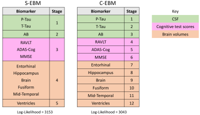 |
S-EBM: generalising event-based modelling of disease
progression for simultaneous events
Christopher Samuel Parker1,
Neil P Oxtoby1,
Daniel C Alexander1,
and Hui Zhang1
1Centre for Medical Image Computing, Department of Computer Science, UCL, London, UK, London, United Kingdom Keywords: Neurodegeneration, Modelling, Disease progression S-EBM generalises the event-based model (EBM) of disease progression for simultaneous events. In synthetic data, S-EBM can tell the difference between simultaneous and non-simultaneous events under a range of experimental conditions. In comparison to conventional EBM, S-EBM avoids artificial serial orderings, permitting more accurate and parsimonious descriptions of disease progression. When applied to real Alzheimer’s disease biomarker data, S-EBM estimates a sequence containing neurologically plausible simultaneous events which more closely explain the data than conventional EBM. S-EBM may be applied to recover novel patterns of disease progression thereby informing our understanding of disease evolution. |
| 13:30 |
0589.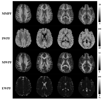 |
Multicompartment Imaging of the Brain Using a Comprehensive
MR Imaging Protocol
James Lo1,2,
Eric Y. Chang1,3,
Jiang Du1,2,3,
Graeme M Bydder1,
and Yajun Ma1
1Department of Radiology, University of California San Diego, San Diego, CA, United States, 2Department of Bioengineering, University of California San Diego, San Diego, CA, United States, 3Radiology Service, Veteran Affairs San Diego Healthcare System, San Diego, CA, United States Keywords: Neurodegeneration, Quantitative Imaging, Myelin In this study, we developed a comprehensive MR imaging protocol to quantify all the major components of the brain. This protocol includes four different kinds of sequences: a magnetization transfer prepared Cones (MT-Cones) for two-pool MT modeling, a short-TR adiabatic inversion-recovery prepared Cones (STAIR-Cones) for myelin water imaging, a proton-density weighted Cones (PDw-Cones) for total water imaging, and a highly T2 weighted Cones (T2w-Cones) for extracellular water imaging. Using a combination of these techniques, we successfully quantified the proton fractions of brain macromolecules, myelin water, intracellular water, and extracellular water components in three healthy volunteers with a 3T clinical scanner. |
| 13:30 | 0590. | WITHDRAWN |
| 13:30 |
0591.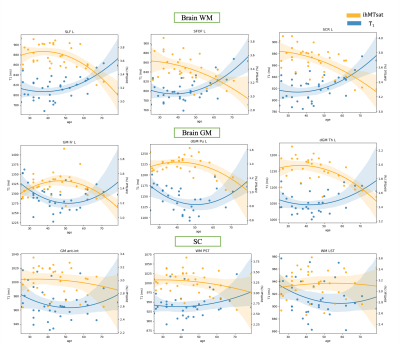 |
Brain and cervical spinal cord myelination and age-related
changes in adulthood: a preliminary study based on ihMTsat
and T1 relaxometry mapping
Arash Forodighasemabadi1,2,3,4,
Lucas Soustelle1,2,
Olivier M. Girard1,2,
Thomas Troalen5,
Jean-Philippe Ranjeva1,2,
Guillaume Duhamel1,2,
and Virginie Callot1,2,4
1Aix-Marseille Univ, CNRS, CRMBM, Marseille, France, 2APHM, Hopital Universitaire Timone, CEMEREM, Marseille, France, 3Aix-Marseille Univ, Université Gustave Eiffel, LBA, Marseille, France, 4iLab-Spine International Associated Laboratory, Montreal, Canada, Marseille, France, 5Siemens Healthcare SAS, Saint-Denis, France Keywords: Neurodegeneration, Aging Inhomogeneous Magnetization Transfer (ihMT) has shown to be a biomarker of myelin with enhanced specificity as compared to conventional MT. To compensate for B1+ and T1 effects, a 3D ihMT-RAGE sequence with an ihMTsat framework has recently been proposed. In this study, ihMTsat was used in combination with an optimized MP2RAGE sequence to study both brain and spinal cord adulthood aging process. The ihMTsat and R1=1/T1 metrics followed an inverse U-shaped evolution with age in different ROIs, from which a maturation age was extracted. A multiparametric (R1,ihMTsat) voxel-wise analysis revealed significant age effect in the microstructure of different cord tracts. |
| 13:30 |
0592.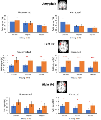 |
Are fMRI findings on emotional aging affected by
cerebrovascular aging?
Loretta Donaldson1,
Parimal Joshi1,
Lincoln Kartchner1,
Sara Akar1,
and Peiying Liu1
1Diagnostic Radiology and Nuclear Medicine, University of Maryland, Baltimore, Baltimore, MD, United States Keywords: Neurodegeneration, fMRI (task based) Age-related changes in emotional circuitry have been studied using BOLD fMRI and revealed age-related increases in the activation of the prefrontal cortex and inconsistent findings in the amygdala. Previous emotional aging studies did not account for vascular aging which causes a reduction in cerebrovascular reactivity (CVR). Using picture viewing task fMRI and gas inhalation MRI, the fMRI signals are calibrated by the vascular measures to improve the inference of neural activity. After accounting for vascular changes, age-invariant activity was seen in the amygdala, and increased age-related activation of prefrontal regions was observed compared to activation before vascular correction. |
| 13:30 |
0593.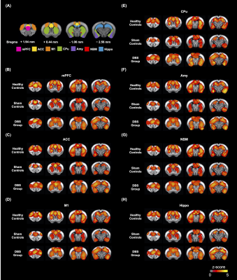 |
MR Imaging Deep Brain Stimulation of the Nucleus Basalis
Meynert Restored Cognitive Function in Alzheimer’s Disease
Model
Ching-Wen Chang1,2,
Yi-Chen Lin1,
Chiung-Yuan Ko3,
Yu-Chun Lo3,
Ting-Chieh Chen1,
Ssu-Ju Li1,
Mu-Hua Wang1,
Tsai-Yu Cho1,
Sheng-Huang Lin4,5,
and You-Yin Chen1,3
1Department of Biomedical Engineering, National Yang Ming Chiao Tung University, Taipei, Taiwan, 2Biomedical Translation Research Center, Academia Sinica, Taipei, Taiwan, 3Ph.D. Program in Medical Neuroscience, Taipei Medical University, Taipei, Taiwan, 4Department of Neurology, Buddhist Tzu Chi Medical Foundation, Hualien, Taiwan, 5Department of Neurology, Tzu Chi University, Hualien, Taiwan Keywords: Neurodegeneration, fMRI, Nucleus Basalis of Meynert Alzheimer’s disease (AD) is an intricate neurodegenerative disease. Nucleus Basalis of Meynert (NBM), a key region of the cholinergic system that provides acetylcholine to cortex, has been shown to be target for deep brain stimulation (DBS) in AD. The 5×FAD mouse model was used to investigate the change of behavioral tasks, resting-state functional MRI, and acetylcholinesterase (AChE) assay were applied in this study. We found that NBM-DBS may play an important role in modulating cognitive function and spatial working memory in 5×FAD mice. Increasing functional connectivity and decreasing AChE activity may be the biomarkers for AD individuals. |
 Back to Meeting Home
 Back to the Program-at-a-Glance
The International Society for Magnetic Resonance in Medicine is accredited by the Accreditation Council for Continuing Medical Education to provide continuing medical education for physicians.
 Back to Meeting Home
 Back to the Program-at-a-Glance
 Full Session Video