Power Pitch
Pitch: Head & Neck
ISMRM & ISMRT Annual Meeting & Exhibition • 03-08 June 2023 • Toronto, ON, Canada

13:45 |
1328.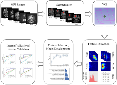 |
Development and Validation of a Radiomics Model in
Differentiating Sinonasal Mucosal Melanomas from Sinonasal
Lymphomas
Shengyong Li1,
Linying Guo2,
Jing Zhang1,
Yang Song3,
Shengjian Zhang4,
Rifeng Jiang5,
Guang Yang1,
and Zuohua Tang2
1Shanghai Key Laboratory of Magnetic Resonance, East China Normal University, Shanghai, China, 2Department of Radiology, Eye & ENT Hospital of Shanghai Medical School, Fudan University, Shanghai, China, 3MR Scientific Marketing, Siemens Healthcare, Shanghai, China, 4Department of Radiology, Shanghai Cancer Center of Shanghai Medical School, Fudan University, Shanghai, China, 5Department of Radiology, Fujian Medical University Union Hospital, FuZhou, China Keywords: Head & Neck/ENT, Multimodal Sinonasal mucosal melanomas (SNMM) are clinically more aggressive than its cutaneous counterpart and presented markedly poor prognosis. To differentiate sinonasal melanomas from sinonasal lymphomas, a radiomics model was built using features from multi-parametric MRI, including T1-weighted imaging (T1WI), T2 weighted imaging (T2WI), DWI and C-T1WI. In this multicenter retrospective study, 189 patients diagnosed with SNMMs or sinonasal lymphoma were enrolled from three institutions. The proposed model achieved AUCs of 0.884 and 0.870 in the internal and external validation set, respectively. |
| 13:45 |
1329.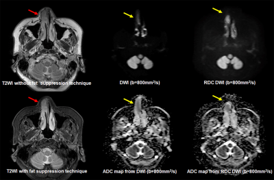 |
Comparison of Image Quality and ADC Measurement between DWIs
with and without Reverse Encoding Distortion Correction in
Head and Neck Tumors
Hirotaka Ikeda1,
Yoshiharu Ohno1,2,
Kaori Yamamoto3,
Maiko Shinohara3,
Masato Ikedo3,
Masao Yui3,
Akiyoshi Iwase4,
Minami Furuta1,
Yuki Obama1,
Hiroyuki Nagata2,
Takahiro Ueda1,
Yoshiyuki Ozawa1,
and Hiroshi Toyama1
1Radiology, Fujita Health University School of Medicine, Toyoake, Japan, 2Joint Research Laboratory of Advanced Medical Imaging, Fujita Health University School of Medicine, Toyoake, Japan, 3Canon Medical Systems Corporation, Otawara, Japan, 4Fujita Health University Hospital, Toyoake, Japan Keywords: Cancer, Head & Neck/ENT We hypothesize that RDC is useful for image quality and diagnostic performance improvements on DWI with b value at 1500 s/mm2 in suspected prostatic cancer patients, although there was little influence of RDC on DWI at in vitro study. The purpose of this study was to determine the influence of RDC for ADC measurement at in vitro study and its’ utility for improving image quality and diagnostic performance of malignant from benign head and neck tumors on DWI as in vivo study. |
| 13:45 |
1330.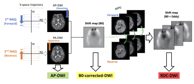 |
Evaluation of reverse encoding distortion correction DWI in
patients with non-functioning pituitary macroadenoma
Shuichi Ito1,
Sachi Okuchi1,
Yasutaka Fushimi1,
Kanae Kawai Miyake2,
Koji Fujimoto3,
Hitomi Numamoto2,
Satoshi Nakajima1,
Akihiko Sakata1,
Takuya Hinoda1,
Sayo Otani1,
Azusa Sakurama1,
Krishna Pandu Wicaksono1,
Hiroshi Tagawa1,
Yang Wang1,
Satoshi Ikeda1,
Miyuki Takiya1,
Hiroki Kondo4,
Rimika Imai4,
Tsuneo Saga2,
and Yuji Nakamoto1
1Department of Diagnostic Imaging and Nuclear Medicine, Graduate School of Medicine, Kyoto University, Kyoto, Japan, 2Department of Advanced Medical Imaging Research, Graduate School of Medicine, Kyoto University, Kyoto, Japan, 3Department of Real World Data Research and Development, Graduate School of Medicine, Kyoto University, Kyoto, Japan, 4Canon Medical Systems Corporation, Otawara, Japan Keywords: Head & Neck/ENT, Diffusion/other diffusion imaging techniques Reverse encoding distortion correction diffusion-weighted imaging (RDC-DWI) is a novel on-console technique to reduce eddy current-induced distortion of motion probing gradient (MPG) images in addition to B0 field inhomogeneity. We compared RDC-DWI, B0-corrected-DWI, and original DWI in patients with unoperated non-functioning macroadenoma or residual pituitary macroadenoma after surgery. RDC-DWI had the best image quality regarding distortion, artifacts and overall tumor visualization, which suggested that RDC-DWI facilitates accurate visualization in the pituitary region on DWI. |
| 13:45 |
1331.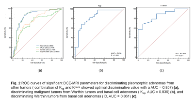 |
Use of diffusion kurtosis imaging and dynamic
contrast-enhanced MRI in differentiating parotid gland
tumors
zijun Liu1,
baohong Wen1,
and yan Zhang1
1The First Affiliated Hospital of Zhengzhou University, Zhengzhou, China Keywords: Head & Neck/ENT, Head & Neck/ENT, Parotid gland tumors This study evaluates the usefulness of combined Diffusion kurtosis imaging (DKI) and dynamic contrast-enhanced MRI (DCE-MRI) in differentiating parotid gland tumors. DKI and DCE-MRI quantitative parameters were analyzed by using the Kruskal-Wallis H test and post hoc test with bonferroni correction or the one-way analysis of variance (ANOVA) with LSD method, and ROC curve. Our research shows that the significant parameters in stepwise diagnosing parotid gland tumors were Kep, Ktrans, and D value. Therefore, the combined use of DKI and DCE-MRI could be used to differentiate various parotid gland tumors, and it may be helpful for differentiating parotid gland tumors. |
| 13:45 | 1332. |
Feasibility study of using time-dependent diffusion MRI to
distinguish parotid polymorphic adenoma from Warthin's tumor
Di Geng1,
Xiance Zhao2,
Yishi Wang3,
Xiaoquan Xu1,
and Feiyun Wu1
1Department of Radiology, First Affiliated Hospital of Nanjing Medical University, Nanjing, China, Nanjing, China, 2Philips Healthcare, Shanghai, China, Shanghai, China, 3Philips Healthcare, Beijing, China, Beijing, China Keywords: Head & Neck/ENT, Head & Neck/ENT IMPULSED (Imaging Microstructural Parameters Using Limited Spectrally Edited Diffusion) imaging method based on time-dependent diffusion magnetic resonance imaging (MRI) could be used to quantify cell sizes. We found that several quantitative parameters derived from IMPULSED including Vin (intracellular volume fraction), d (volume-weighted mean cell size) and cellularity showed significant differences between polymorphic adenoma (PA) and Warthin’s tumor (WT) in the parotid gland. Our findings shed light on the role of IMPULSED-derived quantitative parameters in distinguishing PAs from WTs preoperatively. |
| 13:45 |
1333.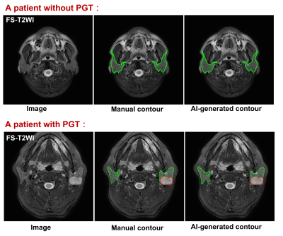 |
Deep learning for automatic detection and contouring of
parotid gland tumors on MRI
Rongli Zhang1,
Qi Yong H. Ai1,2,
Lun M. Wong1,
Qiao Deng1,
and Ann D. King1
1Department of Imaging and Interventional radiology, The Chinese University of Hong Kong, Prince of Wales Hospital, HongKong, China, 2Department of Health Technology and Informatics, The Hong Kong Polytechnic University, Hong Kong, China Keywords: Cancer, Head & Neck/ENT Parotid gland tumors (PGTs) are often asymptomatic and an incidental finding on MRI that can be overlooked. We constructed an accurate artificial intelligence (AI) tool trained on fat-suppressed T2-weighted MRI to automatically identify patients with PGTs with an accuracy of 94.3% (99/105), a sensitivity of 94.0% (47/50) and a specificity of 94.5% (52/55). For identified PGT patients, automatic segmentations of the tumor and gland were performed and achieved dices of 77.2% and 86.3%, respectively. The proposed AI tool may assist radiologists by acting as a second pair of eyes to ensure incidental PGTs on MRI are not missed. |
| 13:45 |
1334. |
A Pilot Evaluation of TSE MVXD based IVIM in Characteristics
and Diagnosis of Nasopharyngeal Carcinoma
Yifen Zhou1,2,
Rui Chen1,2,
Huifen Ye1,2,
Zhigang Wu3,
Yongzhou Xu3,
Zaiyi Liu1,2,
and Guangyi Wang1,2
1Department of Radiology, Guangdong Provincial People’s Hospital, Guangdong Academy of Medical Sciences, Guangzhou, Guangdong Province, China, Guangzhou, China, 2Guangdong Provincial Key Laboratory of Artificial Intelligence in Medical Image Analysis and Application, Guangdong Provincial People's Hospital, Guangdong Academy of Medical Sciences, Guangzhou, China, Guangzhou, China, 3MSC Clinical & Technical Solutions, Philips Healthcare, China, Shenzhen, China Keywords: Diffusion/other diffusion imaging techniques, Contrast Mechanisms, Turbo Spin-echo; Multivane-XD; Intravoxel Incoherent Motion; Contrast-enhanced imaging; Nasopharyngeal carcinoma Diagnosis of Nasopharyngeal Carcinoma (NPC) remains challenge since the contrast-enhanced T1-weighted imaging (CE-T1WI) may lead to harmful impact by the accumulation of contrast agent in patients and its potential adverse reactions. TSE MVXD DWI based IVIM, a considerable alternative, was performed on 32 NPC patients with significant tumors to evaluate the relation with CE-T1WI. As the result showed, the TSE MVXD DWI based IVIM had significantly relation with CE T1WI. It is potentially a promising and valuable non-invasive method in the detection of NPC. |
| 13:45 |
1335. |
Radiomics model based on MRI for early prediction of
radiation encephalopathy in nasopharyngeal carcinoma
Lixuan Huang1,
Zongxiang Yang1,
Hao Ren2,
Yao Hu1,
Cheng Tang1,
Huiting Zhang3,
and Liling Long1
1The First Affliated Hospital of Guangxi Medical University, Nanning, China, 2Guangxi Medical University Kaiyuan Langdong Hospital, Nanning, China, 3MR Scientific Marketing, Siemens Healthineers Ltd, Wuhan, China Keywords: Head & Neck/ENT, Radiomics This study aimed to develop radiomic models based on MRI to investigate the changes of temporal lobe heterogeneity in nasopharyngeal carcinoma (NPC) patients with radiation encephalopathy (REP) during the latent period, and to predict the temporal lobe REP early. Results showed that The AUC of radiomics-clinics combined model was higher than radiomics model and clinics model, with better accuracy. Our study suggested that the radiomics-clinics combined model may be an effective method for the noninvasive prediction of REP in NPC patients after radiotherapy. |
| 13:45 |
1336.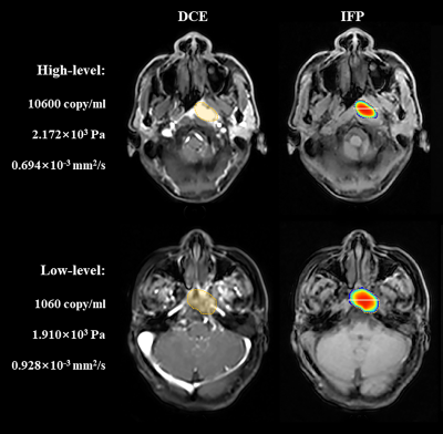 |
Dynamic contrast-enhanced MRI-based interstitial fluid
pressure model in evaluation of nasopharyngeal carcinoma
Guixiao Xu1,
Chuanmiao Xie1,
Hui Li1,
Yun He1,
Liangru Ke1,
Haibin Liu1,
Liyun Zheng2,
and Yongming Dai3
1Department of Radiology, Sun Yat-sen University Cancer Center, State Key Laboratory of Oncology in South China, Collaborative Innovation Center for Cancer Medicine, Guangdong Key Laboratory of Nasopharyngeal Carcinoma Diagnosis and Therapy, Guangzhou, China, 2Shenzhen United Imaging Research Institute of Innovative Medical Equipment, Shenzhen, China, 3MR Collaboration, Central Research Institute, United Imaging Healthcare, Shanghai, China Keywords: Head & Neck/ENT, Head & Neck/ENT Nasopharyngeal carcinoma is cancer arising from the nasopharynx epithelium. The most widely researched etiological factor for nasopharyngeal carcinoma is the Epstein-Barr virus (EBV) infection. Elevated interstitial fluid pressure (IFP) is a significant biomarker for assessing head and neck malignant tumors. This study aimed to use the non-invasive dynamic contrast-enhanced magnetic resonance imaging (DCE-MRI)-based IFP model to evaluate nasopharyngeal carcinoma and distinguish the differences between nasopharyngeal carcinoma patients with low and high EBV-infected levels. As a result, the non-invasive DCE-MRI-based IFP model could be used to evaluate nasopharyngeal carcinoma and differentiate patients with high- and low-level plasma EBV DNA. |
| 13:45 |
1337.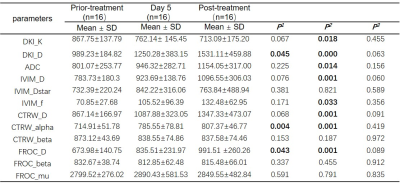 |
Application of IVIM, DKI, FROC and CTRW in vascular
normalization induced by recombinant human endostatin in
nasopharyngeal carcinoma.
Lixuan Huang1,
Hao Ren2,
Zongxiang Yang1,
Yao Hu1,
Huiting Zhang3,
and Liling Long1
1The First Affliated Hospital of Guangxi Medical University, Nanning, China, 2Guangxi Medical University Kaiyuan Langdong Hospital, Nanning, China, 3MR Scientific Marketing, Siemens Healthineers Ltd., Wuhan, China Keywords: Cancer, Diffusion/other diffusion imaging techniques This study explored the vascular normalization induced by recombinant human endostatin (RHES) in nasopharyngeal carcinoma (NPC) based on IVIM, DKI, FROC and CTRW. Results showed that the DKI_D, CTRW_alpha, and FROC_D had a significant difference between prior-treatment and Day 5 in RHES group, FROC_mu in Day 5 and CTRW_alpha in post-treatment were statistically significant in two groups, and DKI_K with decreased trend with the progress of RHES + IC treatment. These results suggest that the four diffusion models can detect the noninvasively monitor the vascular normalization induced by RHES in NPC. |
| 13:45 |
1338.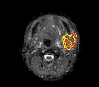 |
Apparent diffusion coefficient histogram analysis of adenoid
cystic carcinoma: correlation with histopathology and the
prediction of survival
TANG WEIQING1,
SONG YANG2,
YING YUAN1,
and TAO XIAOFENG1
1Radiology, Shanghai Ninth People’s Hospital,Shanghai Jiao Tong University School of Medicine, Shanghai, China, 2MR Scientific Marketing, Siemens Healthcare. Shanghai, China, Shanghai, China Keywords: Head & Neck/ENT, Cancer The aim of this retrospective study is to explore the value of histogram analysis of apparent diffusion coefficient (ADC) values for distinguishing different subtypes of adenoid cystic carcinoma (ACC) and prediction of survival. Receiver operating characteristic curve analysis was used to determine the best differentiating parameters. The ADC_10th percentile values achieved highest diagnostic efficacy with an AUC of 0.821. The multivariable Cox proportional hazards model found that radiomic signature and tumor stage were significant predictors in ACC patients. Histogram analysis of ADC values may be helpful for differentiating the subtypes of ACC, leading to improved targeted treatment and reduced morbidity. |
| 13:45 |
1339.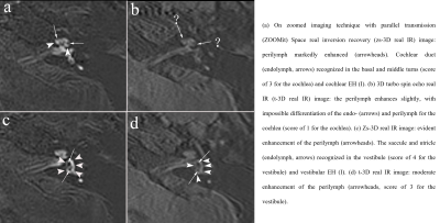 |
A Novel MR Sequence of 3D-ZOOMit Real Inversion Recovery
Imaging Improves Endolymphatic Hydrops Detection in Patients
with Ménière’s Disease
Jinye Li1,
Lixin Sun2,
Na Hu2,
Mengxiao Liu3,
Linsheng Wang2,
and Chuanting Li2
1shangdong provincial ENT hospital, Jinan, China, 2Shandong Provincial ENT Hospital, Jinan, China, 3Siemens healthineers, Shanghai, China Keywords: Head & Neck/ENT, Head & Neck/ENT In order to visualize endolymphatic hydrops in Ménière’s disease better, we compared the conventional 3D real IR and ZOOMit 3D real IR sequence. Results suggest that visualization of the endolymphatic space might be higher by zs-3D real IR compared with t-3D real IR, especially in the cochlea. |
| 13:45 |
1340.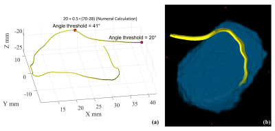 |
Automatic facial nerve tractography in patient with
vestibular schwannoma
Chenxi Lu1,
Qiqi Tong2,
Mantao Chen3,
Xiujue Zheng3,
Jianhui Zhong1,4,
and Hongjian He1,5
1Center for Brain Imaging Science and Technology, College of Biomedical Engineering and Instrument Science, Zhejiang University, Hangzhou, China, 2Research Center for Healthcare Data Science, Zhejiang Lab, Hangzhou, China, 3Department of Neurosurgery, The First Affiliated Hospital, School of Medicine, Zhejiang University, Hangzhou, China, 4University of Rochester, Rochester, NY, United States, 5School of physics, Zhejiang University, Hangzhou, China Keywords: Nerves, Tractography & Fibre Modelling Facial nerve tractography has recently been recognized as a valuable tool for predicting risk before vestibular schwannoma surgery to preserve function. Due to complexity of tissue structure, professional experience is usually required to manually adjust tracking parameters and place exclusion ROIs for optimal tractography. A pipeline for automatic facial nerve tractography will significantly lessen surgeons' workload. Here, we proposed a fiber growing method for adjusting the angle threshold adaptively and removed track outliers. Results show that the proposed method achieves good consistency with the manual method. The pipeline would have great potential for assisting clinic surgeries for vestibular schwannoma. |
| 13:45 |
1341.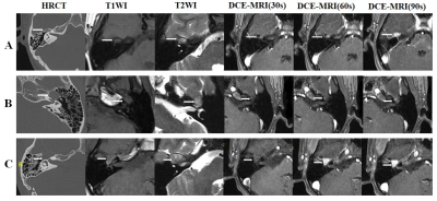 |
Identifiy geniculate ganglion venous malformation from
geniculate ganglion schwannoma using dynamic
contrast-enhanced MRI
Mengda Jiang1,
Yongchuan Chai2,
Yang Song3,
and Xiaofeng Tao1
1Department of Radiology, Shanghai Ninth People’s Hospital, affiliated to Shanghai Jiaotong Universit, ShangHai, China, 2Department of Otolaryngology, Head & Neck Surgery, Shanghai Ninth People’s Hospital, affiliated to Shanghai Jiaotong University School of Medicine, Shanghai, China, 3MR Scientific Marketing, Siemens Healthcare, Shanghai, China Keywords: Head & Neck/ENT, Nerves, Geniculate Ganglion, Facial Nerve To evaluate the CT and MRI findings to identify geniculate ganglion venous malformation (GGVM) and geniculate ganglion schwannoma (GGS). Clinical data, lesion size, involvement of facial nerve (FN) segment, signal intensity, homogeneity, the enhancement pattern on DCE-MRI, characteristics of bone destruction on HRCT were evaluated. Lesion size, involvement of FN segment, T1W and T2W intensity, and homogeneity on DCE-MRI were statistically different between them. For regression model, the “honeycomb” sign and “point-to-side” enhancement pattern were independent risk factor (AUC=0.975, accuracy=97.70%, sensitivity=95%, specificity=100%, PPV=100%, NPV=95.80%). |
| 13:45 |
1342.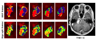 |
Quantitative ΔR1 and PK Mapping Supports Tumor Habitat
Delineation and Anti-Angiogenic Response Prediction of
Vestibular Schwannoma
Xiaoping Zhu1,
Daniel Lewis2,3,4,
Ka-Loh Li1,
William Lloyd1,
Mueez Waqar2,3,5,
Ibrahim Djoukhadar3,6,
David J Coope2,3,4,
Andrew T King2,3,
Timothy Cootes1,
and Alan Jackson1
1DIIDS, University of Manchester, Manchester, United Kingdom, 2Dept. of Neurosurgery, Manchester Academic Health Science Centre, Manchester Academic Health Science Centre, Salford, United Kingdom, 3Geoffrey Jefferson Brain Research Centre, University of Manchester, Salford, United Kingdom, 4Division of Neuroscience and Experimental Psychology, University of Manchester, Manchester, United Kingdom, 5Division of Cancer Sciences, University of Manchester, Manchester, United Kingdom, 6Dept. of Neuroradiology, Manchester Academic Health Science Centre, Manchester, United Kingdom Keywords: Head & Neck/ENT, Tumor In this study we sought to undertake histogram and habitat analyses of tumoural DCE-MRI derived microvascular kinetic parameters and quantitative relaxation rate changes (ΔqR1) in patients with sporadic and neurofibromatosis type 2 (NF2) related vestibular schwannoma (VS). We demonstrate that within imaged VS tumours there is considerable intratumoural heterogeneity in DCE-MRI derived microvascular metrics and ΔqR1, with distinct intratumoural regions or habitats displaying high Ktrans, vp and ΔqR1 values respectively. We furthermore demonstrate within a cohort of NF2-related VS undergoing anti-angiogenic (bevacizumab) therapy that both pre-treatment Ktrans and ΔqR1 are predictive of later tumour volumetric response at 90 days post-treatment. |
| 13:45 |
1343. |
Disrupted within-network segregations and between-network
integrations in age-related hearing loss with cognitive
decline
Zhaopeng Tong1,
Chunhua Xing2,
Xiaomin Xu2,
Jin-Jing Xu2,
Yuanqing Wu2,
Richard Salvi3,
Xindao Yin2,
Yu-Chen Chen2,
and Yuexin Cai1
1Sun Yat-sen Memorial Hospital, Sun Yat-sen University, Guangzhou, China, 2Nanjing First Hospital, Nanjing Medical University, Nanjing, China, 3University at Buffalo, The State University of New York, Buffalo, NY, United States Keywords: Head & Neck/ENT, fMRI (resting state), Brain Connectivity, Degenerative, Dementia, Neuroscience Age-related hearing loss is generally associated with dementia. However, the underlying mechanisms and causal relationship linking ARHL to dementia are poorly understood. Based on resting-state fMRI, the study found that ARHL disrupts specific aspects of resting-state functional connectivity patterns across frontal-parietal regions of the central nervous system; these changes presumably reflect cortical reorganization resulting from auditory sensory deprivation and/or the long-term consequences of effortful listening. The ARHL disruption of network information processing presumably accelerates brain aging and contributes to cognitive decline. |
| 13:45 |
1344. |
Optimisation of an MRI protocol to assess the effects of
noise exposure on the auditory pathway
Rebecca Susan Dewey1,2,3,
Hannah Guest4,5,
Rebecca E Millman4,5,
Garreth Prendergast4,5,
Christopher J Plack4,5,6,
and Susan T Francis1
1Sir Peter Mansfield Imaging Centre, School of Physics and Astronomy, University of Nottingham, Nottingham, United Kingdom, 2National Institute for Health and Care Research (NIHR) Nottingham Biomedical Research Centre, Nottingham, United Kingdom, 3Hearing Sciences, Division of Mental Health and Clinical Neurosciences, School of Medicine, University of Nottingham, Nottingham, United Kingdom, 4Manchester Centre for Audiology and Deafness (ManCAD), University of Manchester, Manchester, United Kingdom, 5National Institute for Health and Care Research (NIHR) Manchester Biomedical Research Centre, Central Manchester University Hospitals NHS Foundation Trust, Manchester, United Kingdom, 6Department of Psychology, Lancaster University, Lancaster, United Kingdom Keywords: Head & Neck/ENT, Nerves, Neurography, diffusion, DTI Recent findings suggest that noise exposure can cause substantial damage to the auditory nerve, without damage to the sensory hair cells or loss of threshold sensitivity. It is unclear which physiological measures are most sensitive to neural damage. Here, a comprehensive MRI protocol (neurography, high-resolution DTI of the auditory nerve and brain, morphometry, T1-myelination mapping, and resting-state functional connectivity) to study the ascending auditory pathway (auditory nerve, auditory brainstem, and cortex) is described. 200 participants will be recruited with varying noise exposure levels, with the aim of identifying diagnostic tests indicative of future hearing loss. |
| 13:45 |
1345.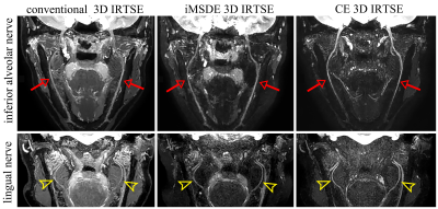 |
Visualization of the extracranial branches of the trigeminal
nerve using iMSDE preparation 3D IR-TSE
Dejun She1,2,3,
Hao Huang1,2,
Peiying You1,2,
Lu Li4,
Xiance Zhao5,
and Dairong Cao1,2,3,6
1Department of Radiology, First Affiliated Hospital of Fujian Medical University, Fuzhou, China, 2Department of Radiology, National Regional Medical Center, Binhai Campus of the First Affiliated Hospital, Fujian Medical University, Fuzhou, China, 3Key Laboratory of Radiation Biology of Fujian higher education institutions, First Affiliated Hospital, Fujian Medical University, Fuzhou, China, 4The School of Medical Imaging, Fujian Medical University, Fuzhou, China, 5Philips Healthcare, Shanghai, China, 6Department of Radiology, Fujian Key Laboratory of Precision Medicine for Cancer, First Affiliated Hospital of Fujian Medical University, Fuzhou, China Keywords: Nerves, Nerves, magnetic resonance neurography; trigeminal nerve Visualization of the anatomy of extracranial the trigeminal nerve (TGN) is crucial to detect nerve pathological alterations and differentiate pathologic causes. As a novel and safe nerve imaging technique in magnetic resonance neurography (MRN), the iMSDE pulse could result in uniform vascular signal suppression without additional contrast agents, which has been demonstrated to improve the visualization of peripheral nerves in several anatomical regions. Our results suggested that the iMSDE 3DIRTSE is a viable alternative to conventional 3DIRTSE and contrast-enhanced 3DIRTSE for MRN of the extracranial branches of TGN in clinical practice. |
| 13:45 |
1346. |
Carotid plaque predicts progression of intracranial
atherosclerosis: A MR imaging-based community cohort study
Miaoxin Yu1,
Dandan Yang2,
Runhua Zhang1,
Yong Jiang1,
Huiyu Qiao3,
Xihai Zhao3,
Gaifen Liu1,
and Yongjun Wang1
1Department of Neurology, Beijing Tiantan Hospital, Beijing, China, 2Department of Radiology, Beijing Geriatric Hospital, Beijing, China, 3Tsinghua University, Beijing, China Keywords: Head & Neck/ENT, Atherosclerosis Intracranial atherosclerotic disease progression is associated with recurrent stroke risk. In the present study, we investigated the association between carotid plaque and intracranial atherosclerosis progression in stroke-free participants using MR vessel wall imaging. In 312 participants recruited from a community cohort, we found that carotid plaque was independently associated with intracranial atherosclerosis progression during around 3-years’s follow-up. Our findings suggest that carotid plaque may be an effective predictor for intracranial atherosclerosis progression. |
| 13:45 |
1347. |
Dose finding and sequence tailoring in sentinel lymph node
detection of tongue cancer using superparagmagnetic
iron-oxide particles
Gijs Heldens1,
Daphne Driessen2,
Tim Dijkema3,
Anne Arens3,
Patrik Zámecnik3,
Sjoert Pegge3,
Willem Weijs4,
Adriana van Engen-van Grunsven5,
Robert Takes1,
Johannes Kaanders2,
and Tom Scheenen3
1Department of Otorhinolaryngology and Head and Neck Surgery, Radboud University Medical Center, Nijmegen, Netherlands, 2Department of Radiation Oncology, Radboud University Medical Center, Nijmegen, Netherlands, 3Department of Medical Imaging, Radboud University Medical Center, Nijmegen, Netherlands, 4Department of Oral- and Maxillofacial Surgery and Head and Neck Surgery, Radboud University Medical Center, Nijmegen, Netherlands, 5Department of Pathology, Radboud University Medical Center, Nijmegen, Netherlands Keywords: Cancer, Head & Neck/ENT To clarify the amount, timing and optimal pulse sequence settings of using interstitial superparamagnetic iron oxide particles (SPIO) as a contrast medium to detect sentinel lymph nodes in head and neck radiology, we injected SPIO peritumorally in six patients with early-stage oral squamous cell carcinoma. Anatomical and T2*-weighted MR images were acquired, and dose was altered after every two patients. Images with different computed echo times were created to determine the signal attenuation effect of SPIO on the sentinel lymph nodes. |
 Back to Meeting Home
 Back to the Program-at-a-Glance
The International Society for Magnetic Resonance in Medicine is accredited by the Accreditation Council for Continuing Medical Education to provide continuing medical education for physicians.
 Back to Meeting Home
 Back to the Program-at-a-Glance
 Full Session Video