Power Pitch
Pitch: MRI of Nerves & the Nervous System
ISMRM & ISMRT Annual Meeting & Exhibition • 03-08 June 2023 • Toronto, ON, Canada

Power Pitch
Pitch: MRI of Nerves & the Nervous System
| 08:15 |
1189.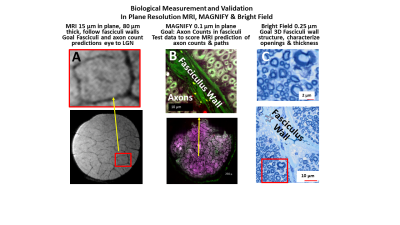 |
Fasciculus Axonal Connective Tissue Multiscale Imaging
(FACTMI) - Connectome Mapping of Optic Nerve with 16 µm MRI
at 14T and 0.1 µm histology
Walter Schneider1,
Yijen Wu1,
Alan Watson1,
Kasia Kedziora1,
Sudhir Pathak1,
Yongxin Zhao2,
Vijay Gorantla3,
Jens Anders4,
Rolf Polman5,
and Klaus Scheffler5
1University of Pittsburgh, Pittsburgh, PA, United States, 2Carnegie Mellon University, Pittsburgh, PA, United States, 3Wake Forest University, Winston-Salem, NC, United States, 4University of Stuttgart, Stuttgart, Germany, 5Max Planck Institute for Biological Cybernetics, Tübingen, Germany Keywords: Quantitative Imaging, Brain Connectivity, Connectome, Histology, Phantom Accurate brain connectome mapping requires tracking fasciculus bundles of axons within tracts. In porcine optic nerve harvested tissue and TAXON diffusion phantom on a 14T magnet with a new linear coil array, we identify fasciculi with 16 µm resolution and follow TAXON fibers over centimeters from eye to LGN. MAGNIFY and bright field optical histology provide 0.1 & 0.25-micron resolution with accurate counts of the 1.2 million axons within fasciculi aligned with MRI and fasciculus wall structure. We use MRI and deep learning to predict the axon paths at each point and axon counts in each fasciculus. |
| 08:15 |
1190.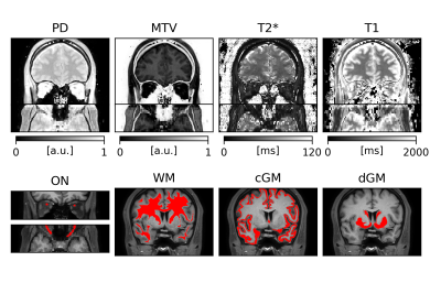 |
In vivo multimodal MRI of the healthy optic nerves
Antonio Ricciardi1,
Marios C. Yiannakas1,
Ratthaporn Boonsuth1,
Rodas Ghilom Bogatsion1,
Claudia A. M. Gandini Wheeler-Kingshott1,2,3,
and Rebecca S. Samson1
1NMR Research Unit, Queen Square Multiple Sclerosis Centre, UCL Queen Square Institute of Neurology, Faculty of Brain Sciences, University College London, London, United Kingdom, 2Department of Brain and Behavioural Sciences, University of Pavia, Pavia, Italy, 3Brain Connectivity Research Center, IRCCS Mondino Foundation, Pavia, Italy Keywords: Nerves, Quantitative Imaging The optic nerve (ON) is implicated in a variety of neurological disorders but is challenging to study through magnetic resonance imaging due to its small size and jittering. In this study, we developed an acquisition protocol and analysis pipeline that enables to produce quantitative proton density (PD), macromolecular tissue volume (MTV), T2* and T1 maps of the ON from a 20-minute scan. Results show good scan-rescan reproducibility of ON values, and agreement with regional brain values. Significant differences in PD, MTV and T1 were observed between left-right ON measures, and left-right fronto-temporal white matter, which warrant further investigation. |
| 08:15 |
1191.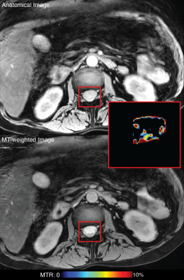 |
Magnetization transfer imaging of patients with
Guillain-Barre Syndrome
Alison R Roth1 and
Richard D Dortch2
1Barrow Neuroimaging Innovation Center, Barrow Neurological Institute, Phoenix, AZ, United States, 2Barrow Neurological Institute, Phoenix, AZ, United States Keywords: Nerves, Magnetization transfer, Guillain-Barre Syndrome New biomarkers are needed in Guillain-Barre Syndrome to predict patient recovery and severe complications and aid in drug development. We propose using magnetization transfer (MT) MRI to measure myelin content to act as such a biomarker. We have shown the ability of the MT imaging to visualize the cauda equina, but further work is needed to confirm these differences and biomarker suitability. |
| 08:15 |
1192. |
Brain PET Synthesis from MRI Using Joint Probability
Distribution of Diffusion Model at Ultrahigh Fields
Xie Taofeng1,2,
Cao Chentao3,
Cui Zhuoxu3,
Li Fanshi3,
Wei Zidong3,
Zhu Yanjie3,
Li Ye3,
Liang Dong3,4,
Jin Qiyu1,
Chen Guoqing1,
and Wang Haifeng3
1Inner Mongolia University, Hohhot, China, 2Inner Mongolia Medical University, Hohhot, China, 3Shenzhen Institutes of Advanced Technology,Chinese Academy of Sciences, shenzhen, China, 4Guangdong Laboratory of Artificial Intelligence and Digital Economy (SZ), shenzhen, China Keywords: Nerves, Multimodal, Ultrahigh Field, Artificial Intelligence, Multimodal MRI and PET are important modalities and can provide complementary information for the diagnosis of brain diseases because MRI can provide structural information of brain and PET can obtain functional information of brain. However, PET is usually missing. Especially, simultaneous PET and MRI imaging is not achievable at ultrahigh field in the current. Thus, synthetic PET using MRI at ultrahigh field is essential. In this paper, we synthetic PET using MRI as a guide by joint probability distribution of diffusion model (JPDDM). Meanwhile, We utilized our model in ultrahigh fields. |
| 08:15 |
1193.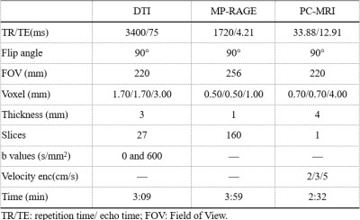 |
Pathogenesis of Chiari Malformation:insights from
morphology,neurobiology and hydrodynamics based on MR
neuroimaging technology
Yishuang Wang1,
Yun sen HE2,
Meining Chen3,
Yuting Wang1,
and Longlin Yin1
1Radiology, Sichuan Provincial People's Hospital, Chengdu, China, 2Department of Neurosurgery, Sichuan Provincial People's Hospital, Chengdu, China, 3MR Scientific Marketing, Siemens Healthcare, Shanghai, China Keywords: Nerves, Nerves The pathogenic mechanism of different clinical and radiologic manifestations of Chiari malformation I (CMI) is still unclear. Using MR morphology, neurobiology, and hydrodynamics techniques allow for a comprehensive assessment of CMI. FA values and CSF flow may well explain the correlation between white matter fiber tract, CMI pain, and compensations at other body parts. MRI technology helps us to better understand the pathogenic mechanism of CMI and can provide more valuable information in the diagnosis and treatment of CMI. |
| 08:15 |
1194.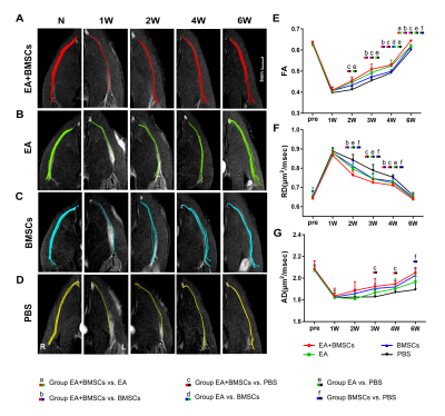 |
MRI Assessment of the Therapeutic Effect of Combined
Electroacupuncture and Stem Cells in Acute Peripheral Nerve
injury
yueyao chen1,
zhongxian pan1,
fanqi meng1,
xuewen yu2,
qian xu1,
leyu huang1,
qiumei liang1,
yanglei wu3,
hanqing lyu1,
and xiaofeng lin4
1Department of radiology, Shenzhen Traditional Chinese Medicine Hospital, Shenzhen, China, 2Department of pathology, Shenzhen Traditional Chinese Medicine Hospital, Shenzhen, China, 3MR Collaboration, SIEMENS Healthineers Ltd., Beijing, China, 4Department of Nuclear Medicine, The Seventh Affiliated Hospital, Sun Yat-sen University, Shenzhen, China Keywords: Nerves, Nervous system, Electroacupuncture, Mesenchymal stem cells We aimed to verify the efficacy of bone mesenchymal stem cells (BMSCs) combined with electroacupuncture (EA) in the treatment of peripheral nerve injury (PNI) using pathology and MRI. In a rat model of sciatic nerve crush damage, we found that BMSCs and EA combined therapy improved axon and myelin regeneration synergistically and significantly decreased post-injury nerve edema, improved axon guiding factor, and expedited motor function recovery. This suggests that a combination of BMSCs and EA can provide both topological and biomolecular guidance to promote axonal extension, myelin regeneration, and functional recovery after PNI. |
| 08:15 |
1195.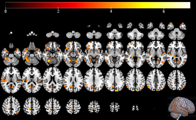 |
Altered white matter diffusion properties and gray matter
volume in classical trigeminal neuralgia
Yang Zhang1,
Wei Su2,
Xiaoyu Du3,
Rui Li4,
Hang Zhao4,
Zhaoping Wang1,
Lei Feng1,
liangjie lin5,
and Kaihua Zhang2
1Department of Neurosurgery, Jining No. 1 People’s Hospital, Jining, China, 2School of Psychology, Shandong Normal University, Ji’nan, China, 3Faculty of Medicine, Dentistry and Health Sciences, The University of Melbourne, Victoria, Australia, 4Department of Radiology, Jining No. 1 People’s Hospital, Jining, China, 5Clinical and Technical Support, Philips Healthcare, Beijing, China Keywords: Nerves, Diffusion Tensor Imaging, Trigeminal neuralgia, trigeminal nerve fiber Investigating microstructural changes in patients with classical trigeminal neuralgia (CTN) has contributed to understanding the pathological neural mechanism of CTN. This study aimed to reveal abnormalities of the trigeminal fiber bundles by combing MR diffusion tensor imaging (DTI) with voxel-based morphometry (VBM). 32 patients with CTN and 32 matched healthy controls were recruited with the main fiber bundle diffusion indices calculated and the whole-brain gray matter volume measured for each subject. Results showed that CTN exhibited microstructural changes in the trigeminal nerve fibers, and the changes might be associated with the pathogenesis of trigeminal neuralgia. |
| 08:15 |
1196.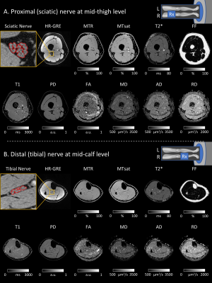 |
Multiparametric Quantitative MRI of Peripheral Nerves: A
Reliability Study
Yongsheng Chen1,
Jacob Baraz1,
Stephanie Yan Xuan1,
Ryan Castoro1,
Yang Xuan2,
Alison Roth3,
Richard D. Dortch3,
and Jun Li1,4
1Department of Neurology, Wayne State University School of Medicine, Detroit, MI, United States, 2Department of Radiology, Wayne State University School of Medicine, Detroit, MI, United States, 3Department of Translational Neuroscience, Barrow Neurological Institute, Phoenix, AZ, United States, 4Department of Neurology, Houston Methodist Hospital, Houston, TX, United States Keywords: Nerves, Nerves This study developed a multiparametric qMRI method to quantify fat fraction of leg muscles, myelin and axonal pathologies of peripheral nerves using MTR, MTsat, T1, PD, T2*, FA, MD, AD, RD, and nerve fascicular volume. The results will be applied in ongoing longitudinal studies to develop monitoring biomarkers in patients with polyneuropathies. |
| 08:15 |
1197.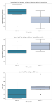 |
Associations Between Central Canal Stenosis, Resting State
Functional Connectivity Networks, and Pain Perception
Jennifer Anne Cummings1,
Madeline Hess1,
Kenneth Gao1,
Upasana Bharadwaj1,
Misung Han1,
Cynthia Chin1,
Salvatore Torrisi1,
Jennifer Townsend1,
An Vu1,
Valentina Pedoia1,
and Sharmila Majumdar1
1Radiology and Biomedical Imaging, University of California, San Francisco, San Francisco, CA, United States Keywords: Nerves, Nervous system Chronic lower back pain remains difficult to characterize with imaging. With this study, we investigate the relationship between imaging-based brain and spine biomarkers in pain perception. We present relationships between resting state functional connectivity networks, automatic spinal canal stenosis grading, and patient-reported pain measures. Patients reporting severe pain show weaker connectivity in the Left Ventral Attention Network and stronger connectivity within the Salience network when compared to those reporting no pain or mild/moderate pain. The severe back pain group also shows a higher spinal canal stenosis grading. |
| 08:15 |
1198.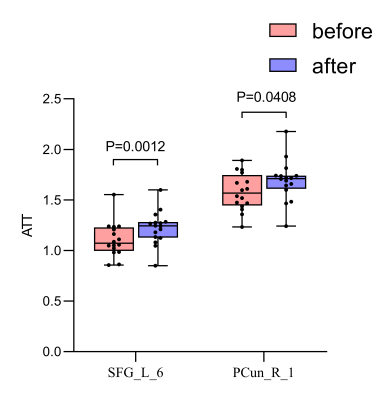 |
Analysis of cerebral perfusion changes after microvascular
decompression in patients with hemifacial spasm
Kuan Lv1,
Aocai Yang1,
Bing Liu1,
Jixin Luan1,
and Guolin Ma1
1China-Japan Friendship Hospital, Beijing, China Keywords: Nerves, Brain We used the eASL technique to explore the mCBF, ATT and CBV alterations after MVD in patients with HFS. And found that the ATT values in the left superior frontal gyrus region 6 and the right precuneus region 1 were significantly increased in the L-HFS group after surgery compared with the preoperative ones. Furthermore, preoperative ATT values in the right precuneus 1 region were negatively correlated with mCBF values in the L-HFS group. It is suggested that these brain regions may play different roles in the underlying pathological mechanisms of HFS. |
| 08:15 | 1199. | WITHDRAWN |
| 08:15 |
1200.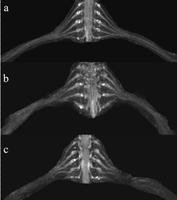 |
MR neurography Aids in Detection of Longitudinal
Brachial Plexus Nerve Root Alterations in Patients With
Amyotrophic Lateral Sclerosis
Shanshan Wang1,
Xiao Man1,
Guangbin Wang1,
Weibo Chen2,
and Avneesh Chhabra3
1Shandong Provincial Hospital Affiliated to Shandong First Medical University, Jinan, China, 2Philips Healthcare, Shanghai, China, 3UT Southwestern Medical Center, Dallas, TX, United States Keywords: Nerves, Nerves We assessed proximal-distal longitudinal signal and size alterations of brachial plexus nerve roots in amyotrophic lateral sclerosis (ALS) patients using 3D nerve-sheath signal increased with inked rest-tissue rapid acquisition of relaxation enhancement imaging (3D SHINKEI) . The conclusion confirmed proximal-distal longitudinal diameters and SNR values decreased significantly for brachial plexus nerve roots in ALS patients with larger differences in slopes compared to the HC. Thus, the size and signal alterations of brachial plexus nerve roots using 3D SHINKEI can be used to supplement clinical diagnosis for ALS patients. |
| 08:15 |
1201.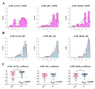 |
Neurovascular decoupling in type 2 diabetes: A meta-analysis
and an independent validation of Neuroimaging
Zeyang zeyang Li1,
Ying Yu1,
Guang-Bin Cui1,
and Lin-Feng Yan1
1Hospital, Fourth Military Medical University, 569 Xinsi Road, Xi’an 710038, Shaanxi, China, Xi’an, China Keywords: Nerves, fMRI (resting state) Disrupted neurovascular (NV) coupling is considered as a potential mechanism of Type 2 diabetes mellitus (T2DM) induced mild cognitive impairment (MCI). The study quantitatively explored whether NV decoupling were associated with cognitive impairment in patients with T2DM by means of neuroimaging meta-analysis and an independent validation. In T2DM, NV uncoupling existed in many brain regions, and the degree of uncoupling was related to cognition. It contributed to a better understanding of the mechanism in T2DM cognitive impairment and would be a promising neuroimaging biomarker. This study combined meta-analysis and independent validation model can be extended to other similar studies. |
| 08:15 |
1202.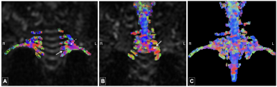 |
Evaluation of Brachial Plexus Injuries in Infants with DTI
and Tractography
Meltem Karatas1,
Kader Karli Oguz1,2,3,
Gulgun Sengul4,
Akin Uzumcugil5,
and Gokcen Coban2
1National Magnetic Resonance Research Center (UMRAM), Bilkent University, Ankara, Turkey, 2Department of Radiology, Hacettepe University, Ankara, Turkey, 3Department of Radiology, University of California, Davis, Sacramento, CA, United States, 4Department of Anatomy, Ege University, Izmir, Turkey, 5Department of Orthopedics and Traumatology, Hacettepe University, Ankara, Turkey Keywords: Nerves, Diffusion Tensor Imaging, Brachial plexus, peripheral nerve tractography Neonatal brachial plexus injury (NBPI) is caused by traction on the neck during birth and presents with flaccid paralysis of the upper extremity. 10-20% of the cases result in neurological sequelae and require surgical intervention; the treatment decisions principally depend on the clinical assessments through the first six months of life. Early stratification of NBPI cases may lead to earlier intervention for severe injuries, better disease outcomes, and higher quality of life. Here, we investigated the use of DTI and tractography for infants with NBPI; our results suggest that this approach might be suitable as a diagnostic and prognostic tool. |
| 08:15 |
1203.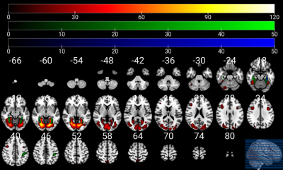 |
Gender differences in brain response to infant emotional
faces
Wei Su1,
Xiaoyu Du2,
Xianling Liu3,
Zhenhua Sun4,
Kaihua Zhang1,
Mengxing Wang5,
and Xiaoxia Du6
1School of psychology, Shandong Normal University, Ji’nan, China, 2Faculty of Medicine, Dentistry and Health Sciences, The University of Melbourne, Victoria, Australia, 3Department of Medicine Imaging, The People's Hospital of Jinan Central District, Jinan, China, 4School of information science and engineering, Linyi University, Linyi, China, 5College of Medical Imaging, Shanghai University of Medicine and Health Sciences, Shanghai, China, 6Department of Psychology, Shanghai University of Sport, Shanghai, China Keywords: Nerves, fMRI (task based), Infant emotional faces, Empathy Exploring the neural processes of recognizing infant stimuli promotes better understandings of the mother-infant attachment mechanisms. Here combining Task-fMRI and resting-state fMRI investigated the effects of infants’ emotional faces on the brain activity of women and men. The task-fMRI showed that the brains of women and men reacted differently to infants’ faces, and these differential areas are in facial processing and empathetic networks. The rs-fMRI further showed that the connectivity of the default-mode network-related regions increased in women than men. These differences might facilitate women to more effective and quick adjustments in behaviors and emotions during the nurturing infant period. |
| 08:15 |
1204.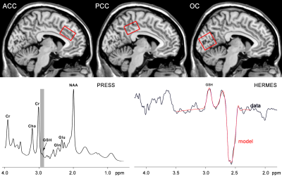 |
Brain glutathione levels decrease with age and correlate
with cognitive function
Xin Hu1,
Min Zhao1,
Jing Yang1,
Longji Xu1,
Weibo Chen2,
Fuxin Ren1,
and Fei Gao1
1Department of Radiology, Shandong Provincial Hospital Affiliated to Shandong First Medical University, Jinan, China, 2Philips Healthcare, Shanghai, China Keywords: Nerves, Aging Cognitive impairment and the improvement of oxidative stress are common in the aged. GSH is a key player to defend oxidative stress and avoid ferroptosis. We aim to explore the variation of brain GSH levels with age including anterior cingulate cortex, posterior cingulate cortex and occipital cortex, and to test whether GSH levels in these regions are associated with cognitive function. Our findings indicate that oxidative stress and ferroptosis abnormality caused by the decreased GSH levels may contribute to cognitive decline of the aged without a regional specificity manner. |
| 08:15 |
1205.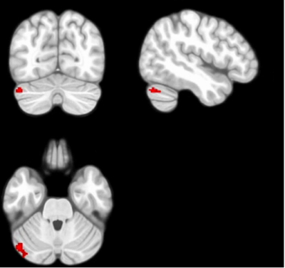 |
Abnormal brain function and connectivity in patients with
convergence insufficiency: A resting-state functional MRI
study
Yuxia Wanng1,
Ye Wu2,
Huaiqiang Sun3,
and Fei Li1
1Huaxi MR Research Center (HMRRC), Department of Radiology, West China Hospital of Sichuan University, Chengdu 610041, Sichuan, P.R. China., Chengdu, Sichuan, China, 2Department of Ophthalmology, Laboratory of Optometry and Vision Sciences, West China Hospital of Sichuan University, Chengdu 610041, Sichuan, P.R. China., Chengdu, Sichuan, China, 3Huaxi MR Research Center (HMRRC), Department of Radiology, West China Hospital of Sichuan University, Chengdu 610041, Sichuan, P.R. China.Huaxi MR Research Center (HMRRC), Department of Radiology, West China Hospital of Sichuan University, Chengdu 610041, Sichuan, P.R. China., Chengdu, Sichuan, China Keywords: Nerves, Brain, Convergence insufficiency; brain function; functional connectivity; fALFF Convergence insufficiency (CI) is the most common binocular vision disorder in optometric clinics relevant to the dysfunction of the specific neural circuit, while the neural mechanism is unknown. Using the fractional amplitude of low-frequency fluctuations fALFF and seed-to-voxel analysis, we found increased fALFF in the left cerebellum crus 1 and increased functional connectivity between the left cerebellum crus 1 and the right cerebellum crus 1 in patients with CI than healthy controls, which showed positive correlations with the severity of symptoms. These findings provide evidence of altered brain function features underlying neurobiological mechanisms of patients with CI. |
| 08:15 |
1206.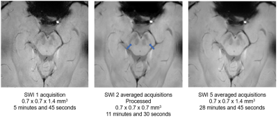 |
A brainstem-dedicated approach to assess the N1 sign using a
3T SWI
Sabrina Houidef1,
Germain Arribarat1,
and Patrice Péran1
1ToNIC, Toulouse NeuroImaging Center, University of Toulouse, Inserm, Toulouse, France Keywords: Parkinson's Disease, Parkinson's Disease, Neurodegeneration Because of its anatomical situation, the brainstem is difficult to image. With whole brain acquisitions, we end up with artifactual images. Our team developed a brainstem-dedicated T2*-weighted MRI acquisition method. Using this, we imaged the brain of sixteen healthy volunteers. This sequence was repeated 5 times for each volunteer. Our MRI protocol also consisted of a three-dimensional T1-weighted whole-brain sequence. The aim was to reduce the number of acquisitions required to visualize the nigrosome-1 by using image processing methods. We quantified the quality of the images by using image quality assessment indices and had one criterion: the visualization of nigrosome-1. |
| 08:15 |
1207.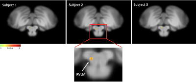 |
Functional brainstem imaging of sympathetic drive using MSNA
coupled fMRI and SSNA coupled fMRI at ultra-high field.
Rebecca Glarin1,
Luke Henderson2,
Donggyu Rim3,
and Vaughan Macefield3
1University of Melbourne, Parkville, Australia, 2University of Sydney, Sydney, Australia, 3Baker Heart and Diabetes Institute, Prahran, Australia Keywords: Nerves, High-Field MRI, Brainstem Muscle sympathetic nerve activity (MSNA) is responsible for blood pressure control and skin sympathetic nerve activity (SSNA) for thermoregulation. This activity can be recorded through a process of microneurography. This nerve activity can be temporally coupled with fMRI to image the brainstem. With the use of ultra high field MRI we are able to image the subcortical and cortical regions responsible for these sympathetic nervous system processes. Brainstem imaging using this technique for the first time at high field, has identified with high specificity the rostral ventrolateral medulla (RVLM) when using MSNA coupled fMRI. |
 Back to Meeting Home
 Back to the Program-at-a-Glance
The International Society for Magnetic Resonance in Medicine is accredited by the Accreditation Council for Continuing Medical Education to provide continuing medical education for physicians.
 Back to Meeting Home
 Back to the Program-at-a-Glance
 Full Session Video