Power Pitch
Pitch: Brain, Spinal Cord & White Matter Injury
ISMRM & ISMRT Annual Meeting & Exhibition • 03-08 June 2023 • Toronto, ON, Canada

Power Pitch
Pitch: Brain, Spinal Cord & White Matter Injury
| 08:15 |
0451.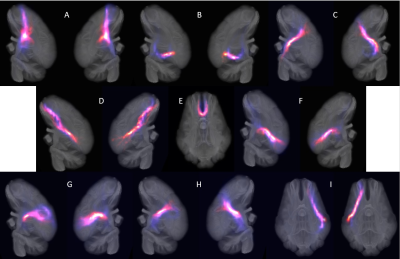 |
Affinity of Structural White Matter Tracts between Infant
and Adult Pigs
Wenwu Sun1,
Ishfaque Ahmed1,
Stephanie Dubrof2,
Franklin West3,
Hea Jin Park2,
and Qun Zhao1
1Department of Physics and Astronomy, Franklin College of Arts and Sciences, University of Georgia, Athens, GA, United States, 2College of Family and Consumer Sciences, University of Georgia, Athens, GA, United States, 3Regenerative Bioscience Center, University of Georgia, Athens, GA, United States Keywords: Brain Connectivity, Diffusion/other diffusion imaging techniques, Tractography Affinity of structural white matter tracts during development is critical for longitudinal studies of many neurological diseases and brain injuries. Diffusion weighted imaging (DWI) data was collected from 3-week-old piglets. Data-driven tractography analysis was applied to profile white matter tracts for the piglets, which were then compared to recently reported 27 adult pig white matter tracts. Among the 27 tracts, 17 were found with consistent high spatial correlations between infant and adult pigs. This result provides possibilities to further study white matter during development, and to evaluate how different interventions (e.g., TBI) alter the brain myelination trajectory. |
| 08:15 | 0452. | WITHDRAWN |
| 08:15 |
0453.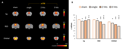 |
Repetitive Mild Traumatic Brain Injury Induces Significant
Changes on Functional Connectivity with Different Impact
Numbers in Rats
Yi-Han Kao1,
Chia-Feng Lu1,
Bao-Yu Hsieh2,3,
and Yu-Chieh Jill Kao1
1Department of Biomedical Imaging and Radiological Sciences, National Yang-Ming Chiao-Tung University, Taipei, Taiwan, 2Department of Medical Imaging and Radiological Sciences, Chang Gung University, Taoyuan, Taiwan, 3Department of Medical Imaging and Intervention, Chang Gung Memorial Hospital at Linkou, Taoyuan, Taiwan Keywords: Traumatic brain injury, Animals, rsfMRI In the current study, we discussed about changes of brain connectivity through rsfMRI and behavioral performances following repetitive mild traumatic brain injury (rmTBI) with different impact numbers and intervals. In rats with larger impact number but longer inter-injury interval, may show anxiety-like behavior with the significant reduction of connectivity in their DMN. |
| 08:15 |
0454.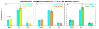 |
Simultaneous Measurements of GABA, Glx and GSH in the
Thalamus in Patients with Mild Traumatic Brain Injury: A
Preliminary Study
Xiao Liang1,
Muhammad G Saleh1,
Rosy Linda Njonkou Tchoquessi1,
Alexa G. Colinco1,
Steve Roys1,
Prashant Raghavan1,
Rao P Gullapalli1,
and Jiachen Zhuo1
1Department of Diagnostic Radiology and Nuclear Medicine, University of Maryland School of Medicine, Baltimore, MD, United States Keywords: Traumatic brain injury, Spectroscopy, Thalamus, HERMES In this study, we report the preliminary results of simultaneous measurements of GABA, Glx (glutamate + glutamine), and GSH in the thalamus using HERMES in the mTBI patients. HERMES was acquired in the thalamus for patients in acute, subacute, and chronic stages, and control subjects as reference using optimized acquisition and processing. Metabolite fitting were performed in Gannet with spectral alignment. The results demonstrate that HERMES can maintain the spectral and fitting quality in the mTBI patients versus the control subjects. Differences in the metabolite levels warrant accruing a larger number of patients for a more definite evaluation. |
| 08:15 |
0455.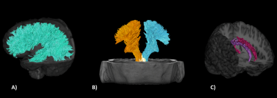 |
Exploring whether differences in brain diffusion MRI metrics
distinguish symptom phenotypes in athletes exposed to
repetitive head impacts.
Joshua P McGeown1,2,
Maryam Tayebi1,3,
Matthew A McDonald1,4,
Paul Condron1,
Samantha Holdsworth1,5,
Leigh Potter1,6,
Davidson Taylor1,7,
Patrick McHugh1,8,
Miao Qiao9,
Jerome Maller10,
Justin Fernandez3,
Vickie Shim1,3,
Mangor Pedersen11,
and Eryn E Kwon1,3,5
1Mātai Medical Research Institute, Tairāwhiti-Gisborne, New Zealand, 2Department of Anatomy and Medical Imaging, Faculty of Medical and Health Sciences, University of Auckland, Auckland, New Zealand, 3Auckland Bioengineering Institute, University of Auckland, Auckland, New Zealand, 4Faculty of Medical and Health Sciences, University of Auckland, Auckland, New Zealand, 5Faculty of Medical and Health Sciences & Centre for Brain Research, University of Auckland, Auckland, New Zealand, 6Ngāti Porou, Ngāti Kahungunu, Rongomaiwahine, Rongowhakaata, Tairāwhiti-Gisborne, New Zealand, 7Ngai Tāmanuhiri, Rongowhakaata, Ngāti Porou, Tairāwhiti, New Zealand, 8Turanga Health, Tairāwhiti-Gisborne, New Zealand, 9School of Computer Science, Faculty of Science, University of Auckland, Auckland, New Zealand, 10General Electric Healthcare, Victoria, Australia, 11Department of Psychology and Neuroscience, Auckland University of Technology, Auckland, New Zealand Keywords: Traumatic brain injury, Diffusion Tensor Imaging, Symptomology It is important to account for the heterogeneity of clinical presentation when studying mild traumatic brain injury (mTBI) with advanced brain imaging. We used post-season symptom data from a cohort of rugby players exposed to repetitive head impacts, and we applied unsupervised learning to cluster athletes into clinically distinct groups. We explored whether these clusters demonstrated post-season group differences in white matter tracts compared to controls. This analysis framework suggests that group differences of diffusion metrics in athletes exposed to repetitive head impacts may be associated with clinical presentation rather than generalisable across all participants. |
| 08:15 |
0456.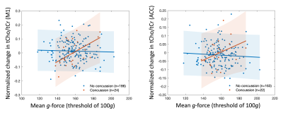 |
Association between brain metabolites and head impact
exposure measured with MRS in a cohort of high school
American football athletes
Zexuan Liu1,
Jonathan A. Dudley2,
Nadine Ahmed3,
Jed A. Diekfuss4,5,6,
David A. Edmondson2,7,
Kim M. Cecil2,7,
Weihong Yuan2,7,
Taylor M. Zuleger4,5,6,8,
Alexis B. Slutsky-Ganesh4,5,6,9,
Kim D. Barber Foss4,5,6,
Gregory D. Myer4,5,6,10,
and Candace C. Fleischer1,11
1Department of Biomedical Engineering, Georgia Institute of Technology and Emory University, Atlanta, GA, United States, 2Imaging Research Center, Cincinnati Children's Hospital Medical Center, Cincinnati, OH, United States, 3Department of Neuroscience, Georgia Institute of Technology, Atlanta, GA, United States, 4Emory Sports Performance And Research Center (SPARC), Flowery Branch, GA, United States, 5Emory Sports Medicine Center, Atlanta, GA, United States, 6Department of Orthopedics, Emory University School of Medicine, Atlanta, GA, United States, 7Department of Radiology, University of Cincinnati College of Medicine, Cincinnati, OH, United States, 8Neuroscience Graduate Program, University of Cincinnati College of Medicine, Cincinnati, OH, United States, 9Department of Kinesiology, University of North Carolina at Greensboro, Greensboro, NC, United States, 10The Micheli Center for Sports Injury Prevention, Waltham, MA, United States, 11Department of Radiology and Imaging Sciences, Emory University School of Medicine, Atlanta, GA, United States Keywords: Traumatic brain injury, Spectroscopy Diagnosis and prognosis of sports-related concussion is challenging. In this prospective controlled clinical trial of 215 high school American football athletes, we evaluated relationships between brain metabolite and head impacts as a function of concussion diagnosis and wearing a jugular vein compression (JVC) collar. Changes in total choline between pre- and post-season, measured with MR spectroscopy, were positively correlated with mean g-force thresholds above 80g in athletes diagnosed with a concussion, suggesting choline may be a key metric of injury. Metabolite alterations were minimally affected by the JVC collar and only at mean g-force thresholds of 100, 110 and 120g. |
| 08:15 |
0457.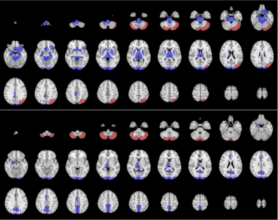 |
Abnormal spontaneous brain fluctuations present in retired
football players
Ethan Danielli1,2,3,
Bhanu Sharma3,4,
Cameron E Nowikow2,3,
and Michael D Noseworthy2,3,4,5
1KITE, Toronto Rehabilitation Institute, Toronto, ON, Canada, 2School of Biomedical Engineering, McMaster University, Hamilton, ON, Canada, 3Imaging Research Centre, St. Joseph's Healthcare Hamilton, Hamilton, ON, Canada, 4Electrical and Computer Engineering, McMaster University, Hamilton, ON, Canada, 5Radiology, McMaster University, Hamilton, ON, Canada Keywords: Traumatic brain injury, fMRI (resting state), ALFF Novel methods are required to understand the scale of potential brain changes in collision sport athletes. with many former athletes having developed neurocognitive deficits or neurological disorders. This study used resting state functional MRI (rsfMRI) to examine if retired professional football players (n=18) had functional brain abnormalities based on a personalized amplitude of low-frequency fluctuations (ALFF) and Z-scoring approach. Brain injuries were identified if the ALFF Z-score exceeded 3 standard deviations from the healthy control mean. Thirty regions were abnormal in more than half of the retired athletes. Cerebellar and central, sub-cortical brain regions were most often seriously abnormal. |
| 08:15 |
0458.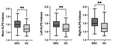 |
MRI Detection of Glymphatic Function in athletes after
Sports-related Concussion
Laiyang Ma1,
Wanjun Hu1,
Jing Zhang1,
Wenjing Huang1,
and Yuhui Xiong2
1Department of Magnetic Resonance, Lanzhou University Second Hospital, Lanzhou, China, 2MR Research, GE Healthcare MR Research, Beijing, China Keywords: Traumatic brain injury, Diffusion/other diffusion imaging techniques Diffusion tensor image analysis along the perivascular space (DTI-ALPS) as a non-invasive method for evaluating the activity of the glymphatic system in human brain by using diffusion images. In this study, DTI-ALPS was used to evaluate the activity of the human lymphatic system in patients with Sports-related concussion (SRC). We found SRC has a higher ALPS index compared to the healthy controls. Our findings suggested that abnormal glymphatic function in brain might be a potential biomarker for explaining the cognitive function decline of SRC. |
| 08:15 |
0459. |
Classifying patients with chronic mild traumatic brain
injury: a rs-fMRI study and support vector machine analysis
Faezeh Vedaei1,
Najmeh Mashhadi2,
George Zabrecky1,
Daniel Monti1,
Emily Navarreto1,
Chloe Hriso1,
Nancy Wintering1,
Andrew B. Newberg1,
and Feroze B. Mohamed1
1Thomas Jefferson University, Philadelphia, PA, United States, 2University of California-Santa Cruz, Santa Crus, CA, United States Keywords: Traumatic brain injury, fMRI (resting state) Machine learning classification of patients with chronic mild traumatic brain injury using rs-fMRI metrics |
| 08:15 |
0460. |
MRE-based assessment of impairment of mechanical isolation
at the skull-brain interface resulting from sports-related
head impact exposure
Xiang Shan1,
Matthew C. Murphy1,
Yi Sui1,
Keni Zheng1,
Armando Manduca2,
Richard L. Ehman1,
John Huston III1,
and Ziying Yin1
1Radiology, Mayo Clinic, Rochester, MN, United States, 2Physiology and Biomedical Engineering, Mayo Clinic, Rochester, MN, United States Keywords: Traumatic brain injury, Elastography, Repeated head impact Increasing recognition of the effect of repeated head impacts (RHI) in leading to neurologic impairment and higher risk of subsequent traumatic head injury, has motivated for developing technology to assess the status of the mechanism of mechanical isolation provided by the structures within the skull-brain interface, known as the pia-arachnoid complex (PAC). To evaluate the RHI-induced PAC alternations, we compared MR elastography-based measures (rotational transmission ratio [Rtr] and cortical normalized octahedral shear strain [NOSS]) between healthy and sports-related RHI subjects. Significantly higher Rtr and cortical NOSS were found among RHI individuals, showing their potential in assessing the effect of RHI. |
| 08:15 |
0461.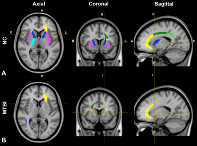 |
Influence of White Matter Microstructure on Interhemispheric
Processing Speed After Mild Traumatic Brain Injury Using
Advanced Diffusion MRI
Sohae Chung1,2,
Tamar Bacon3,
Joseph F. Rath4,
Alaleh Alivar1,2,
Santiago Coelho1,2,
Prin X. Amorapanth4,
Els X. Fieremans1,2,
Dmitry S. Novikov1,2,
Steven R. Flanagan4,
Joshua H. Bacon3,
and Yvonne W. Lui1,2
1Center for Advanced Imaging Innovation and Research (CAI2R), Department of Radiology, New York University Grossman School of Medicine, New York, NY, United States, 2Bernard and Irene Schwartz Center for Biomedical Imaging, Department of Radiology, New York University Grossman School of Medicine, New York, NY, United States, 3Department of Neurology, New York University Grossman School of Medicine, New York, NY, United States, 4Department of Rehabilitation Medicine, New York University Grossman School of Medicine, New York, NY, United States Keywords: Traumatic brain injury, Diffusion/other diffusion imaging techniques The corpus callosum (CC) is especially vulnerable to mild traumatic brain injury (MTBI). Since it connects left and right cerebral hemispheres, damage to the CC or neighbor white matter (WM) pathways may specifically disrupt interhemispheric communication. Here we employ a mediation framework to study the collaboration between tissue microstructure of the CC and neighbor WM pathways that may influence interhemispheric processing. Our results show different patterns in individuals with recent MTBI compared to healthy controls in terms of the relationships between callosal microstructure and interhemispheric communication mediated by other pathways, highlighting tracks specifically related to primary visual and language processes. |
| 08:15 |
0462.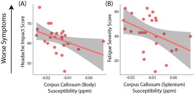 |
Quantitative Susceptibility Mapping in Patients with
Persistent-Post Concussion Symptoms
Tiffany K Bell1,2,3,
Muhammad Ansari4,
Leah Mercier2,5,
David G Gobbi6,
Richard Frayne1,2,5,6,
Chantel Debert2,5,
and Ashley D Harris1,2,3
1Department of Radiology, University of Calgary, Calgary, AB, Canada, 2Hotchkiss Brain Institute, University of Calgary, Calgary, AB, Canada, 3Alberta Children's Hospital Research Institute, University of Calgary, Calgary, AB, Canada, 4Department of Biomedical Engineering, University of Calgary, Calgary, AB, Canada, 5Department of Clinical Neuroscience, University of Calgary, Calgary, AB, Canada, 6Calgary Image Processing and Analysis Centre, University of Calgary, Calgary, AB, Canada Keywords: Traumatic brain injury, Quantitative Susceptibility mapping, "Persistent Post-Concussion Symptoms" Twenty percent of people experience symptoms months to years following a concussion, known as persistent post concussive symptoms (PPCS). We used quantitative susceptibility mapping (QSM) to measure tissue susceptibility in white matter tracts of patients with PPCS. We provide preliminary evidence for alterations in white matter susceptibility in patients with PPCS and show that susceptibility levels are correlated with symptom severity. We hypothesise altered susceptibility is related to gliosis, resulting in sustained inflammation. Further, this study demonstrates the potential application of QSM to provide novel information on the pathophysiology of PPCS. |
| 08:15 |
0463.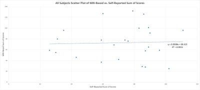 |
Comparing self-reporting concussion assessments with an
objective Diffusion Tensor Imaging (DTI) and resting state
MRI (rsMRI) based measure
Nicholas M Simard1,
Michael D Noseworthy1,2,3,4,
Dinesh A Kumbhare5,6,
Stephan Ulmer7,8,
and Ethan Danielli2
1Electrical and Computer Engineering, McMaster University, Hamilton, ON, Canada, 2School of Biomedical Engineering, McMaster University, Hamilton, ON, Canada, 3Imaging Research Centre, St. Joseph's Healthcare Hamilton, Hamilton, ON, Canada, 4Radiology, McMaster University, Hamilton, ON, Canada, 5Toronto Rehabilitation Institute, Toronto, ON, Canada, 6Medicine, University of Toronto, Toronto, ON, Canada, 7neurorad.ch, Zurich, Switzerland, 8Radiology & Neuroradiology, University hospital of Schleswig-Holstein, Kiel, Germany Keywords: Traumatic brain injury, Brain Connectivity, Gray Matter, White Matter Traditional concussion self-reporting has significant limitations due to its subjectivity and inconsistency, therefore a more objective MRI-based approach is proposed in this research. A concussion population was assessed using the traditional PCSS method versus DTI anisotropy analysis and rsMRI complexity analysis. Concussed patients were compared to large age and sex matched datasets and a Z-transform was used on white and gray matter ROIs to identify injured areas in the cerebrum. A weighted mean equation was then used to compare Z-scores to the 7-point PCSS. Results confirmed sex differences in self reporting and shows promise as a future objective assessment tool. |
| 08:15 |
0464.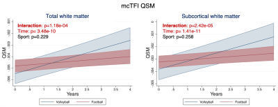 |
Longitudinal QSM imaging reveals disrupted subcortical white
matter maturation in football vs volleyball college athletes
Marios Georgiadis1,
Mahta Karimpoor1,
Pascal Spincemaille2,
Alexey Dimov2,
Brian Mills1,
Maged Goubran1,
Hossein Moein Taghavi1,
Nicole Mouchawar1,
Sohrab Sami1,
Max Wintermark1,
Gerald Grant1,
David Camarillo1,
Yi Wang2,
and Michael Zeineh1
1Stanford University School of Medicine, Stanford, CA, United States, 2Weill Cornell Medical College, New York, NY, United States Keywords: Traumatic brain injury, Quantitative Susceptibility mapping Head impacts in sports may cause long-term brain changes. Here, we assessed quantitative susceptibility mapping (QSM) changes over multiple seasons in high-contact American football vs low-contact volleyball college athletes using the multi-echo complex total field inversion (mcTFI) method. We found widespread changes over time (likely developmental) in all athletes, while time-independent sports differences were detected by R2*. mcTFI revealed an altered QSM trajectory in the white matter (total and subcortical) between sports: QSM increased in volleyball athletes but changed minimally in football, likely indicating disrupted subcortical white matter maturation in football. QSM can sensitively detect longitudinal changes in contact sports. |
| 08:15 |
0465.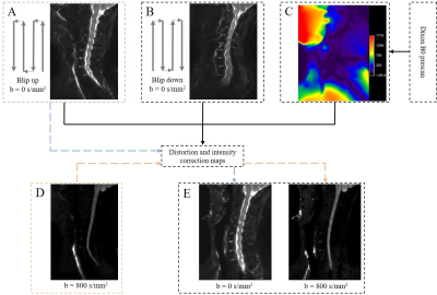 |
Distortion-corrected cervical spine diffusion-weighted
imaging at 3.0T
Zheng Sun1,
Peng Wu2,
Xiance Zhao2,
and Kan Deng3
1Shanghai Sixth People's Hospital, Shanghai, China, 2Philips Healthcare, Shanghai, China, 3Philips Healthcare, Guangzhou, China Keywords: Head & Neck/ENT, Spinal Cord Diffusion-weighted imaging (DWI) could benefit the detection and evaluation of the spine and spinal cord pathologies. Traditional single-shot EPI (SS-EPI) DWI suffers from a large geometry distortion due to the inhomogeneous B0 field and the low bandwidth in the phase encoding direction. This work demonstrated the application of an EPI geometry correction method for the cervical spine which can improve the geometry accuracy of the DWI images. |
| 08:15 |
0466.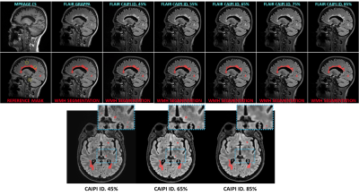 |
Effect of iterative denoising in automated white matter
hyperintensities segmentation using accelerated FLAIR
sequences
Ricardo Alberto Corredor-Jerez1,2,3,
Mathilde Carrière4,
Thierry Chaptal5,
Bénédicte Maréchal1,2,3,
Tobias Kober1,2,3,
Xavier Ayrignac6,
Nicolas Menjot de Champfleur4,5,
Thomas Troalen7,
and Emmanuelle Le Bars4,5
1Advanced Clinical Imaging Technology, Siemens Healthineers International AG, Lausanne, Switzerland, 2Department of Radiology, Lausanne University Hospital and University of Lausanne, Lausanne, Switzerland, 3LTS5, École Polytechnique Fédérale de Lausanne, Lausanne, Switzerland, 4Department of Neuroradiology, Hospital and University of Montpellier, Montpellier, France, 5Institut d'Imagerie Fonctionnelle Humaine, I2FH, Hospital and University of Montpellier, Montpellier, France, 6Department of Neurology, Gui de Chauliac Montpellier University Hospital, Montpellier, Switzerland, 7Siemens Healthcare SAS, Saint-Denis, France Keywords: Multiple Sclerosis, Segmentation, Fast MR Protocols Acquisition time in brain MR protocols can be reduced using acceleration techniques like CAIPIRINHA. The inherent increase of noise due to their undersampling schemes can be mitigated with additional processing methods. Iterative denoising has shown good performance filtering images in k-space while preserving image details. This work evaluates the impact of iterative denoising on automated segmentation of white matter hyperintensities using CAIPIRINHA 3D FLAIR and compressed sensing 3D MPRAGE. Reliable segmentations were generated across different denoising levels (45% to 85%); small structures presented lower detection rates with stronger denoising (≥75%). These findings are revelant for designing optimized brain imaging protocols. |
| 08:15 |
0467.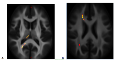 |
Brain white matter alterations in military service members
after a remote mild traumatic brain injury
Ping-Hong Yeh1,
Chihwa Song1,
Rujirutana Srikanchana1,
Cheng Guan Koay1,
Wei Liu1,
Grant Bonavia1,
and John Ollinger1
1National Intrepid Center of Excellence,Walter Reed National Military Medical Center, Bethesda, MD, United States Keywords: Traumatic brain injury, Traumatic brain injury In this study, we applied non-Gaussian diffusion MRI, including bi-tensor diffusion tensor imaging, diffusion kurtosis imaging, neurite orientation dispersion and density imaging and fixel-based analysis, to assess white matter disruption after a remote brain injury. The findings of both increased and decreased orientation distribution index over the forceps major and forceps minor of the corpus callous, along with increased fiber density and fiber cross-sectional area of fiber bundle over the anterior frontal region suggests a mixed pattern of white matter alterations, e.g. loosely white matter such as gliosis along with neuroplasticity and brain repair after a remote mild traumatic brain injury. |
| 08:15 |
0468. |
White matter changes detected based on multi-component MR
Fingerprinting in Multiple Sclerosis
Martijn Nagtegaal1,2,
Ingo Hermann1,3,
Sebastian Weingärtner1,
Eloy Martines-Heras4,
Elisabet Solano4,
Sare Llufriu4,
Achim Gass5,
Dirk H. J. Poot6,
Matthias J.P. van Osch2,
Frans M. Vos1,6,
and Jeroen de Bresser7
1Department of Imaging Physics, Delft University of Technology, Delft, Netherlands, 2C.J. Gorter MRI Center, Radiology Department, Leiden University Medical Center, Leiden, Netherlands, 3Computer Assisted Clinical Medicine, Medical Faculty Mannheim, Heidelberg University, Mannheim, Germany, 4Neuroimmunology and Multiple Sclerosis Unit and Laboratory of Advanced Imaging in Neuroimmunological Diseases (ImaginEM), Hospital Clinic Barcelona, Institut d'Investigacions Biomediques August Pi i Sunyer (IDIBAPS) and Universitat de Barcelona, Barcelona, Spain, 5Department of Neurology, Medical Faculty Mannheim, Heidelberg University, Mannheim, Germany, 6Department of Radiology and Nuclear Medicine, Erasmus MC, Rotterdam, Netherlands, 7Department of Radiology, Leiden University Medical Center, Leiden, Netherlands Keywords: Multiple Sclerosis, MR Fingerprinting White matter hyperintensities are an MRI biomarker of Multiple Sclerosis (MS). However, not all white matter changes are visible on conventional, qualitative MRI. We applied a multi-component MR Fingerprinting protocol to identify potential white matter abnormalities based on increased $$$T_2^*$$$-values. FLAIR and MRF scans were performed in 44 MS patients and 12 healthy control subjects. Significant differences were found in the volume of MRF components with 500ms<$$$T_1, T_2^*$$$<2.5s. This volume correlated moderately with white matter damage on structural MR images. The MRF approach identified larger abnormal tissue volumes than those visible on the structural scans. |
| 08:15 |
0469. |
Structural covariance in subcortical regions in MS and
NMOSD: An MRI-based study with automated brain volumetry
Yan Xie1,
Yan Zhang1,
Chengxia Liu1,
and Wenzhen Zhu1
1Tongji Hospital, Tongji Medical College, Huazhong University of Science and Technology, Wuhan, China Keywords: Multiple Sclerosis, Multiple Sclerosis MS and NMOSD, as two major demyelinating diseases of the CNS, both could cause brain structural volume changes. Moreover, the synergistic volume changes between brain regions can reflect the intrinsic connection network between these regions, which is helpful to further explore the underlying pathophysiological changes of the disease. Our study identified volumetric changes and structural covariance in subcortical regions in MS and NMOSD patients. Furthermore, MS and NMOSD patients had distinct patterns of anatomical connection in brain regions, which reflected the different underlying damage to brain structures in the two diseases. |
 Back to Meeting Home
 Back to the Program-at-a-Glance
The International Society for Magnetic Resonance in Medicine is accredited by the Accreditation Council for Continuing Medical Education to provide continuing medical education for physicians.
 Back to Meeting Home
 Back to the Program-at-a-Glance
 Full Session Video