Power Pitch
Pitch: RF Strategies at High & Ultra-High Field
ISMRM & ISMRT Annual Meeting & Exhibition • 03-08 June 2023 • Toronto, ON, Canada

Power Pitch
Pitch: RF Strategies at High & Ultra-High Field
| 13:45 |
0199.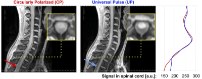 |
Universal pulses for the cervical spinal cord at 7T: a
feasibility study
Daniel Papp1,
Nicolas Boulant2,
Aurelien Massire3,
Frank Mauconduit2,
Vincent Gras2,
and Julien Cohen-Adad1,4,5,6
1NeuroPoly Lab, Institute of Biomedical Engineering, Polytechnique Montreal, Montreal, QC, Canada, 2University of Paris-Saclay, CEA, NeuroSpin, CNRS, BAOBAB, Gif sur Yvette, France, 3Siemens Healthcare SAS, Saint-Denis, France, 4Mila - Quebec AI Institute, Montreal, QC, Canada, 5Functional Neuroimaging Unit, Centre de recherche de l'Institut universitaire de gériatrie de Montréal, Montreal, QC, Canada, 6Centre de recherche du CHU Sainte-Justine, Université de Montréal, Montreal, QC, Canada Keywords: Parallel Transmit & Multiband, Spinal Cord While the Universal Pulse approach to mitigate the RF field inhomogeneity effect has been successfully deployed in brain imaging, adoption in spinal cord imaging is lacking. Here, we demonstrate the feasibility of designing Universal Pulses for the cervical spinal cord at 7T, and show a marked improvement for signal intensity in the upper thoracic cord. |
| 13:45 |
0200. |
Multidimensional RF pulse design in spin-domain using
auto-differentiation for 3D refocusing pulse
Jiayao Yang1,
Jon-Fredrik Nielsen2,
and Yun Jiang2,3
1Department of Electrical Engineering and Computer Science, University of Michigan, Ann Arbor, MI, United States, 2Department of Biomedical Engineering, University of Michigan, Ann Arbor, MI, United States, 3Department of Radiology, University of Michigan, Ann Arbor, MI, United States Keywords: RF Pulse Design & Fields, RF Pulse Design & Fields This study proposed a novel algorithm for designing multidimensional RF in spin-domain using auto-differentiation in PyTorch. Our algorithm uses spin-domain description of rotations to formulate the RF pulse design into optimization problems. We designed 3D refocusing pulses and verified the performance using spin-echo based sequences in phantom at 1.5T and in vivo in the brain at 3T. |
| 13:45 |
0201.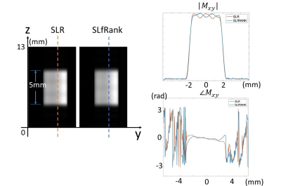 |
Validation and Application of SLfRank RF Pulse Design
zheng zhong1,
Congyu Liao2,
Janhavi Singhal3,
Frank Ong2,
Shreyas S. Vasanawala2,
and John M. Pauly4
1Radiology, Stanford University, Palo Alto, CA, United States, 2Radiology, Stanford University, Stanford, CA, United States, 3University of California Santa Barbara, Santa Barbara, CA, United States, 4Electrical Engineering, Stanford University, Stanford, CA, United States Keywords: RF Pulse Design & Fields, RF Pulse Design & Fields SLfRank is a novel RF pulse design framework proposed to improve upon the well-established Shinnar Le-Roux (SLR) design algorithm. Numerical experiments demonstrated promising effectiveness of SLfRank, prompting a need to validate its experimental feasibility. An experimental comparison of SLR and SLfRank is achieved by measuring slice profiles, multiband excitation, and spin-echo pulses on phantom and human brain. SLfRank produced images of comparable quality to the SLR algorithm, but with reduced RF energy and greater control over RF pulse phase. SLfRank’s numerical feasibility aligned closely with its effectiveness experimentally thus proving its technical feasibility for applications in this work and beyond. |
13:45 |
0202. |
A Versatile Toolbox for Rapid, Joint Design of pTx RF and
Gradient Pulses Using Pytorch's Autodifferentiation
Dario Bosch1,2,
Jonas Bause1,
and Klaus Scheffler1,2
1High Field MR Center, MPI for Biological Cybernetics, Tuebingen, Germany, 2Department for Biomedical Magnetic Resonance, University Hospital Tübingen, Tuebingen, Germany Keywords: Parallel Transmit & Multiband, RF Pulse Design & Fields, Python, PyTorch, Optimization Traditional pTx pulse design focuses on optimizing RF pulses with a fixed or parametrized gradient waveform. Utilizing fast GPUs for autodifferentiation, the optimization process becomes sufficiently efficient that RF pulses and the underlying gradient waveforms can be freely optimized concurrently within short time. This allows for the time-efficient creation of pTx pulses without prior knowledge about suitable gradient trajectories. Still, restrictions e.g. due to hardware limitations may be included into the optimization process.We provide a pulse design toolbox and demonstrate its ability to generate universal small-FA excitation pulses as well as large-FA pulses. |
| 13:45 |
0203.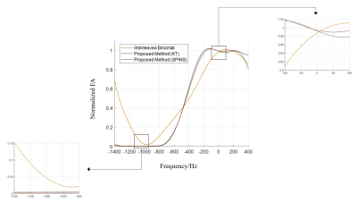 |
Parallel transmit spatial spectral pulse design with
specific absorption rate control: demonstration for robust
water excitation at 7 Tesla
Xin Shao1,
Xiaodong Ma2,
Hua Guo1,
Kamil Ugurbil3,
and Xiaoping Wu3
1Center for Biomedical Imaging Research, Department of Biomedical Engineering, School of Medicine, Tsinghua University, Beijing, China, 2Department of Radiology and Imaging Sciences, University of Utah, Salt Lake City, UT, United States, 3Center for Magnetic Resonance Research, Radiology, Medical School, University of Minnesota, Minneapolis, MN, United States Keywords: Parallel Transmit & Multiband, RF Pulse Design & Fields There has been an increasing interest in designing parallel transmit spatial spectral (pTx SPSP) RF pulses for reducing transmit B1 inhomogeneity while achieving water selective excitation. In this study, we propose a new pTx SPSP pulse design method with explicit SAR control with nearly complete fat suppression. Our results based on human calibration data acquired at 7T suggest that our method is equally applicable to both kT-point and SPINS pulse design, outperforming an existing method based on combining pTx with binomial pulse design approach. We believe our method will have a utility for high-resolution, whole-brain functional MRI at ultrahigh field. |
13:45 |
0204.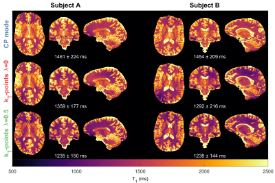 |
Hybrid-PUSH pulses for controlled MT bias in T1 mapping at
7T
David Leitão1,
Raphael Tomi-Tricot2,3,
Pip Bridgen1,
Patrick Liebig4,
Rene Gumbrecht4,
Dieter Ritter4,
Jo Hajnal1,3,
and Shaihan Malik1,3
1Biomedical Engineering & Imaging Sciences, King's College London, London, United Kingdom, 2MR Research Collaborations, Siemens Healthcare, Frimley, United Kingdom, 3Centre for the Developing Brain, King's College London, London, United Kingdom, 4Siemens Healthcare GmbH, Erlangen, Germany Keywords: RF Pulse Design & Fields, Magnetization transfer While standard pulse design methods control the rotation of magnetization (i.e. flip angle), the recently proposed PUSH method aims to control the root-mean-squared B1, to control magnetization transfer (MT) effects. General RF pulses create both effects simultaneously: this is important in T1 mapping, where incidental MT effects introduce bias. We show that using standard pulse design methods to improve flip angle uniformity can actually worsen the bias in T1 measurement by producing uncontrolled spatially variable MT effects. We propose a ‘hybrid-PUSH’ method optimizing both flip angle and B1rms, and demonstrate this improves quality of T1 estimation in-vivo at 7T. |
| 13:45 |
0205.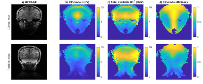 |
RF shimming for improved B1+ in the carotid arteries using
parallel transmission (pTx) head coils at 7T
Matthijs H.S. de Buck1,
Aaron T. Hess1,
and Peter Jezzard1
1Wellcome Centre for Integrative Neuroimaging, FMRIB, Nuffield Department of Clinical Neurosciences, University of Oxford, Oxford, United Kingdom Keywords: Parallel Transmit & Multiband, High-Field MRI, Neurovascular Cerebrovascular imaging methods suffer from a rapid drop in B1+ into the neck when using typical transmit head coils at 7T. Custom RF shims on regular pTx head coils could improve the B1+ magnitude in the major feeding arteries in the neck region over standard CP transmit mode. We found that this can improve the B1+ magnitude in the carotid arteries by 36% while also improving the RF homogeneity. This can be achieved using universal, phase-only RF shims, facilitating easy implementation in existing sequences and without requiring custom hardware. |
| 13:45 |
0206.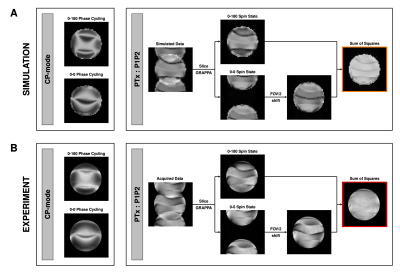 |
Eliminating Banding Artifacts in bSSFP using Parallel
Transmission
Chia-Yin Wu1,2,3,
Jin Jin2,4,
Markus Barth1,2,3,
and Martijn Cloos1,2
1Centre for Advanced Imaging, University of Queensland, St Lucia, Australia, 2ARC Training Centre for Innovation in Biomedical Imaging Technology, University of Queensland, St Lucia, Australia, 3School of Information Technology and Electrical Engineering, University of Queensland, St Lucia, Australia, 4Siemens Healthcare Pty Ltd, Brisbane, Australia Keywords: Parallel Transmit & Multiband, RF Pulse Design & Fields In this work, we demonstrate a parallel transmit implementation of the bSSFP sequence at 7 Tesla. The bSSFP sequence is one of the most efficient acquisition strategies however highly sensitive to B0 inhomogeneity which creates undesirable banding artefacts. A tailored pair of pTx pulses can be used to induce the desired steady-state magnetisation behaviour at all spatial locations regardless of the off-resonance frequency due to B0 inhomogeneity. Shown through simulation and experimental validation, we were able to capture signal in one acquisition with significant mitigation of banding artifacts and improvement in signal uniformity without SNR loss and time penalty. |
| 13:45 |
0207.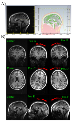 |
Experimental Assessment of the Effects of Subject Motion on
Local SAR and pTx Pulse Performance at 7T
Sydney Nicole Williams1,
Paul McElhinney1,
Belinda Ding1,2,
Sarah Allwood-Spiers3,
David A. Porter1,
Shajan Gunamony1,4,
and Wyger Brink5
1Imaging Centre of Excellence, University of Glasgow, Glasgow, Scotland, 2Siemens Healthcare Ltd., Frimley, United Kingdom, 3NHS GGC MRI Physics, NHS Greater Glasgow & Clyde, Glasgow, Scotland, 4MR CoilTech Ltd., Glasgow, Scotland, 5Magnetic Detection & Imaging Group, University of Twente, TechMed Centre, Netherlands Keywords: Parallel Transmit & Multiband, Safety, RF Pulse Design, RF Coils Previous simulation studies of motion in parallel transmission (pTx) have identified large increases in specific absorption rate (SAR) and pulse performance error. This abstract extends this by evaluating the effects of motion on RF shimming and dynamic pTx in vivo at 7T. The T1w image of a healthy volunteer was segmented to create a body model for electromagnetic simulation in a custom pTx coil. The model was simulated at six head positions matched to experimental measurements. Motion had a detrimental effect on pTx pulse performance and also caused local SAR changes, though not as severe as seen previously in simulation. |
|
13:45 |
0208.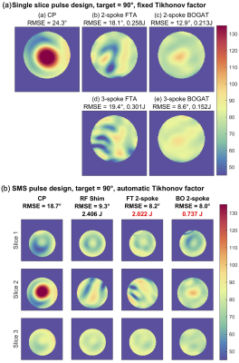 |
Online gradient optimisation for spokes parallel transmit –
the BOGAT method
Minghao Zhang1 and
Christopher T. Rodgers1
1Wolfson Brain Imaging Centre, University of Cambridge, Cambridge, United Kingdom Keywords: Parallel Transmit & Multiband, Parallel Transmit & Multiband Spokes parallel transmit pulses improve the homogeneity of ultra-high field imaging. Typically the gradient trajectory (i.e. spoke kt-space positions) is determined using the iterative Fourier transform approach (FTA). We introduce “BOGAT” (Bayesian Optimisation of GrAdient Trajectory) to efficiently determine globally optimal gradient trajectories.
We evaluated BOGAT in phantoms for single-band and multi-band excitation, and retrospectively in 9 volunteers using existing B0 and B1+ maps.
BOGAT improves flip angle homogeneity (by 12.8% vs FTA, P<0.001) and reduces SAR (17.2%, P<0.001). Calculations take ~10s extra for a set of multiband pulses, making it feasible to use for online per-subject optimisation. |
| 13:45 |
0209.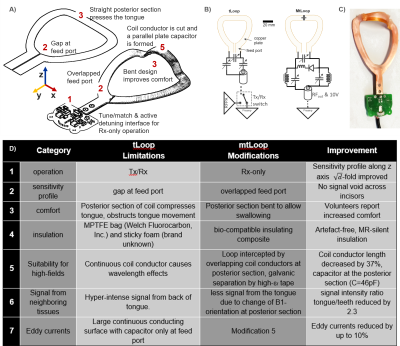 |
An Improved Intraoral Transverse Loop Coil Design for High
Resolution Dental MRI
Ali Caglar Özen1,
Serhat Ilbey1,
Feng Jia1,
and Michael Bock1
1Division of Medical Physics, Department of Radiology, University Medical Center Freiburg, Freiburg, Germany Keywords: Non-Array RF Coils, Antennas & Waveguides, New Devices, dental MRI MRI can simultaneously image soft and hard tissues such as glands, teeth, gum, nerves and bone, thus could be valuable for diagnosis of dental pathologies and implant planning. Intraoral coils have been proved essential to dental MRI due to superior sensitivity compared to external coils. In this study, we introduce a modified transverse loop coil design, which provides improved sensitivity, homogeneity, comfort and safety. We also introduce a bio-compatible, artefact-free and MR-silent coating for intraoral coils. Phantom and in vivo dental MR images demonstrate the advantages of the new intraoral coil design. |
| 13:45 |
0210.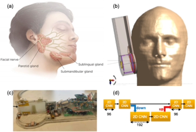 |
A 3D Surface Coil with Deep Learning Based Noise Reduction
for Parotid Gland Imaging
Sayim Gokyar1,
Chenyang Zhao1,
Shajan Gunamony2,3,
Jiaruo Yan2,
Jonathan West4,
Niels Kokot4,
and Danny JJ Wang1,5
1USC Stevens Neuroimaging and Informatics Institute, Los Angeles, CA, United States, 2MR Coiltech Limited, Glasgow, UK, Glasgow, United Kingdom, 3Imaging Centre of Excellence, University of Glasgow, Glasgow, United Kingdom, 4Department of Otolaryngology - Head and Neck Surgery, LAC+USC Medical Center, Keck Medicine of USC, Los Angeles, CA, United States, 5Department of Neurology, Keck School of Medicine, University of Southern California, Los Angeles, CA, United States Keywords: New Devices, Neuro Parotid salivary gland neoplasms occur in conspicuous locations near the facial nerve and have a risk for malignant transformation – making early diagnosis and treatment essential. Here we present a novel three-dimensional surface coil (3D Coil) architecture and a deep learning-based noise reduction method that offers twice higher signal to noise ratio compared to single channel surface coil for parotid gland imaging at 7T. |
| 13:45 |
0211.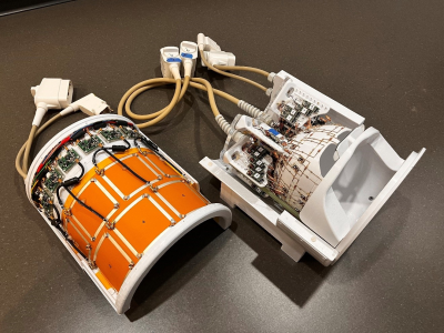 |
A self-decoupled 16-channel transmit, 80-channel receive
array for 10.5 Tesla human head imaging
Matt Waks1,
Russell Lagore1,
Edward Auerbach1,
Andrea Grant1,
Lance DelaBarre1,
Steve Jungst1,
Nader Tavaf1,
Jerahmie Radder1,
Pierre-Francois Van de Moortele1,
Alireza Sadeghi-Tarakameh1,
Yigitcan Eryaman1,
Gregor Adriany1,
and Kamil Ugurbil1
1Center for Magnetic Resonance Research, University of Minnesota, Minneapolis, MN, United States Keywords: RF Arrays & Systems, RF Arrays & Systems We present a splittable 16-channel self-decoupled (SD)1 transmit/receive (Tx/Rx) loop array combined with a 64-channel receive-only (Rx) loop array to generate a 80Rx/16Tx array for human head imaging at 10.5 Tesla. Compared to the previously presented SD transmitter, we designed, miniaturized, and integrated the MR system interface, including custom transmit/receive switches and preamplifiers, into the coil housing. We also implemented our new custom 128 receiver system, which supported this combined 80 channel receive configuration. Experimental MR results demonstrate advantages over our previous 16-channel transmit-only SD array and substantially increased central SNR with the 80-channel compared to the 64-Rx only configuration. |
| 13:45 |
0212. |
Dynamic dipole receive coils for improved 3D parallel
imaging at ultra-high magnetic field
Felix Glang1,
Anton V. Nikulin1,2,
Nikolai Avdievich1,
Dario Bosch1,2,
Theodor Steffen1,
and Klaus Scheffler1,2
1High-field Magnetic Resonance Center, Max Planck Institute for Biological Cybernetics, Tübingen, Germany, 2Department of Biomedical Magnetic Resonance, Eberhard Karls University Tübingen, Tübingen, Germany Keywords: RF Arrays & Systems, High-Field MRI Parallel imaging with electronically modulated time-varying receive sensitivities is a novel concept for improved reconstruction quality and reduced noise amplification. Previously, it was demonstrated for 2D imaging using reconfigurable surface loop receive elements. In the present work, we extend the approach to 3D imaging and for that introduce a reconfigurable single-row dipole receive array. By using PIN diodes to switch between capacitive and inductive impedance in the dipole arms, spatially distinct sensitivity profiles are formed that can be rapidly modulated. This is shown to enable parallel imaging acceleration along the dipole’s axis and improve reconstruction quality compared to static sensitivities. |
| 13:45 |
0213.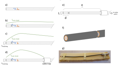 |
Using the end of the feeding cable directly as a flexible
antenna at 7T: the coax monopole antenna
Lyanne M I Budé1,2,
Bart R Steensma3,
Irena Zivkovic1,
and Alexander J E Raaijmakers2,3
1Electrical Engineering, Eindhoven University of Technology, Eindhoven, Netherlands, 2Biomedical Engineering, Eindhoven University of Technology, Eindhoven, Netherlands, 3Division of Imaging and Oncology, UMC Utrecht, Utrecht, Netherlands Keywords: RF Arrays & Systems, High-Field MRI This work introduces the coax monopole antenna. The antenna consists of a continuation of the feeding cable and uses only one inductor at the distal side of the antenna to achieve matching. Creating a radiative antenna is done by introducing one interruption in the shield of the coaxial cable, and the antenna length is enforced using a cable trap. Simulations and measurements show that the performance of this antenna is comparable to the fractionated dipole antenna, while introducing many advantages like easy cable routing, proper loading of the coil due to its flexibility, and easy construction. |
| 13:45 |
0214.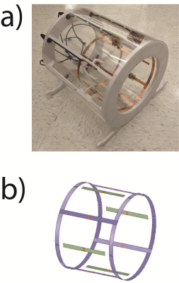 |
Combined triple tuned X-nuclei (2H, 23Na, 31P) birdcage and
1H 4 channel dipole array for head imaging at 7T
Jan Paska1,2,
Bili Wang1,2,
Anna Chen1,2,
Guillame Madelin1,2,
and Ryan Brown1,2
1Center for Advanced Imaging Innovation and Research (CAI2R), NYU School of Medicine, New York, NY, United States, 2Center for Biomedical Imaging, Department of Radiology, NYU School of Medicine, New York, NY, United States Keywords: RF Arrays & Systems, RF Arrays & Systems One method to achieve multiple coil resonances involves the use of diode or microelectromechanical system (MEMS) switches to toggle between resonances at proton and x-nuclei frequencies. While this approach provides a straightforward means to activate or deactivate coils corresponding to the desired nucleus, it does not allow simultaneous multinuclear data acquisition hat is required to eliminate temporal disparities. In this work we present a triple tuned birdcage for truly simultaneous imaging of X-nuclei combined with a proton dipole array at 7T. We show initial phantom imaging experiments and compare SNR with existing RF coils. |
| 13:45 |
0215.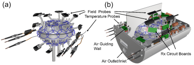 |
Design of a 64-Channel ex vivo Brain Rx Array Coil with
field monitoring and temperature control for DWI at 3T
Alina Scholz1,
Mirsad Mahmutovic1,
Mona Alem1,
Roland Müller2,
Torsten Schlumm2,
Harald E Möller2,
Anastasia Yendiki3,4,
Gabriel Ramos-Llorden3,4,
Lawrence L Wald3,4,5,
Susie Y Huang3,4,5,
and Boris Keil1,6
1Institute of Medical Physics and Radiation Protection (IMPS), TH-Mittelhessen University of Applied Sciences, Giessen, Germany, 2Max Planck Institute for Human Cognitive and Brain Sciences, Leipzig, Germany, 3A. A. Martinos Center for Biomedical Imaging, Department of Radiology, Massachusetts General Hospital, Charlstown, MA, United States, 4Harvard Medical School, Boston, MA, United States, 5Harvard-MIT Division of Health Sciences and Technology, Cambridge, MA, United States, 6Center for Mind, Brain and Behavior (CMBB), Marburg, Germany Keywords: RF Arrays & Systems, Brain Long-durational diffusion weighted MRI scans with high gradient strength and high slew rate experiences in addition to the generally low signal-to-noise-ratio several problems, such as image artifacts due to eddy currents and the gradual increase of the sample temperature. Combining a high-density anatomically shaped receive coil with field monitoring and temperature control can overcome these limitations. Therefore, we designed and constructed a 64-channel whole human ex vivo brain Rx coil with integrated field monitoring and temperature control system. First SNR measurements confirm the receive capability with high SNR. |
| 13:45 |
0216.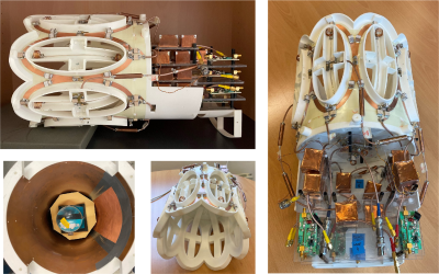 |
Degenerate Birdcage Coil Mixed with Bent Dipole Antennas to
Enhance Central SNR in Phosphorus MRI/MRS of Human Brain at
7T
Daniel Wenz1,2,
Thomas Dardano1,2,
Mark Widmaier1,3,
Songi Lim1,2,
Zhiwei Huang1,2,
and Lijing Xin1,2
1CIBM Center for Biomedical Imaging, Lausanne, Switzerland, 2Animal Imaging and Technology, Ecole Polytechnique Federale de Lausanne (EPFL), Lausanne, Switzerland, 3Laboratory of functional and metabolic imaging, Ecole Polytechnique Federale de Lausanne (EPFL), Lausanne, Switzerland Keywords: RF Arrays & Systems, RF Arrays & Systems Dipole antennas can be combined with loop elements to increase a central SNR in human brain MRI at 7T (300MHz). However, this approach was not investigated at lower frequencies. In this study a degenerate birdcage coil was combined with a pair of bent dipole antennas to increase a central SNR for phosphorus MRS of human brain at 7T (120MHz). Simulations showed that this approach could provide a 1.6-fold central SNR gain vs. degenerate birdcage-only. The RF coil was constructed and successfully tested at the bench. In the next step, an experimental validation of the proposed strategy will be performed. |
| 13:45 |
0217.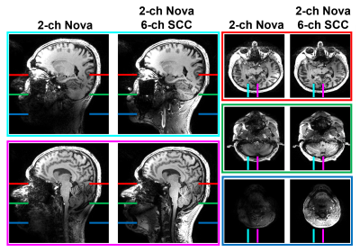 |
A head and neck array add-on to the two-channel Nova coil
using six SCC elements and Nova 32 channel receive array at
7T-MRI
Sadri Güler1,2,
Vitaliy Zhurbenko1,3,
Irena Zivkovic1,4,
Hanne Christensen5,
Sverre Rosenbaum5,
Inger Birgitte Havsteen6,
and Esben Thade Petersen1,2
1Section for Magnetic Resonance, DTU Health Tech, Technical University of Denmark, Kgs Lyngby, Denmark, 2Danish Research Centre for Magnetic Resonance, Centre for Functional and Diagnostic Imaging and Research, Copenhagen University Hospital - Amager and Hvidovre, Copenhagen, Denmark, 3Department of Electrical Engineering, Technical University of Denmark, Kgs Lyngby, Denmark, 4Electrical Engineering Department, Technical University of Eindhoven, Eindhoven, Netherlands, 5Department of Neurology, Copenhagen University Hospital - Bispebjerg and Frederiksberg, Copenhagen, Denmark, 6Department of Radiology, Copenhagen University Hospital - Bispebjerg and Frederiksberg, Copenhagen, Denmark Keywords: RF Arrays & Systems, RF Arrays & Systems The advantage of low coupling ratios of shielded-coaxial-cable coils (SCCs) are used to obtain an eight-channel head-neck transmit array for 7T-MRI. The six SCCs are combined with the two-channel Nova coil by taking the advantage of low coupling between SCCs and the Nova coil. Low inter element coupling along with the independent radiation regions of the SCCs and the Nova coil made it possible to do a straightforward B1-shimming resulting in a relatively homogeneous magnetic field in both head and neck region. Structural images are acquired with large coverage down to the bifurcation while high SNR is preserved. |
| 13:45 |
0218.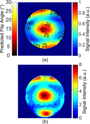 |
A Metamaterial Liner Body Coil for Wide-bore 3T MRI
Leo Rémillard1,
Adam Mitchell Maunder1,
Fraser Robb2,
Ashwin K. Iyer3,
and Nicola De Zanche1
1Oncology, Medical Physics, University of Alberta, Edmonton, AB, Canada, 2GE Healthcare, Aurora, OH, United States, 3Electrical and Computer Engineering, University of Alberta, Edmonton, AB, Canada Keywords: Hybrid & Novel Systems Technology, Body, metamaterial The transmit field suffers from standing wave inhomogeneities at field strengths ≥3T, which degrade image quality and create specific absorption rate (SAR) hotspots. We present the first experimental demonstration of a whole-body metamaterial liner (metaliner) that enables traveling wave excitation at a lower frequency than achievable otherwise. The simulated transmit efficiencies for a comparable birdcage and metaliner were 2.43μT⁄√kW±23.8% and 1.80μT⁄√kW±25.5% respectively, and the maximum 10g local SAR was reduced by 18% with the metaliner. This shows that the metaliner is an attractive alternative to the BC with greater safety thanks to the lower localized SAR. |
 Back to Meeting Home
 Back to the Program-at-a-Glance
The International Society for Magnetic Resonance in Medicine is accredited by the Accreditation Council for Continuing Medical Education to provide continuing medical education for physicians.
 Back to Meeting Home
 Back to the Program-at-a-Glance
 Full Session Video