Power Pitch
Pitch: MR Safety: Everything's Under Control!
ISMRM & ISMRT Annual Meeting & Exhibition • 03-08 June 2023 • Toronto, ON, Canada

Power Pitch
Pitch: MR Safety: Everything's Under Control!
| 13:30 |
0594.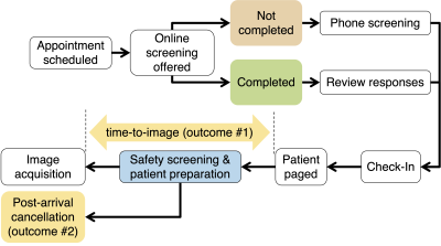 |
Impact of Online Safety Screening on Outpatient MRI Workflow
Sheena Chu1,2,
Elizabeth Briel2,
John W Garrett2,
Scott B Reeder1,2,3,4,5,
and Ali Pirasteh1,2
1Department of Medical Physics, University of Wisconsin-Madison, Madison, WI, United States, 2Department of Radiology, University of Wisconsin-Madison, Madison, WI, United States, 3Department of Medicine, University of Wisconsin-Madison, Madison, WI, United States, 4Department of Biomedical Engineering, University of Wisconsin-Madison, Madison, WI, United States, 5Department of Emergency Medicine, University of Wisconsin-Madison, Madison, WI, United States Keywords: Safety, MR Value MRI safety screening is required for all patients prior to their MRI exam. We aim to determine the impact of online MRI safety screening on delays and post-arrival cancellations. Time-to-image is the time between when the patient is paged in the waiting area and their first image acquisition. Post-arrival cancellations occur when the patient arrives but does not complete their MRI due to an unexpected safety or other issues (e.g., claustrophobia). We conclude that post-arrival cancellations and overall time-to-image decreased as a result of online MRI safety screening. Keywords: Physics & Engineering: Safety, New Devices, MR Value |
13:30 |
0595.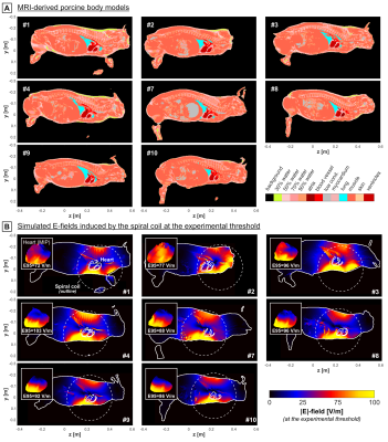 |
Comparison of measured and simulated cardiac
magnetostimulation thresholds in eight pigs
Valerie Klein1,2,3,
Livia Vendramini1,
Mathias Davids1,2,
Natalie G. Ferris1,4,
Lothar R. Schad3,
David E. Sosnovik1,2,4,5,
Christopher T. Nguyen6,7,8,
Lawrence L. Wald1,2,4,
and Bastien Guérin1,2
1A. A. Martinos Center for Biomedical Imaging, Department of Radiology, Massachusetts General Hospital, Charlestown, MA, United States, 2Harvard Medical School, Boston, MA, United States, 3Computer Assisted Clinical Medicine, Medical Faculty Mannheim, Heidelberg University, Mannheim, Germany, 4Harvard-MIT Division of Health Sciences and Technology, Cambridge, MA, United States, 5Cardiovascular Research Center, Cardiology Division, Massachusetts General Hospital, Charlestown, MA, United States, 6Cardiovascular Innovation Research Center, Heart Vascular & Thoracic Institute, Cleveland Clinic, Cleveland, OH, United States, 7Cardiovascular Imaging, Imaging Institute, Cleveland Clinic, Cleveland, OH, United States, 8Biomedical Engineering, Lerner Research Institute, Cleveland Clinic, Cleveland, OH, United States Keywords: Safety, Bioeffects & Magnetic Fields We use a combined electromagnetic-electrophysiological modeling framework to predict cardiac stimulation (CS) thresholds in individualized porcine body models and compare those simulations to thresholds measured in eight pigs using strong dB/dt pulses. For all pigs, the simulated and measured thresholds agree within 30%, and no significant differences between simulations and measurements were detected (p<0.05, paired t-test). The threshold model uncertainty was found to be ~25% in a sensitivity analysis of the relevant model parameters. A well-validated model may help inform appropriate safety limits for MRI gradients to protect patients from CS without overly restricting gradient performance. |
13:30 |
0596.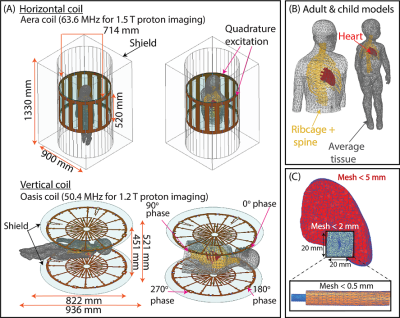 |
Vertical MRI systems may offer a safer platform with
substantially reduced RF heating in adult and pediatric
patients with CIEDs
Jasmine Vu1,2,
Fuchang Jiang1,
Bhumi Bhusal2,
Pia Sanpitak2,
Gregory Webster3,
Giorgio Bonmassar4,
and Laleh Golestanirad1,2
1Biomedical Engineering, Northwestern University, Evanston, IL, United States, 2Radiology, Northwestern University, Chicago, IL, United States, 3Cardiology, Ann and Robert H. Lurie Children's Hospital of Chicago, Northwestern University Feinberg School of Medicine, Chicago, IL, United States, 4Athinoula A. Martinos Center for Biomedical Engineering, Charlestown, MA, United States Keywords: Safety, Cardiovascular, Implants MRI is restricted for patients with cardiac implantable electronic devices (CIEDs)—especially children with epicardial systems—due to potential radiofrequency (RF) heating of the tissue around the lead. Here, we present the first assessment of RF heating of epicardial and endocardial CIEDs with varying lead lengths in a vertical MRI system, where we found up to a 78-fold reduction in the simulated maximum specific absorption rate (SAR) compared to a horizontal coil. Reduction in the maximum 0.1g-averaged SAR in the vertical coil was consistent for leads with various internal wire geometries and electrical lengths. |
| 13:30 |
0597. |
Ultra-low-field Magnetization Transfer Imaging with Low SAR
Shi Su1,2,
Yujiao Zhao1,2,
Vick Lau1,2,
Linfang Xiao1,2,
Ye Ding1,2,
Jiahao Hu1,2,
Junhao Zhang1,2,
Christopher Man1,2,
Alex T. L. Leong1,2,
and Ed X. Wu1,2
1Laboratory of Biomedical Imaging and Signal Processing, the University of Hong Kong, Hong Kong, China, 2Department of Electrical and Electronic Engineering, the University of Hong Kong, Hong Kong, China Keywords: Low-Field MRI, Contrast Mechanisms At high field, magnetization transfer (MT) imaging suffers from the high specific absorption ratio (SAR) issue due to the usage of high power off-resonance MT RF pulses, and on-resonance saturation caused by B0 field inhomogeneity. At ultra-low-field (ULF), the low SAR and low absolute B0 inhomogeneity (in Hz) greatly facilitates the application of strong and versatile MT pulses without the confounding on-resonance saturation in practice. We demonstrate brain MT imaging at ULF first the first time using a 0.055 Tesla MRI platform with an extremely low SAR. |
| 13:30 |
0598. |
Simulated radiation patterns of MRI without a shielded room
from 0.5 to 7 Tesla
Ehsan Kazemivalipour1,2,
Bastien Guerin1,2,
and Lawrence L. Wald1,2,3
1A. A. Martinos Center for Biomedical Imaging, Department of Radiology, Massachusetts General Hospital, Charlestown, MA, United States, 2Harvard Medical school, Boston, MA, United States, 3Harvard-MIT Division of Health Sciences Technology, Cambridge, MA, United States Keywords: Safety, Safety Far-field electromagnetic radiation patterns and levels were simulated on the 10m radius regulatory sphere for conventional MRI scanners at 0.5T, 1.5T, 3T, and 7T operated without an RF shielded room. The levels and patterns were strongly affected by the symmetry of the load. With a body load, the peak E-fields on a 10m radius surface rose with roughly the square of the frequency and far exceeded regulatory limits even for 0.5T. With the body in the bore, the radiated patterns also take on a highly asymmetric pattern not present for more symmetric loads. |
| 13:30 |
0599.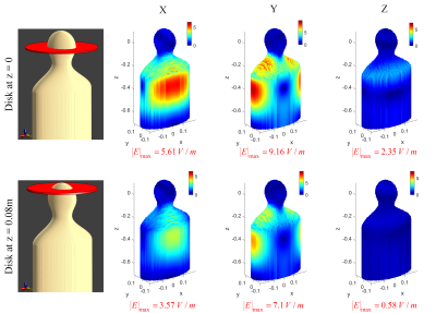 |
Minimum Electric Field Gradient Array Body Coil with
Adjustable Regions of Linearity
Reza Babaloo1,2,
Manouchehr Takrimi2,
and Ergin Atalar1,2
1Department of Electrical and Electronics Engineering, Bilkent University, Ankara, Turkey, 2National Magnetic Resonance Research Center (UMRAM), Bilkent University, Ankara, Turkey Keywords: Safety, Safety A large region of linearity in whole-body gradient coils exposes large body areas to switching magnetic fields, inducing high electric fields, which may cause peripheral nerve stimulation. A body gradient array coil (made up of multiple coil elements) can produce linear gradients in different region of linearity shapes by optimizing the feeding currents, which can minimize the induced E-fields. |
| 13:30 |
0600.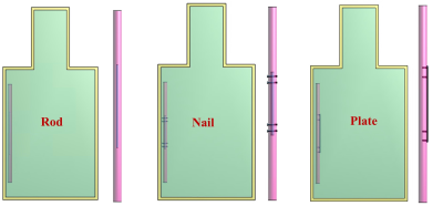 |
Impacts of bone on the worst-case configuration for
orthopedic implants under 1.5T and 3T MRI
xiaolin Yang1,
Jianfeng Zheng1,
Ran Guo1,
Wolfgang Kainz2,
and Ji Chen1
1Univ of Houston, Houston, TX, United States, 2HPC for MRI Safety, Jasper, GA, United States Keywords: Safety, Bone Due to the differences in both electrical and thermal properties, the RF-induced heating from implantable devices under MRI can have different behaviors inside muscle-like tissue and bone. A locally modified ASTM phantom with bone tissue was developed in the study. Simulations were used in the study to determine the worst-case heating configurations in the original and the modified ASTM phantoms. Based on our study, it was observed that the worst-case heating configuration can be altered when the devices are implanted in/near bone tissues. Consequently, additional in-vivo modeling would be required to understand the clinically relevant RF-induced heating. |
| 13:30 |
0601.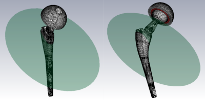 |
A method to assess the orientation of the maximum heating of
an arbitrarily shaped object inside a homogeneous magnetic
field
Umberto Zanovello1,
Mario Chiampi1,
Oriano Bottauscio1,
Alessandro Arduino1,
and Luca Zilberti1
1Istituto Nazionale di Ricerca Metrologica, Torino, Italy Keywords: Safety, Gradients The ISO/TS 10974 standard proposes a test method to assess the heating of a metallic AIMD due to switched gradient magnetic fields. Aiming at achieving a conservative evaluation, the standard suggests to evaluate the heating for the worst orientation of the AIMD inside a homogeneous harmonic magnetic field. The abstract presents a strategy to assess this orientation for an arbitrarily shaped metallic object. The strategy has been successfully tested numerically against expectations with a disk object and it has been applied to a realistic hip and knee implant. |
| 13:30 |
0602.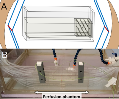 |
Simulated and experimental approaches to perfusion cooling
in Sim4Life: verification and applications to RF heating of
implants
Amgad Louka1,
Blaine Chronik1,2,
and William Handler2
1Medical Biophysics, University of Western Ontario, London, ON, Canada, 2Physics & Astronomy, University of Western Ontario, London, ON, Canada Keywords: Safety, Safety, RF heating, perfusion, Experimental, verification Perfusion cooling of implants is an emergent field that has the potential to greatly improve patient access to MRI for those living with implants that previously failed the radiofrequency heating test (ASTM F2182). Many implants fail by a small margin, meaning they would likely be safe inside the body when perfusion is considered. Here, we present initial steps for the experimental verification of perfusion simulations in Sim4Life, which showed reasonable agreement and provided some insight on future experimental perfusion platforms. From there, a perfusion cooling factor can be quantified for use during regulatory approval of these implants. |
| 13:30 |
0603.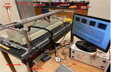 |
Lead Insulation and Wavelength Effects on Active Implantable
Medical Device Heating under MRI at 1.5T and 3T using
Transfer Function (TF) Modeling
Ananda Kumar1,
Ji Chen2,
Md Zahidul Islam2,
and Hongbae Jeong1
1CDRH, Food and Drug Administration, Silver Spring, MD, United States, 2Department of Electrical and Computer Engineering, University of Houston, Houston, TX, United States Keywords: Safety, Safety The influence of the thickness, loss tangent value of the lead insulation material along with lead length effects on the heating of an active implantable medical device (AIMD) lead electrode is analyzed using full-wave EM simulations. Decreasing the thickness of the insulation decreases heating of the electrode significantly when lead lengths are less than or equal to wavelengths in the medium. Increasing the loss tangent of the insulation material has a moderate effect in reducing heating at the electrode. Standing wave distortions affect lead electrode heating at lead lengths greater than wavelengths in the medium. |
|
13:30 |
0604.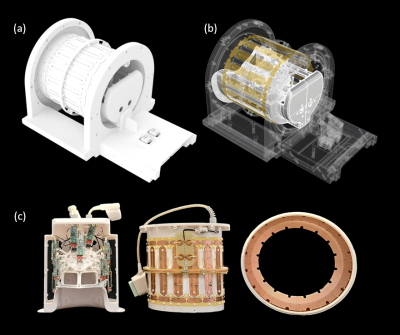 |
An Implant-Friendly Coil System for Imaging Deep Brain
Stimulation at 3T MRI
Nicolas Kutscha1,
Bhumi Bhusal2,
Mirsad Mahmutovic1,
Chaimaa Chemlali1,
Jasmine Vu2,
Sam-Luca Hansen1,
Laleh Golestanirad2,
and Boris Keil1
1Institute of Medical Physics and Radiation Protection, TH Mittelhessen University of Applied Sciences, Giessen, Germany, 2Department of Radiology and Department of Biomedical Engineering, Northwestern University, Chicago, IL, United States Keywords: Safety, Safety A prototype of an adjustable DBS-friendly Tx/Rx 3T coil system was built and evaluated with measurements and numerical simulations characterizing its image quality and SAR profile. The work envisions the use of novel MRI hardware that will make 3T MRI fully accessible to patients with DBS implants. |
| 13:30 |
0605.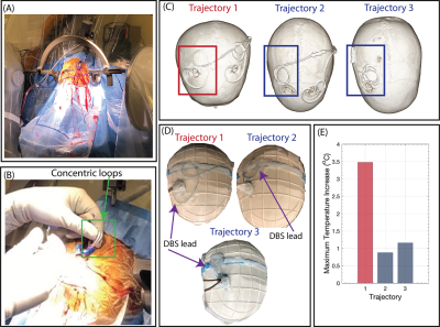 |
Surgical modification of deep brain stimulation lead
trajectories reduces RF heating during 3 T MRI: From
phantoms to implementation in patients
Jasmine Vu1,2,
Bhumi Bhusal2,
Joshua Rosenow3,
and Laleh Golestanirad1,2
1Biomedical Engineering, Northwestern University, Evanston, IL, United States, 2Radiology, Northwestern University, Chicago, IL, United States, 3Neurosurgery, Northwestern University, Chicago, IL, United States Keywords: Safety, Brain, Translational studies MRI at 3 T is restricted for patients with deep brain stimulation (DBS) systems due to potential radiofrequency (RF) heating. Here, we present the first large-scale, systematic study to determine how trajectory-related parameters affect RF heating and quantify the extent of RF heating reduction. Introducing concentric loops close to the surgical burr hole substantially reduced RF heating. Increasing the number of loops correlated well with decreased heating. Recommendations based on the results from phantom experiments were easily adopted during the surgical procedure within 30 seconds. Subsequent replication of the trajectories based on postoperative computed tomography images confirmed low RF heating. |
| 13:30 |
0606.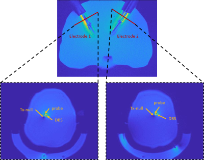 |
Temperature Prediction for Bilateral Deep Brain Stimulation
Electrodes Undergoing MRI
Nur Izzati Huda Zulkarnain1,
Alireza Sadeghi-Tarakameh1,
Jeromy Thotland1,
Noam Harel1,
and Yigitcan Eryaman1
1Center for Magnetic Resonance Research (CMRR), University of Minnesota, Minneapolis, MN, United States Keywords: Safety, Neuro We utilized a previously proposed workflow to predict heating around the contacts of bilateral deep brain stimulation (DBS) electrodes undergoing MRI. Phantom experiments demonstrated a quantitative agreement with the simulated and experimentally measured temperature for different electrode trajectories and excitations. |
| 13:30 |
0607.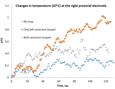 |
Assessment of MRI-related heating with excess deep brain
stimulation extension wires at 3 tesla
Anupa Vijayakumari1,
Mark Lowe1,
Benjamin Walter1,
Joseph Sakai2,
Jody Tanabe2,
Richard Wojcik3,
and Pallab Bhattacharyya1
1Cleveland Clinic Foundation, CLEVELAND, OH, United States, 2University of Colorado Anschutz Medical Campus, Aurora, CO, United States, 3University of Colorado Denver, Denver, CO, United States Keywords: Safety, Safety When implanting a DBS system, the excess amount of extension wires is looped and placed in different regions (near the skull burr holes or in the chest), depending on the neurosurgeon/institution. In this study, we tested three different DBS configurations with excess extension wires looped behind the IPG. A phantom with a DBS device (lead model B33005, IPG Percept B35200, and Extension wire model B34000) was used to perform this study. We observed that the temperature rise was higher with one loop placed behind the IPG compared to two or no loops. |
| 13:30 |
0608.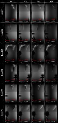 |
A geometrical approach to rapidly evaluate and optimize pTx
transmission vectors for imaging and RF safety of implants
Berk Silemek1,
Frank Seifert1,
Bernd Ittermann1,
and Lukas Winter1
1Physikalisch-Technische Bundesanstalt (PTB), Braunschweig and Berlin, Germany Keywords: Safety, Safety, Implant safety, RF safety, implants, Deep brain stimulator A simple geometrical approach is presented to rapidly calculate safe pTx excitation vectors for implants, while further optimizing imaging performance. The proposed approach does not require any additional imaging protocols to be performed and pTx vector optimizations can be calculated solely e.g. based on low-cost sensors embedded in implants. The methodology was tested on a custom-built sensor-equipped wireless implant in 3T MRI experiments and realistic DBS lead configurations. |
| 13:30 |
0609.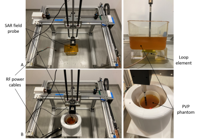 |
RF coil safety validation with a 3D SAR measurement setup
Nur Izzati Huda Zulkarnain1,
Mert Ates2,
Grace Cole3,
Alireza Sadeghi-Tarakameh1,
Steve Jungst1,
Lance DelaBarre1,
Gregor Adriany1,
and Yigitcan Eryaman1
1Center for Magnetic Resonance Research (CMRR), University of Minnesota, Minneapolis, MN, United States, 2Department of Electrical and Electronics Engineering, Bilkent University, Ankara, Turkey, 3Bethel University, St Paul, MN, United States Keywords: Safety, Validation We implemented a framework to validate the RF safety of MRI coils experimentally using a phantom in our RF safety lab which supports coil excitation using up to 16 individually-controlled power amplifiers. A 3D measurement setup and a dosimetric probe were used to map the spatial distribution of the specific absorption rate (SAR). To demonstrate the accuracy of the framework, we compared the simulated and measured 10 g averaged SAR of a rectangular loop element and an 8-channel transmit/receive head coil. We obtained quantitative agreements with 7.8% and 11.6% root-mean-squared error for the loop element and head coil respectively. |
| 13:30 |
0610.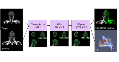 |
Tuning and validation of an image registration procedure for
patient-specific SAR simulation
Eros Montin1,2,
Giuseppe Carluccio1,
Christopher M Collins1,
and Riccardo Lattanzi1,2,3
1Center for Advanced Imaging Innovation and Research (CAI2R) Department of Radiology, Radiology Department, New York University Grossman School of Medicine, New York, New York, USA, New York, NY, United States, 2Bernard and Irene Schwartz Center for Biomedical Imaging, Department of Radiology, New York University Grossman School of Medicine, New York, New York, USA, New York, NY, United States, 3Vilcek Institute of Graduate Biomedical Sciences, New York University Grossman School of Medicine, New York, New York, USA, New York, NY, United States Keywords: Safety, Whole Body In this article, we build an automatic pipeline for patient-specific assessment of SAR. Given a target MR image and an atlas comprising a reference image and a model, an MR-shaped model can be obtained by registering the AIM with the target MR and applying the resulting displacement fields to the corresponding ABM. The results of the analysis on simulated data showed that the proposed automatic pipeline was reliable and accurate both in terms of SAR distribution and the similarity of the patient-specific models. |
| 13:30 |
0611.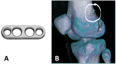 |
7 Tesla MRI of the knee joint after anterior cruciate
ligament graft reconstruction: safety discussion and image
quality study
Oliver Kraff1,
Jens M Theysohn2,
Jana Theisejans3,
and Harald H Quick1,4
1Erwin L Hahn Institute for MRI, University Duisburg-Essen, Essen, Germany, 2Department of Diagnostic and Interventional Radiology and Neuroradiology, University Hospital Essen, Essen, Germany, 3General Psychology: Cognition and Center for Behavioral Addiction Research (CeBAR), University Duisburg-Essen, Duisburg, Germany, 4High-Field and Hybrid MR Imaging, University Hospital Essen, Essen, Germany Keywords: Safety, Artifacts This study presents the MR safety discussion for imaging subjects with metallic fixation buttons after reconstruction of the anterior cruciate ligament, which have not been labeled MR conditional at 7T by the implant vendor. In addition, image quality and artifacts are evaluated in a knee imaging protocol consisting of both gradient- and spin echo sequences. Two imaging cases are presented: one with 7T imaging before and after surgery, and another case with comparative imaging between 1.5T and 7T. Artifact sizes from metallic fixation buttons at the femur did not impair the image quality and diagnostic evaluation of the knee joint. |
| 13:30 |
0612.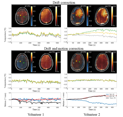 |
MR thermometry of RF heating in the human brain at 7T using
the harmonical initialized multi-echo model with SVD-based
motion correction
Mathijs W.I. Kikken1,
Bart R. Steensma1,
Cornelis A.T. van den Berg1,
and Alexander J.E. Raaijmakers1,2
1Center for Image Sciences - Computational Imaging Group, UMC Utrecht, Utrecht, Netherlands, 2Biomedical Engineering - Medical Imaging Analysis, Eindhoven University of Technology, Eindhoven, Netherlands Keywords: Safety, Safety A multi-echo signal model was presented to measure RF-induced temperature rise in the brain at 7T. The proposed method corrects drift fields based on near-harmonic 2D reconstruction and is complemented by an SVD-based motion-correction scheme. The method was tested in 2 volunteers, showing a maximum temperature increase of 0.25 °C with a precision of 0.12 °C. The reliability of the results was strengthened by measurements in which a heating pad was placed on the forehead of one volunteer. This measurement and thermal simulations indicated that the heatpad induced considerably more heating in the brain (3.5 °C) than SAR-constrained RF exposure. |
| 13:30 |
0613.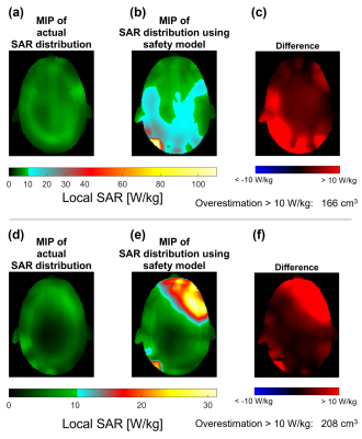 |
All-inclusive Safety Models Limit Imaging Performance at 7T
Emre Kopanoglu1
1CUBRIC, School of Psychology, Cardiff University, Cardiff, United Kingdom Keywords: Safety, High-Field MRI, RF Pulse Design & Fields; Parallel Transmit & Multiband Safety models on scanners are patient-position unaware and may include multiple patient positions to ensure safety. This may lead to overestimation of the specific absorption rate (SAR) and limit scanning performance. This study investigates the effect of an all-inclusive safety model on SAR estimation for parallel-transmit and quadrature-excitation at 7T. Results show that more than 4-fold SAR overestimation can be commonly observed. RF shimming suffered the most, with 11-fold overestimation at the worst-case and more than 4-fold overestimation in 37% of cases. RF shimming also offered the lowest peak local SAR and may be unnecessarily penalized by overconservative safety models. |
 Back to Meeting Home
 Back to the Program-at-a-Glance
The International Society for Magnetic Resonance in Medicine is accredited by the Accreditation Council for Continuing Medical Education to provide continuing medical education for physicians.
 Back to Meeting Home
 Back to the Program-at-a-Glance
 Full Session Video