Power Pitch
Pitch: Novel Quantitative Imaging Methods: Reconstruction & Analysis
ISMRM & ISMRT Annual Meeting & Exhibition • 03-08 June 2023 • Toronto, ON, Canada

Power Pitch
Pitch: Novel Quantitative Imaging Methods: Reconstruction & Analysis
| 15:45 |
1089.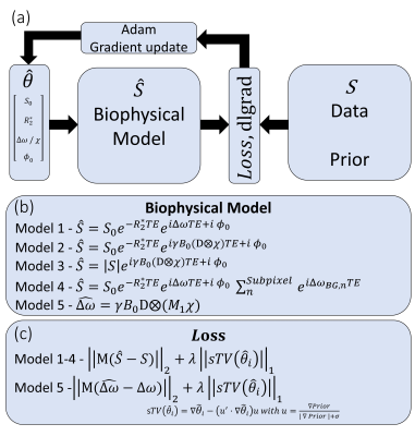 |
Improved R2* and QSM mapping for dummies - ask Adam
José P. Marques1,
Dennis Den Hollander1,
David G Norris1,
and Kwok-Shing Chan1
1Donders Institute for Brain Cognition adn Behaviour, Radboud University, Nijmegen, Netherlands Keywords: Quantitative Imaging, Quantitative Susceptibility mapping In this abstract we demonstrate a simple framework that builds up on deep learning infrastructure to perform quantitative susceptibility mapping and correct for susceptibility related effects on R2* maps, taking the advantage of high GPU computational efficiency. Asking the Adam optimizer to point you in the right direction results in a gradient descent method that can perform field mapping, background field removal, QSM and reduce macroscopic intravoxel dephasing artifacts in R2* maps with most operations being performed in under 60 seconds even for 0.8mm isotropic whole brain multi-echo data. |
| 15:45 |
1090.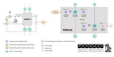 |
Diving into Extended Phase Graph-based Deep Learning for
accurate T2 mapping with PENGUIN
Catarina N. Carvalho1,
Teresa M. Correia2,3,
and Rita G. Nunes1,4
1Institute for Systems and Robotics - Lisboa and Department of Bioengineering, Instituto Superior Técnico – Universidade de Lisboa, Lisbon, Portugal, Lisbon, Portugal, 2School of Biomedical Engineering and Imaging Sciences, King’s College London, United Kingdom, London, United Kingdom, 3Center of Marine Sciences - CCMAR, Faro, Portugal, Faro, Portugal, 4Centre for the Developing Brain, School of Biomedical Engineering and Imaging Sciences, King's College, London, UK, London, United Kingdom Keywords: Quantitative Imaging, Relaxometry Model-based deep learning approaches have shown promising results to accelerate T2 relaxometry, but most adopt a pure exponential curve to model the signal, which does not account for indirect and stimulated echoes. A PhasE graph sigNal and Gradients QUantitative Inference MachiNe (PENGUIN) is proposed, which implements a dictionary of pre-calculated echo-modulation curves following the Extended Phase Graph (EPG) formulation and respective gradients as the inputs of a Recurrent Inference Machine to perform accurate T2 mapping from the reconstructed images. PENGUIN is 25-fold faster than a pattern recognition approach with a T2 dictionary step of 2 ms. |
| 15:45 |
1091.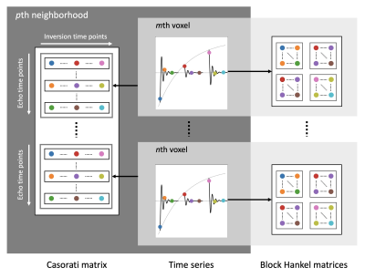 |
QRAGE - Multi-Echo MPnRAGE and Model-Based Reconstruction
for Quantitative MRI of Water Content, T1, T2* and Magnetic
Susceptibility at 7T
Markus Zimmermann1,
Zaheer Abbas1,
Yannic Sommer1,
Alexander Lewin1,
Shukti Ramkiran1,
Ana-Maria Oros-Peusquens1,
Seong Dae Yun1,
and N. Jon Shah1,2,3,4
1Institute of Neuroscience and Medicine 4, INM-4, Forschungszentrum Jülich, Germany, Jülich, Germany, 2Institute of Neuroscience and Medicine 11, INM-11, JARA, Forschungszentrum Jülich, Germany, Jülich, Germany, 3JARA - BRAIN - Translational Medicine, Aachen, Germany, Aachen, Germany, 4Department of Neurology, RWTH Aachen University, Aachen, Germany, Aachen, Germany Keywords: Sparse & Low-Rank Models, Image Reconstruction, Brain, Relaxometry, Quantitative Susceptibility mapping The development of fast, accurate, and robust methods for multiparametric quantitative MRI (qMRI) at ultrahigh field strength remains an important topic of research. Here, we present a novel qMRI technique for the simultaneous quantification of water content, T1, T2*, and magnetic susceptibility, termed QRAGE. The proposed method combines a highly undersampled multi-echo MPnRAGE sequence with a model-based reconstruction approach. It acquires 171 different contrasts with full brain coverage and 1 mm isotropic resolution within 7:20 min from which the parametric maps are estimated. The accuracy and precision of QRAGE are demonstrated by comparison to gold-standard reference methods. |
| 15:45 |
1092.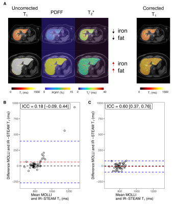 |
An empirical approach to determine water T1 from
multiparametric MR images of the liver
Filippo Carlo Michelotti1,
Yuliya Kupriyanova1,
Tim Mori1,
Thomas Küstner2,
Geronimo Heilmann1,
Maria Bombrich1,
Clara Moeser1,
Martin Schön1,
Michael Roden1,
and Vera Schrauwen-Hinderling1
1Institute for Clinical Diabetelogy, German Diabetes Center (DDZ) Leibniz Center for Diabetes Research, Düsseldorf, Germany, 2Medical Image and Data Analysis (MIDAS.lab), University Hospital of Tübingen, Diagnostics and Interventional Radiology, Tübingen, Germany Keywords: Quantitative Imaging, Liver, Machine Learning, Segmentation This work focuses on an empirical approach to determine water T1 from multiparametric MR images, including T1, PDFF and T2* maps. To this end, a multiple linear regression model was fit to describe the deviation in MOLLI T1 based on PDFF and T2* values, which were measured in phantoms built at increasing lipids and iron content. This method was validated on a cohort of healthy volunteers and diabetes subjects (n=45). Further investigations were conducted to elucidate the relationship between MOLLI T1 values, before and after correction for hepatic lipid and iron content, and liver stiffness measured by MR elastography. |
| 15:45 |
1093.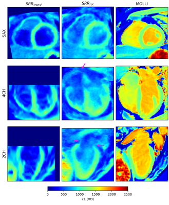 |
Towards isotropic 3D whole-heart T1 mapping using
model-based motion-corrected super-resolution reconstruction
Simone Hufnagel1,
Patrick Schuenke1,
Jeanette Schulz-Menger2,3,4,
Tobias Schaeffter1,5,6,
and Christoph Kolbitsch1
1Physikalisch-Technische Bundesanstalt (PTB), Braunschweig and Berlin, Germany, 2Charité Medical Faculty University Medicine, Berlin, Germany, 3Working Group on Cardiovascular Magnetic Resonance, Experimental and Clinical Research Center (ECRC), Charité Humboldt University Berlin, DZHK partner site Berlin, Berlin, Germany, 4Department of Cardiology and Nephrology, HELIOS Klinikum Berlin Buch, Berlin, Germany, 5School of Biomedical Engineering and Imaging Sciences, King’s College London, London, United Kingdom, 6Department of Biomedical Engineering, Technical University of Berlin, Berlin, Germany Keywords: Quantitative Imaging, Heart, Super-Resolution Reconstruction Cardiac T1 mapping provides valuable information for the diagnosis of a variety of heart diseases. However, due to SNR and scan time limitations, often only 2D imaging with a low through-plane resolution covering a few slices of the left ventricle is possible. In this work, a super-resolution reconstruction approach is presented aiming towards whole-heart 1.3 mm isotropic T1 mapping within less than three minutes acquisition time. The proposed approach provided a whole-heart cardiac T1 map including the atria and the right ventricle with improved visualization of small structures and overall image quality. |
| 15:45 |
1094.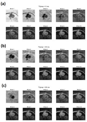 |
Simultaneous T1-T2 mapping, CINE and Multi-contrast
Anatomical 3D whole-heart MRI
Nicolás Garrido1,2,
Andrew Phair3,
Ronal Coronado1,4,
Haikun Qi5,
Claudia Prieto1,3,4,
and René M Botnar1,2,3
1Millennium Institute for Intelligent Healthcare Engineering (iHEALTH), Santiago, Chile, 2Institute for Biological and Medical Engineering, Pontificia Universidad Católica de Chile, Santiago, Chile, 3King's College London, London, United Kingdom, 4Electrical Engineering Department, Pontificia Universidad Católica de Chile, Santiago, Chile, Santiago, Chile, 5ShanghaiTech University, Shanghai, China Keywords: Quantitative Imaging, Myocardium, Motion Correction Cardiac T1-T2 mapping provides information about focal and diffuse fibrosis and inflammation of the myocardium. A recently proposed free-running 3D mapping technique allows time efficient and simultaneous whole-heart T1-T2 mapping within a single scan, with retrospective respiratory motion correction. However, this approach loses the information about the temporal contrast evolution and does not reconstruct multi-contrast 3D whole-heart images, which may carry useful clinical information in patients with myocardial infarction and, acute and subacute thrombus. In this work, we propose to extend this approach to enable joint T1-T2 mapping, CINE, and multi-contrast 3D whole-heart imaging from a single free-running scan. |
| 15:45 |
1095.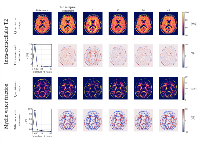 |
Is linear subspace constraint reconstruction suitable for
multi-compartment T2 imaging? Evaluation and guidelines.
Nadège Corbin1,2,
Trotier J. Aurélien 1,
Laurent Petit3,
Silvio Sarubbo4,
Sylvain Miraux1,
and Emeline J. Ribot1
1Centre de Résonance Magnétique et Systèmes Biologiques, UMR5536, CNRS, University of Bordeaux, Bordeaux, France, 2Wellcome Center for Human Neuroimaging, UCL Queen Square Institute of Neurology, University Coleege of London, London, United Kingdom, 3Groupe d’Imagerie Neurofonctionnelle, Institut des Maladies Neurodégénératives UMR 5293, CNRS, CEA, University of Bordeaux, Bordeaux, France, 4Department of Neurosurgery, Azienda Provinciale per i Servizi Sanitari, Santa Chiara Hospital, Trento, Italy Keywords: Quantitative Imaging, Image Reconstruction, myelin water fraction Multi-compartment T2 imaging suffers from long acquisition time. Undersampling the k-space combined with advanced iterative reconstructions could be beneficial to reach a reasonable scan duration. This work investigates the suitability of linear subspace-based reconstruction for myelin water fraction and intra-extracellular T2 mapping. Our findings suggest that subspace-based reconstruction for intra-extra cellular T2 and myelin water fraction mapping can be reliably used in combination with spatial regularization enforcing sparsity. The temporal basis can be built from extended phase graph simulations and should include at least 12 components, especially for myelin water fraction mapping. |
| 15:45 |
1096.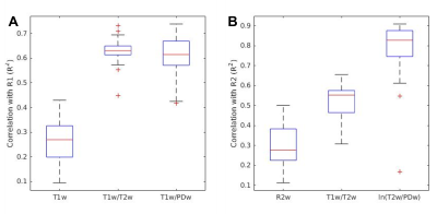 |
New semi-quantitative contrasts can approximate R1 and R2 in
clinical setting
Shachar Moskovich1,
Oshrat Shtangel1,
and Aviv Mezer1
1The Hebrew University, Jerusalem, Israel Keywords: Quantitative Imaging, Signal Representations Weighted MRI images are widely used in both clinical and open-source datasets, while quantitative mapping is not always feasible. The ratio of T1 and T2 weighted images was previously suggested as a semi-quantitative measurement. We propose two additional weighted ratios T1w/PDw and ln(T2w/PDw), as semi-quantitative proxies for R1 and R2, which we tested on phantom and human data. We found that the new ratios accurately represent the quantitative parameters in both datasets. |
| 15:45 |
1097.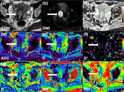 |
Separation of type and grade in cervical tumors using MOLLI
T1 mapping and non-mono-exponential models
diffusion-weighted MR imaging
Shujian Li1,
Jieliang Lin2,
and Jingliang Cheng1
1the First Affiliated Hospital of Zhengzhou University, Zhengzhou, China, 2Advanced Technical Support, Philips Healthcare, Beijing, China Keywords: Quantitative Imaging, Microstructure This study conducted an initial investigation of the feasibility of MOLLI-based T1 mapping and DWI by using mono-exponential, bi-exponential, and DKI models for the noninvasive preoperative evaluation of cervical cancer. Our findings indicated that both T1 mapping and non-mono-exponential model DWI can be used to discriminate cervical cancer from normal cervical tissue and adenocarcinoma from SCC. Our results also achieved a significant information gain for identifying SCC grade by combining native T1 and MKmean. Moreover, the maximum or minimum values of diffusion parameters within the whole lesion had advantages over the mean values in the prediction of SCC grade. |
| 15:45 |
1098.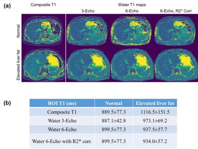 |
Water-Specific T1 Mapping of the Liver: The Influence of R2*
Estimation and Number of Echoes
Zhitao Li1,
Ding Xia2,3,
Shreyeas Vasanawala1,
and Li Feng2,3
1Department of Radiology, Stanford University, Stanford, CA, United States, 2BioMedical Engineering and Imaging Institute (BMEII), Icahn School of Medicine at Mount Sinai, New York, NY, United States, 3Department of Radiology, Icahn School of Medicine at Mount Sinai, New York, NY, United States Keywords: Quantitative Imaging, Liver, fat, Dixon Fat represents a major confounding factor for T1 mapping of the liver. Recently, a number of novel magnetization-prepared multiecho imaging approaches have been proposed to obtain water-only T1 mapping, and they all hold great potential for clinical applications. However, several questions have raised along with this this trend, such as (1) whether concurrent estimation of R2star and the number of echoes is necessary and whether it would affect the quantification of water T1; and (b) whether the number of echoes would affect T1 estimation. This work aimed to investigate these two questions with both numerical simulation and in-vivo experiments. |
| 15:45 |
1099.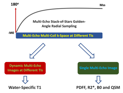 |
Initial Demonstration of Simultaneous Estimation of
Water-Specific T1, PDFF, R2*, and QSM in the Liver Using
Free-Breathing GraspT1-Dixon MRI
Jingjia Chen1,
Ding Xia2,
Kai Tobias Block3,
Chunlei Liu1,
and Li Feng2
1Electrical Engineering and Computer Sciences, University of California, Berkeley, Berkeley, CA, United States, 2Biomedical Engineering and Imaging Institute and Department of Radiology, Icahn School of Medicine at Mount Sinai, New York, NY, United States, 3Center for Advanced Imaging Innovation and Research (CAI2R), NYU Grossman School of Medicine, New York, NY, United States Keywords: Quantitative Imaging, Multi-Contrast This work demonstrates the feasibility of simultaneous estimation of fat/water-separated T1, proton density fat fraction (PDFF), R2*, and quantitative susceptibility mapping (QSM) of the liver using free-breathing GraspT1-Dixon MRI from a single rapid acquisition with an inversion-recovery (IR)-prepared multi-echo stack-of-stars sequence. For fat/water-separated T1 mapping, water-only images are generated from multi-echo images at different inversion times (TIs), from which a water-specific T1 map is estimated. For other parameters, acquired data from all TIs are averaged to generate a single set of multi-echo images, from which PDFF, R2*, and QSM are estimated. |
| 15:45 |
1100.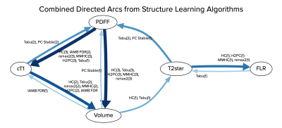 |
Bayesian Networks Reveal the Interplay Between Quantitative
Liver MRI Metrics
Yi-Chun Wang1,2,
Roberto Salvati2,
John Connell2,
Natali Van Zijl1,
Tom Waddell2,
Daniel Bulte1,
and Michael Brady2
1University of Oxford, Oxford, United Kingdom, 2Perspectum, Oxford, United Kingdom Keywords: Quantitative Imaging, Liver, cT1, T2*, PDFF, volume, future liver remnant Understanding the interplay between quantitative MRI metrics is crucial for reliable clinical assessment of liver health. This study utilised Bayesian networks to visualise hidden relationships between cT1, T2*, proton density fat fraction (PDFF), volume and future liver remnant (FLR). Analysing the directionality between Bayesian networks on a pre-operative dataset with 130 participants and a post-operative dataset with 90 participants, clear causal relationships from PDFF to cT1 and from PDFF to volume were found, which are supported by published literature. An additional discovery is the potential for correlation between metrics to help strengthen the clinical utility of cT1 after surgery. |
| 15:45 |
1101.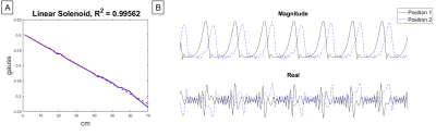 |
Selective Encoding through Nutation and Fingerprinting
(SENF) using Quadratic RF Phase Modulation and the
Bloch-Siegert Shift
Christopher Elliot Vaughn1,2,
N Reid Bolding3,
Mark A Griswold4,
and William A Grissom1,2
1Biomedical Engineering, Vanderbilt University, Nashville, TN, United States, 2Vanderbilt University Institute of Imaging Science, Vanderbilt University, Nashville, TN, United States, 3Physics, Case Western Reserve University, Cleveland, OH, United States, 4Radiology, Case Western Reserve University, Cleveland, OH, United States Keywords: Quantitative Imaging, Data Acquisition We demonstrate a new extension of Selective Encoding through Nutation and Fingerprinting (SENF) which is a RF spatial encoding technique that also encodes quantitative information. This SENF method uses quadratic RF phase modulation sequence with an off-resonant Bloch-Siegert pulse to induce the Bloch-Siegert shift to simultaneously encode spatial and quantitative information. We validate this method on a 47.5mT low-field scanner and in simulation. |
| 15:45 |
1102.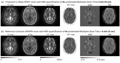 |
Nine-Fold Acceleration of Multi-Parametric Imaging of the
Brain through Joint Sparsity regularized Wave-SPIRiT
reconstruction
Sen Jia1,
Lixian Zou1,
Zhilang Qiu2,
Yongquan Ye3,
Haifeng Wang1,
Chao Zou1,
Ye Li1,
Jian Xu3,
Xin Liu1,
Hairong Zheng1,
and Dong Liang1,4
1Paul C. Lauterbur Research Center for Biomedical Imaging, Shenzhen Institutes of Advanced Technology, Shenzhen, China, 2Biomedical Engineering, Case Western Reserve University, Cleveland, OH, United States, 3UIH America, Houston, TX, United States, 4Medical AI Research Center, Shenzhen Institutes of Advanced Technology, Shenzhen, China Keywords: Quantitative Imaging, Relaxometry, Susceptibility The MULTIPLEX technique could quantify the T1/T2*/PD/Susceptibility maps in a single 3D scan but leads to a long scan time due to the dual-TR, dual-flip angle, and multi-echo signal acquisition strategy. Wave-CAIPI acceleration with SENSE reconstruction is limited by noise amplification at high acceleration factors and is susceptible to artifacts from inaccurate coil sensitivity maps. This work develops a L1 regularized Wave-SPIRiT reconstruction to achieve 9-fold accelerated MULTIPLEX imaging in 3 minutes. The L1 regularized coil-by-coil reconstruction also benefits the Multi-Dimensional Integration (MDI) quantification to achieve comparable accuracy and robustness as the reference scan with 55% reduction of scan time. |
| 15:45 |
1103.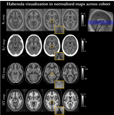 |
In vivo Multi-Parameter Mapping of the Habenula using MRI
Giorgia Milotta1,
Isobel Green2,
Jonathan Roiser3,
and Martina Callaghan1
1Wellcome Centre for Human Neuroimaging, University College London, London, United Kingdom, 2Harvard Medical School, Boston, MA, United States, 3Institute of Cognitive Neuroscience, University College London, London, United Kingdom Keywords: Quantitative Imaging, Tissue Characterization, Multi-Parameter Mapping The habenula has attracted much interest in neuroscience studies because it plays an important role in the reward circuitry of the brain and is implicated in psychiatric conditions. However, imaging the habenula remains challenging due to its sub-cortical location and small size, with few reports analysing its microstructural composition in vivo. To address this gap in the literature, we performed a multi-parametric characterisation of the microstructure of the habenula by quantifying relaxation rates (R1, R2*), water content (PD) and a marker of macromolecular content (MTsat), most notably myelin, in a cohort of 20 healthy participants. |
| 15:45 |
1104.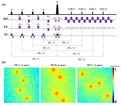 |
Improving single-shot multi-parametric mapping via
multi-slice information sharing based on multiple
overlapping-echo detachment imaging
Chenyang Dai1,
Jiechao Wang1,
Qizhi Yang1,
Zhigang Wu2,
Congbo Cai1,
and Shuhui Cai1
1Department of Electronic Science, Fujian Provincial Key Laboratory of Plasma and Magnetic Resonance, Xiamen University, Xiamen, China, 2MSC Clinical & Technical Solutions, Philips Healthcare, Shenzhen, China Keywords: Quantitative Imaging, Brain, Multi-slice information sharing; modulation pattern; overlapping-echo detachment imaging Multi-parametric quantitative magnetic resonance imaging (mqMRI) has important applications in clinic. Multiple overlapping-echo detachment (MOLED) imaging can achieve single-shot mqMRI. However, the existing methods mainly focus on single-slice reconstruction. To improve the reconstruction quality of parametric maps by exploring data redundancy among adjacent slices, we proposed a multi-slice information sharing method via multiple modulation patterns of MOLED k-space and deep neural network. The results show that our method can effectively utilize the correlation information among adjacent slices and improve the reconstruction quality compared to the single-slice reconstruction method. |
15:45 |
1105.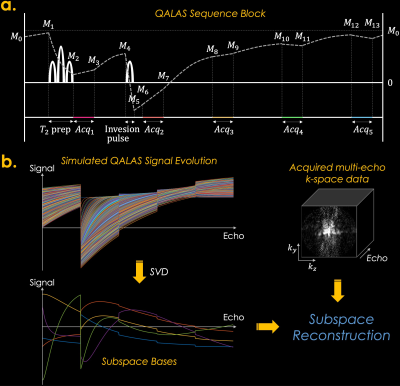 |
Zero-DeepSub: Zero-Shot Deep Subspace Reconstruction for
Multiparametric Quantitative MRI Using QALAS
Yohan Jun1,2,
Yamin Arefeen3,
Jaejin Cho1,2,
Xiaoqing Wang1,2,
Michael Gee2,4,
Borjan Gagoski2,5,
and Berkin Bilgic1,2,6
1Athinoula A. Martinos Center for Biomedical Imaging, Charlestown, MA, United States, 2Department of Radiology, Harvard Medical School, Boston, MA, United States, 3Massachusetts Institute of Technology, Cambridge, MA, United States, 4Department of Radiology, Massachusetts General Hospital, Boston, MA, United States, 5Fetal-Neonatal Neuroimaging & Developmental Science Center, Boston Children’s Hospital, Boston, MA, United States, 6Harvard/MIT Health Sciences and Technology, Cambridge, MA, United States Keywords: Quantitative Imaging, Quantitative Imaging The 3D-quantification using an interleaved Look-Locker acquisition sequence with T2 preparation pulse (3D-QALAS) has been developed and used for acquiring high-resolution T1, T2, and PD maps from five measurements within each repetition time. However, it assumes that each k-space data is acquired instantly at the first echo train length index neglecting T1 and T2 relaxation during the acquisition, which might cause blurring and biases in the reconstructed maps. In this study, we propose to reconstruct accurate quantitative T1 and T2 maps with reduced blurring compared to the conventional QALAS method using our proposed zero-shot deep subspace reconstruction method (i.e., Zero-DeepSub). |
| 15:45 |
1106.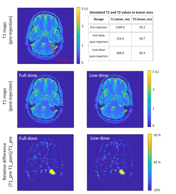 |
MR-STAT for fast contrast agent uptake quantification
Fei Xu1,
Hongyan Liu1,
Stefano Mandija1,
Oscar van den Heide1,
Edwin Versteeg1,
Miha Fuderer1,
Cornelis A.T. van den Berg1,
and Alessandro Sbrizzi1
1Computational Imaging Group for MR diagnostics & therapy, Center for Image Sciences, UMC Utrecht, Utrecht, Netherlands, Utrecht, Netherlands Keywords: Quantitative Imaging, Contrast Agent, MR-STAT; Contrast Enhancement Imaging Gadolinium-based contrast agents (GBCAs) uptake has the ability to facilitate disease diagnosis. In this work, we implement MR-STAT for fast contrast agent uptake quantification by applying keyhole acquisition and regularized reconstruction. We first analyze the accuracy of the proposed method on gadolinium-doped gel phantoms and observe that accurate T1 mapping of post-injection can be achieved with a keyhole factor of 25%. Furthermore, we investigate the impact of lower (20% vs 100%) GBCA dose administration on simulated clinical data. The quantification of pathologic T1 change for low-dose administration was comparable to that for full-dose. |
| 15:45 |
1107.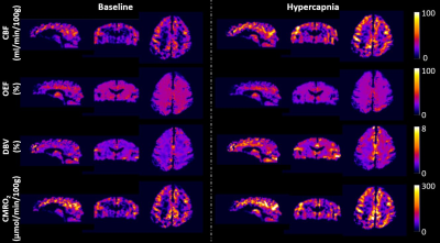 |
Validation of Constrained qBOLD-Based 3D CMRO2 Mapping With
Repeatability Test and Hypercapnic Challenge
Hyunyeol Lee1,2,
Jing Xu2,
and Felix W Wehrli2
1School of Electronics Engineering, Kyungpook National University, Daegu, Korea, Republic of, 2Department of Radiology, University of Pennsylvania, Philadelphia, PA, United States Keywords: Quantitative Imaging, Metabolism In the original qBOLD, it is challenging to separate deoxyhemoglobin’s contribution to R2' from other sources modulating the voxel signal. Further, extracting DBV and Yv from measured R2' is a nontrivial task. It was recently shown that the constrained qBOLD method was able to properly separate the several confounding factors, yielding the expected contrast for both Yv and DBV maps across the entire brain, and, together with a separate measurement of CBF, leading to whole-brain 3D CMRO2 maps within physiologically plausible ranges. Here, we validated the new 3D qBOLD method with respect to repeatability and hypercapnic gas breathing challenges. |
| 15:45 |
1108.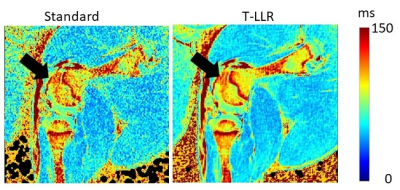 |
Locally low-rank denoising in transform domains.
Steen Moeller1,
Casey P. Johnson1,2,
Erick O. Buko1,2,
Ferenc Toth2,
Greg Metzger1,
Silvia Mangia1,
Shalom Michaeli1,
Sara Ponticorvo1,
Antonietta Canna1,
Kamil Ugurbil1,
and Mehmet Akcakaya1,3
1Radiology, University of Minnesota, Minneapolis, MN, United States, 2Veterinary Clinical Sciences, University of Minnesota, Minneapolis, MN, United States, 3Electrical and Computer Engineering, University of Minnesota, Minneapolis, MN, United States Keywords: Quantitative Imaging, Data Processing The concept of transform processing domain with locally low rank denoising is proposed as T-NORDIC and demonstrated for MSK and brain applications. The improvements on quantitative maps may be leveraged for faster acquisitions by relaxing the number of averages needed to obtain sufficient SNR for high resolution acquisitions and for application of low rank denoising to common clinical acquisitions. |
 Back to Meeting Home
 Back to the Program-at-a-Glance
The International Society for Magnetic Resonance in Medicine is accredited by the Accreditation Council for Continuing Medical Education to provide continuing medical education for physicians.
 Back to Meeting Home
 Back to the Program-at-a-Glance
 Full Session Video