Traditional Poster
Body
ISMRM & ISMRT Annual Meeting & Exhibition • 03-08 June 2023 • Toronto, ON, Canada

| Booth # | |||
|---|---|---|---|
5351.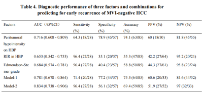 |
Predictive models for early recurrence of hepatocellular
carcinoma without microvascular invasion in patients after
hepatectomy
Qi Qu1,
Tao Zhang1,
Xue-Qin Zhang1,
Meng-Tian Lu1,
Lei Xu1,
and Xian-Ce Zhao2
1Affiliated Nantong Hospital 3 of Nantong University, Nantong Third People’s Hospital, Nantong, Jiangsu, China, Nantong, China, 2Philips Healthcare, Shanghai, China, Shanghai, China Keywords: Liver, Cancer To assess the predictive value of preoperative gadoxetic acid (GA)-enhanced magnetic resonance imaging (MRI) features and postoperative histopathological grading for early recurrence of hepatocellular carcinoma (HCC) without microvascular invasion (MVI) after curative hepatectomy. The results of the present demonstrated that our predictive model incorporating postoperative Edmondson-Steiner grade and preoperative imaging features including peritumoral hypointensity on HBP and RIR on HBP (Model-2) represents a promising model to assess the risk of early recurrence after resection of MVI-negative HCC. This predictive model may help clinicians formulate more aggressive and personalised treatment plans way earlier to improve patient prognosis and reduce early recurrence. |
||
5352.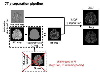 |
A feasibility study of susceptibility source separation via
chi-separation in amyotrophic lateral sclerosis patients at 7T
Jiye Kim1,
Hyeong-Geol Shin2,3,
Sooyeon Ji1,
Hwihun Jeong1,
Hongjun An1,
Cheol-Ho Sohn4,5,
Sohyun Han6,
Huijin Song7,
Ju-Hee Chae8,
Seok-Jin Choi9,
Jung-Joon Sung9,
and Jongho Lee1
1Department of Electrical and Computer Engineering, Seoul National University, Seoul, Korea, Republic of, 2Department of Radiology, Johns Hopkins University School of Medicine, Baltimore, MD, United States, 3F.M. Kirby Research Center for Functional Brain Imaging, Kennedy Krieger Institute, Baltimore, MD, United States, 4Institute of Radiation Medicine, Seoul National University Medical Research Center, Seoul, Korea, Republic of, 5Department of Radiology, Seoul National University Hospital, Seoul, Korea, Republic of, 6Center for Neuroscience Imaging Research, Institute for Basic Science (IBS), Suwon, Korea, Republic of, 7Biomedical Research Institute, Seoul National University Hospital, Seoul, Korea, Republic of, 8Department of Neurology, College of Medicine, Jeonbuk National University, Jeonju, Korea, Republic of, 9Department of Neurology, College of Medicine, Seoul National University, Seoul, Korea, Republic of Keywords: Susceptibility, Susceptibility The in-vivo imaging of iron and myelin concentrations of the motor cortex in amyotrophic lateral sclerosis (ALS) patients has significance in advancing knowledge about the degeneration progress of the disease. Here, we explored the feasibility of applying 𝜒-separation to ALS patients in-vivo at 7T. When the susceptibility values in hand knobs are examined, ALS patients have higher positive susceptibility values than healthy controls, confirming the histological finding of iron accumulation in ALS. |
||
5353.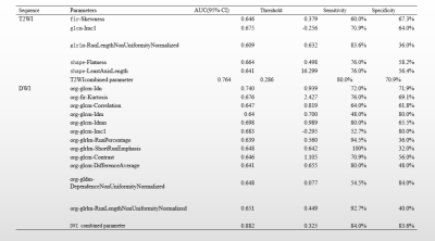 |
Differentiation of stage IA and IB endometrial cancer using
texture analysis based on T2 -weighted and diffusion-weighted
imaging
Meiyu Sun1 and
Shuli Cui1
1Department of Radiology, The First Affiliated Hospital of Dalian Medical University, Dalian, China Keywords: Contrast Mechanisms, Uterus, Endometrial Cancer In our study, Imagingomics texture analysis in DWI and T2WI showed the correlation and inverse gap of glcm, glrlm-RunLengthNonUniformityNormalized and shortRunEmphasis are more effective in identifying stage IA and IB EC . And the diagnostic efficacy combined parameter analysis were higrer and bettter than the single parameter analysis. |
||
5354.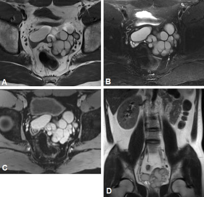 |
Imaging features and differential diagnosis of Zinner's
syndrome: what radiologists should know
Yue Zhao1,
Xiuhong Guan2,
Yongzhou Xu3,
Xinqing Jiang2,
and Ruimeng Yang2
1Guangzhou First People’s Hospital, Guangzhou, China, 2Department of Radiology, Guangzhou First People’s Hospital, Guangzhou, China, 3Philips Healthcare, Guangzhou, China Keywords: Urogenital, Pelvis Zinner's syndrome is a rare congenital urogenital abnormality. Since this disease is rare, knowledge about its imaging manifestations by clinical surgeons and radiologists is inadequate. In this study, data of 11 cases of Zinner's syndrome were retrospectively analyzed, and their morphological characteristics on imaging changes were investigated, aiming to improve the knowledge and differential diagnosis of this disease. The relevant images of Zinner's syndrome show characteristic manifestations, so the seminal vesicle cysts and isolateral kidney development must be carefully observed. Comprehensive judgement should be made according to urogenital mutation or deformity and systemic development situation. |
||
5355.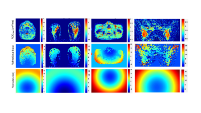 |
Standardized QC procedure for vendor-implemented ADC correction
of gradient nonlinearity bias in multi-center clinical trials
Thomas L Chenevert1,
Yuxi Pang1,
Debosmita Biswas2,
Ramesh Paudyal3,
Amaresh Konar3,
Jiachao Liang4,
Lisa J Wilmes4,
Nastaren Abad5,
Luca Marinelli5,
Humera Tariq1,
Ajit Devaraj6,
Dallas Turley7,
Johannes M Peeters8,
Nola M Hylton4,
David C Newitt4,
Savannah C Partridge2,
Amita Shukla-Dave3,9,
and Dariya Malyarenko1
1Radiology, University of Michigan Health System, Ann Arbor, MI, United States, 2Radiology, University of Washington, Seattle, WA, United States, 3Medical Physics, Memorial Sloan Kettering Cancer Center, New York, NY, United States, 4Radiology and Biomedical Imaging, University of California San Francisco, San Francisco, CA, United States, 5GE Research Center, Niskayuna, NY, United States, 6Clinical Science, Philips Healthcare, Highland Heights, OH, United States, 7Philips Healthcare, Bothell, WA, United States, 8Clinical Science, Philips, Best, Netherlands, 9Radiology, Memorial Sloan Kettering Cancer Center, New York, NY, United States Keywords: Cancer, Diffusion/other diffusion imaging techniques, ADC measurement accuracy, system gradient nonlinearity correction, multi-center oncology imaging trials Gradient nonlinearity (GNL) induces spatial bias in diffusion b-value that confounds apparent diffusion coefficient (ADC) measurements for anatomy offset from MRI scanner isocenter. For emerging vendor-provided GNL correction (GNC) a standardized quality control (QC) procedure is desired to streamline GNC application for multi-site imaging trials that utilize ADC for tumor monitoring and therapy response assessment. This QC procedure was developed and tested on four MRI scanner systems with vendor-provided on-line ADC GNC for trial-specific phantoms and patient scans for head-and-neck, breast, and myelofibrosis cancers. |
||
5356.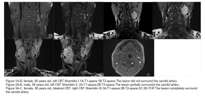 |
Comprehensive evaluation of carotid body tumor complicated with
carotid plaque by high resolution vascular wall imaging in
plateau area
Xindong Sun1 and
Haihua Bao1
1Affiliated Hospital of Qinghai University, Xining, China Keywords: Vessels, Tumor This study investigated risk factors for Carotid body tumor(CBT) in plateau areas, compared the incidence of CBT at high altitude group and low altitude group, and investigated the relationship between CBT and ipsilateral carotid plaque.The results showed that the incidence of CBT was higher at high altitude group than at low altitude group, and CBT will increase the incidence of ipsilateral carotid plaque.high resolution magnetic resonance vascular wall imaging(HR-VWI) has more advantages in the diagnosis of carotid body tumor than conventional MR Imaging. |
||
5357.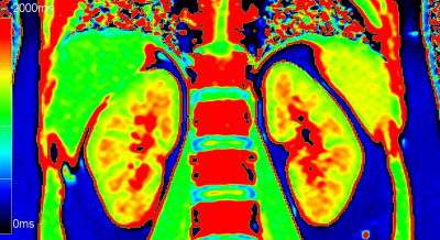 |
Evaluation of renal injury in chronic kidney disease by
Look-Locker T1 mapping
Wei Mao1,
Xiaoqiang Ding1,
Caixia Fu2,
Yuqin Ding1,
Dominik Nickel3,
Mengsu Zeng1,
and Jianjun Zhou1
1Zhongshan Hospital, Fudan University, Shanghai, China, 2MR Application Development, Siemens Shenzhen Magnetic Resonance Ltd., Shenzhen, China, 3MR Application Predevelopment, Siemens Healthcare GmbH, Erlangen, Germany Keywords: Kidney, fMRI, chronic kidney disease;T1 mapping magnetic resonance imaging;renal injury Accurate assessment of renal injury in chronic kidney disease (CKD) is significant for delaying the progression of CKD to end-stage renal disease. Previous studies have demonstrated the feasibility of Look-Locker T1 mapping in the assessment of renal function in healthy adults, but its application in the assessment of renal injury in CKD is still in the exploratory stage. Therefore, the purpose of this study was to investigate the value of T1 mapping in evaluating renal injury in CKD. |
||
5358.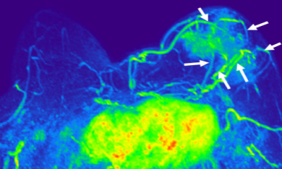 |
Preliminary Evaluation of the Tumor Vessels Based on Ultrafast
DCE MRI in Differential Diagnosis of Breast Tumors (BI-RADS 4)
hongbing liang1,
lina zhang1,
ning ning1,
nan zhang2,
qi wu1,
zhuo wang1,
qingwei song1,
ailian liu1,
and yinghua guo3
1First Affiliated Hospital of Dalian Medical University, Dalian, CHINA, Dalian, China, 2zhongshan Hospital of Fudan University, Shanghai, CHINA., Shanghai, China, 3Clinical & Technical Support, Philips Healthcare, Beijing, China, Beijing, China Keywords: Breast, Blood vessels The occurrence and development of the breast cancer are closely related to the growth and infiltration of blood vessels.This study mainly analyzed the correlation between the maximum diameter of benign and malignant breast tumors, the number of peripheral blood vessels and the first appearance of vascular phase. It is concluded that the number of blood vessels had a certain value in distinguishing benign and malignant breast tumors. There existed a certain correlation between the maximum diameter of malignant tumor and the number of blood vessels. |
||
 Back to Meeting Home
 Back to the Program-at-a-Glance
The International Society for Magnetic Resonance in Medicine is accredited by the Accreditation Council for Continuing Medical Education to provide continuing medical education for physicians.
 Back to Meeting Home
 Back to the Program-at-a-Glance
 View Presentation Video