Traditional Poster
Cardiovascular
ISMRM & ISMRT Annual Meeting & Exhibition • 03-08 June 2023 • Toronto, ON, Canada

| Booth # | |||
|---|---|---|---|
5359.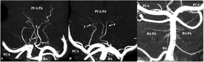 |
Observation of the Cerebral Perforating Arteries around Circle
of Willis Using Compressed Sensing Time-of-Flight at 7T
Qingle Kong1,
Zhe Zhang2,3,
and Jing Jing2,3,4
1MR Collaboration, Siemens Healthineers Ltd., Beijing, China, 2Tiantan Neuroimaging Center of Excellence, Beijing, China, 3Beijing Tiantan Hospital, Capital Medical University, China National Clinical Research Centre for Neurological Diseases, Beijing, China, 4Beijing Tiantan Hospital, Capital Medical University, Neurology Department, Beijing, China Keywords: Atherosclerosis, Vessels, perforating arteries, ultra high resolution The impairment of microvessels can lead to neurologic diseases such as stroke and vascular dementia. Perforating arteries imaging requires an extremely high resolution due to their small caliber size. In this study, for the first time, the feasibility of noninvasive visualization of perforators around Circle of Willis in vivo was demonstrated by using 7T compressed sensing TOF. The number of stems and branches of the perforators was then calculated. The origin and anatomical distribution of the perforating arteries are described. This work revealed that ultra-high-resolution CS TOF might be a promising method for detecting microvasculopathies of cerebral vascular diseases. |
||
5360.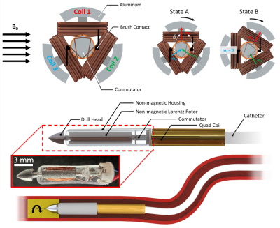 |
MRI-Driven Lorentz Force-based Mechanical Removal of Arterial
Occlusions
Martin Francis Phelan1 and
Metin Sitti1,2,3,4
1Physical Intelligence, Max Planck Institute for Intelligent Systems, Stuttgart, Germany, 2Mechanical Engineering, Carnegie Mellon University, Pittsburgh, PA, United States, 3Institute for Biomedical Engineering, ETH Zurich, Zurich, Switzerland, 4College of Engineering and School of Medicine, Koç University, Istanbul, Turkey Keywords: Atherosclerosis, Atherosclerosis, Mechanical Thrombectomy MR angiography provides high resolution visualization of blood vessels for analyzing arterial occlusions. However, these conditions require the use of a guidewire/catheter for treatment. Catheters integrated with microcoils for actuation under the high (3-7 Tesla), uniform magnetic field within magnetic resonance (MR) scanners have enabled mechanical removal of arterial occlusions. This work introduces an electromagnetic rotablation design, allowing direct tip torque control for both steering and drilling. Results demonstrate in vitro rotational thrombectomy with torque outputs up to 12 mN·m under MR guidance. These results indicate the high MRI external field can provide rotablation for difficult-to-reach areas of the vasculature. |
||
5361.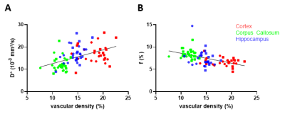 |
Validation of Intravoxel Incoherent Motion (IVIM) as a surrogate
marker of vascular density in a rat model of co-morbidities
Bram Callewaert1,2,
Willy Gsell2,
Marleen Lox1,
Elizabeth Jones1,3,
and Uwe Himmelreich2
1Department of Cardiovascular Sciences, University of Leuven, Leuven, Belgium, 2Department of Imaging and Pathology, University of Leuven, Leuven, Belgium, 3Department of Cardiology, Maastricht University, Maastricht, Netherlands Keywords: Vessels, Blood vessels We assessed whether Intravoxel Incoherent Motion (IVIM) MRI measurements can detect differences in cerebral vascular density obtained by quantitative immunohistochemistry. In vivo IVIM measurements were correlated with invasive measurements of the microvascular density in a rat model of vascular comorbidities. Our results indicate that IVIM can be used to indirectly measure difference in the vascular density of different brain regions |
||
5362.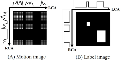 |
Automatic detection method for the stationary period of the
coronary arteries for whole-heart coronary MR angiography using
deep learning
Shigehide Kuhara1,
Remina Kasai2,
Yuta Endo1,
Sanae Takahashi1,
Haruna Shibo1,
Kuninori Kobayashi1,
and Makoto Amanuma1
1Department of Medical Radiological Technology, Faculty of Health Sciences, Kyorin University, Mitaka-shi, Tokyo, Japan, 2Radiology Department, Tokyo Teishin Hospital, Chiyoda-ku, Tokyo, Japan Keywords: Vessels, Cardiovascular, Coronary MRA We developed a new method using a convolutional neural network to obtain the stationary periods of the coronary arteries for whole-heart coronary magnetic resonance angiography. A time-domain segmentation method using U-net was proposed. Two motion curves, which were obtained from the motion of the coronary arteries between cine frames, were vertically arranged, converted to motion images, and used to extract the stationary periods. The results demonstrate that the proposed method can accurately determine the stationary period of the coronary arteries in humans, and it is expected to be a fully automatic determination method for the stationary period. |
||
5363.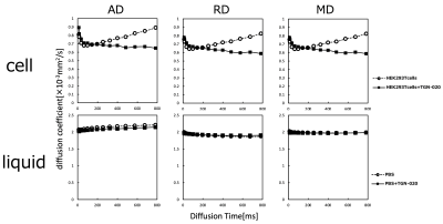 |
Diffusion Time Spectrum Analysis Observation of Aquaporin
Functional Dynamics
Ayano Oku1,2,
Junichi Hata1,2,3,
Naoya Hayashi1,2,
Hinako Oshiro1,2,
Kanako Muta1,
Yawara Haga2,3,
Taeko Ito2,
Ryousuke Nakajima2,3,
Noriyuki Kishi2,3,
and Hideyuki Okano2,3
1Tokyo Metropolitan University, Tokyo, Japan, 2RIKEN Center for Brain Science, Saitama, Japan, 3Keio University School of Medicine, Tokyo, Japan Keywords: Diffusion/other diffusion imaging techniques, Diffusion Tensor Imaging, diffusion time The function of water molecule exchange on the plasma membrane by aquaporins was evaluated using HEK293T cultured cells. Time-dependent diffusion magnetic resonance imaging (MRI), in which diffusion time (DT) was varied in 14 steps from 13 to 784 ms, was used to observe differences in water molecules and the permeability of the cell membrane with and without aquaporin inhibition. The diffusion coefficient increased with increasing DT in normal cells, but not in aquaporin 4-inhibited cells. These results suggest that time-dependent diffusion MRI may capture the function of water molecule exchange at the plasma membrane. |
||
5364.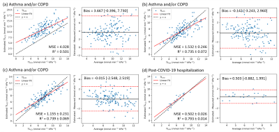 |
Modelling Whole-Lung and Regional Carbon Monoxide Transfer
Factor with Hyperpolarized 129Xe MRI in Asthma and COPD Patients
Jemima H Pilgrim-Morris1,
Laurie J Smith1,
Joshua R Astley1,2,
Laura C Saunders1,
Guilhem J Collier1,
Alberto M Biancardi1,
Bilal A Tahir1,2,3,
Helen Marshall1,
Latife Hardaker4,
Titti Fihn-Wikander5,
Rod Hughes6,
Roger Thompson1,
Neil J Stewart1,
and Jim M Wild1,3
1POLARIS, Department of Infection, Immunity and Cardiovascular Disease, University of Sheffield, Sheffield, United Kingdom, 2Department of Oncology and Metabolism, University of Sheffield, Sheffield, United Kingdom, 3Insigneo Institute for in silico Medicine, University of Sheffield, Sheffield, United Kingdom, 4Priory Medical Group, York, United Kingdom, 5Evidence Delivery, BioPharmaceuticals Medical, BioPharmaceuticals Business Unit, AstraZeneca, Gothenburg, Sweden, 6Clinical Development, Research and Early Development, Respiratory & Immunology, AstraZeneca, Cambridge, United Kingdom Keywords: Hyperpolarized MR (Gas), Lung, Modelling Hyperpolarized 129Xe ventilation and dissolved-phase MRI metrics were used to predict the carbon monoxide transfer factor (TLCO) in the lungs of patients with asthma and/or Chronic Obstructive Pulmonary Disease (COPD) using two linear regression models: (1) an existing analytical model based on physiology and (2) a machine learning model, which also included patient age and sex in the prediction. The machine learning model was further extended to create TLCO maps, providing a regional visualization of this gold standard measure of gas transfer. |
||
5365.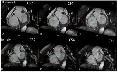 |
An exploratory study to investigate the effect of light music on
whole-heart coronary MRI
Weiwei Wang1,
Yang Wu1,
Peng Sun2,
Haixia Li2,
Yanyan Jiang1,
and xiaojing Ma1
1MRI, Wuhan asia general hospital, Wuhan,HUBEI, China, 2Philips Healthcare,Beijing, Beijing, China Keywords: Vessels, Cardiovascular, compressed sensing Whole-heart coronary magnetic resonance angiography (MRA) is a noninvasive, contrast-free, and radiation-free technique for evaluating the origin, morphology, and stenosis of coronary arteries. Nonetheless, a patient's anxious mood results in a longer scan time or lower imaging quality. Previous research has shown that listening to music can alleviate anxiety in patients with coronary heart disease (CHD). In this study, we found no statistical difference in scanning time and imaging quality with CS acceleration factors (AFs) of 2,4,6 in the music and no-music groups. Music may not be necessary during coronary MRA scanning with the CS technique. |
||
 Back to Meeting Home
 Back to the Program-at-a-Glance
The International Society for Magnetic Resonance in Medicine is accredited by the Accreditation Council for Continuing Medical Education to provide continuing medical education for physicians.
 Back to Meeting Home
 Back to the Program-at-a-Glance
 View Presentation Video