Traditional Poster
Neuro
ISMRM & ISMRT Annual Meeting & Exhibition • 03-08 June 2023 • Toronto, ON, Canada

| Booth # | |||
|---|---|---|---|
5320.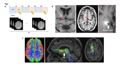 |
Characterizing the Network Effects of Deep Brain Stimulation for
Obsessive-Compulsive Disorder using Structural and Functional
Imaging
Natalya Slepneva1,
Tenzin Norbu2,
Melanie Morrison2,
and Andrew Moses Lee3
1Psychiatry, University of California San Francisco, Vallejo, CA, United States, 2Radiology, University of California San Francisco, San Francisco, CA, United States, 3Psychiatry, University of California San Francisco, San Francisco, CA, United States Keywords: Psychiatric Disorders, Multimodal, brain stimulation Deep brain stimulation (DBS) is a treatment for severe, refractory OCD. We conducted post-operative DTI imaging to characterize structural connectivity from DBS electrodes. We also conducted fMRI during stimulation ON/OFF studies to elucidate the impact of DBS on functional networks in therapeutic and non-therapeutic configurations. In preliminary analysis, we find that therapeutic contacts are structurally connected to components of the OCD circuit in the anterior cingulate cortex (ACC), caudate, and thalamus. We also identified suppression in this cortico-striato-thalamo-cortical circuit during therapeutic DBS ON vs OFF, suggesting DBS therapy operates by inhibiting the OCD network. |
||
5321.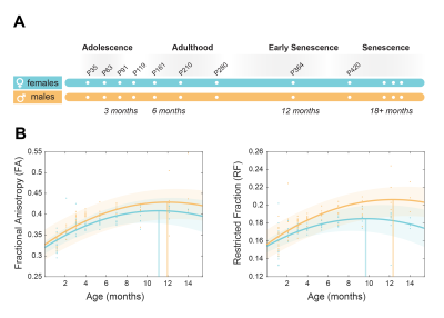 |
Imaging sexual dimorphism in white matter across the lifespan: A
rat model of healthy ageing
Antonio Cerdán Cerdá1,
Patricia Martínez-Tazo1,
Santiago Canals2,
and Silvia De Santis1
1Molecular Neurobiology and Neuropathology, Instituto de Neurociencias de Alicante, San Joan d'Alacant, Spain, 2Cellular and Systems Neurobiology, Instituto de Neurociencias de Alicante, San Joan d'Alacant, Spain Keywords: Neurodegeneration, Aging White matter microstructure evolves across the lifespan due to maturation and degeneration. Recently, MR imaging in humans uncovered region- and sex-specific trajectories of microstructural integrity decline, suggesting prolonged neoteny in the female brain. Here, using advanced diffusion-weighted MRI, we characterize the contribution of different cell-specific white matter biomarkers in a longitudinal rat model of healthy ageing, from puberty to early senescence. Our results validate the use of a rat model of healthy ageing for dissecting longitudinal brain ageing patterns, replicate the sexual dimorphism found in humans and points at myelin as the neurobiological substrate driving the observed sex difference. driving the observed sex difference. |
||
5322.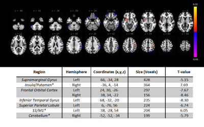 |
Effects of MR-Guided Focused Ultrasound Pallidotomy on
Dyskinesia and Cerebral Regional Homogeneity in Parkinson’s
Disease
Andrew Furman1,
Li Jiang1,
Justin Schumacher2,
Ziachen Zhuo1,
Howard Eisenberg3,
Paul Fishman4,
Dheeraj Ghandi1,
and Rao Gullapalli1
1Diagnostic Radiology and Nuclear Medicine, University of Maryland, School of Medicine, Baltimore, MD, United States, 2University of Maryland, School of Medicine, Baltimore, MD, United States, 3Neurosurgery, University of Maryland, School of Medicine, Baltimore, MD, United States, 4Neurology, University of Maryland, School of Medicine, Baltimore, MD, United States Keywords: Parkinson's Disease, Focused Ultrasound MR-guided Focused Ultrasound (MRgFUS) pallidotomy is a promising, non-invasive neurosurgical approach for treating the primary and secondary symptoms, such as levodopa-induced dyskinesia, of Parkinson’s Disease (PD). While the behavioral effects of MRgFUS pallidotomy in PD have been previously established, its impacts on brain circuity and how these changes relate to symptom resolution are currently unclear. In the current study, we compared measures of Regional Homogeneity (ReHo), a metric of spatiotemporal correlation thought to reflect the integrity and modularity of mesoscopic circuity, before and after FUS lesion of the Globus Pallidus internus (GPi). |
||
5323.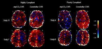 |
Comparing reliability of BOLD cerebrovascular reactivity of
end-tidal pCO2 versus mean cerebellar signal regression in
pediatric moyamoya.
Srivats Srinivasan1,2,
Laura L Lehman1,
Julie Swanson1,
Darren B Orbach1,
and Jeffrey N Stout1
1Cerebrovascular Surgery and Interventions Center, Boston Children's Hospital, Boston, MA, United States, 2University of Texas Southwestern Medical Center, Dallas, TX, United States Keywords: Blood vessels, Pediatric, Cerebrovascular, reactivity, CVR, moyamoya We compared the reliability of BOLD CVR (cerebrovascular reactivity) between two regression approaches – etpCO2 signal and average cerebellar signal – in pediatric moyamoya patients. We estimated CVR using a lagged optimized GLM model, conducted cortical parcellation, and compared left-right hemispheric CVR differences to clinical and imaging reports. We found that etpCO2 had poorer fits during regression compared to the cerebellar approach (p<0.0001) and incorrectly identified disease laterality in 2/8 patients. These effects were strongly observed in subjects with poor breath-hold task compliance, indicating that the cerebellar approach is more reliable for studies of young pediatric moyamoya patients. |
||
5324.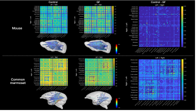 |
Evaluation of the effect of microbiota on brain neural fiber
bundles in germ-free common marmosets by structural connectome
analysis
Chika Tokisugi1,2,
Fumiko Seki2,
Yuji Komaki2,
Takashi Inoue2,
and Junichi Hata1,2
1Tokyo Metropolitan University, Tokyo, Japan, 2Central Institute for Experimental Animals, Kawasaki, Japan Keywords: Brain Connectivity, Animals, microbiota This study aimed to investigate the brain–gut correlation in common marmosets. We examined germ-free (GF) common marmoset in terms of brain connectivity between brain regions using magnetic resonance imaging. We performed three-dimensional diffusion-weighted imaging, generated whole-brain tractography, and created connectome weighted by mean fractional anisotropy (FA). We compared the difference between GF and control. In GF mice, mean FA between regions showed little difference; however, these were reduced in common marmoset compared with conventional ones. Studying the effect of microbiota on common marmoset brain might capture primate-specific characteristics and would help in exploring the microbiota effects on the human brain. |
||
5325.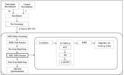 |
Feasibility of a multimodal MR Imaging and Spectroscopy approach
in understanding Myalgic Encephalomyelitis/Chronic Fatigue
Syndrome
Raminder Kaur1,2,
Kashish Mehta1,2,
Alexander Ciok1,2,
Brian Greeley1,
Kati Debelic3,
Hilary Robertson3,
Todd Nelson1,2,
Melody Tsai4,
Lan Xin Zhang1,5,
Margit Glashutter1,2,
Travis Boulter4,
Luis Nacul*4,6,
and Xiaowei Song*1,2
1Clinical Research, Fraser Health Authority, Surrey, BC, Canada, 2Department of Biomedical Physiology Kinesiology, Simon Fraser University, Burnaby, BC, Canada, 3Patient partner in Research, Community member, Vancouver, BC, Canada, 4BC Women's Hospital & Health Centre, Vancouver, BC, Canada, 5Medical Biophysics, University of Toronto, Toronto, ON, Canada, 6University of British Columbia, Vancouver, BC, Canada Keywords: Head & Neck/ENT, Multimodal This first multimodal MRI/MRS study aiming to investigate whether an extensive multimodal neuroimaging approach is feasible for patients with ME/CFS. The study compared female ME/CFS participants to aged matched female healthy controls. The study showed successful completion of the entire protocol that included: anatomical imaging; functional magnetic resonance imaging (fMRI) along with a working memory task; single-voxel magnetic resonance spectroscopy (MRS) to quantify metabolites at three locations (ACC, BS, and l-DLPC); and a hand-grip strength to observe whether fatigue is induced due to the scanning protocol. It also suggested a minimum impact of the MRI session on grip strength. |
||
5326. |
Quantitative susceptibility mapping of hippocampal iron relates
to pattern separation and completion in older adults at risk for
Alzheimer's
Jing Zhou1,
Alfie Wearn1,
Julia Huck2,
Giulia Baracchini1,
Jennifer Tremblay-Mercier3,
Judes Poirier3,4,
Sylvia Villeneuve3,4,5,6,
Christine Tardif 4,6,
Ana Daugherty7,
Claudine Gauthier2,8,
Gary R Turner9,
and R Nathan Spreng6,10,11,12
1Montreal Neurological Institute, McGill University, Montreal, QC, Canada, 2PERFORM Centre, Concordia University, Montreal, QC, Canada, 3StoP-AD Centre, Douglas Mental Health Institute Research Centre, Montreal, QC, Canada, 4McGill University, Montreal, QC, Canada, 5McGill Centre for Integrative Neuroscience, McGill University, Montreal, QC, Canada, 6McConnell Brain Imaging Center, McGill University, Montreal, QC, Canada, 7Department of Psychology and Institute of Gerontology, Wayne State University, Detroit, MI, United States, 8Physics Department, Concordia University, Montreal, QC, Canada, 9Department of Psychology, York University, Toronto, ON, Canada, 10Department of Neurology and Neurosurgery, Montréal Neurological Institute, Montreal, QC, Canada, 11Douglas Mental Health University Institute, Montreal, QC, Canada, 12Departments of Psychiatry and Psychology, McGill University, Montreal, QC, Canada Keywords: Alzheimer's Disease, Quantitative Susceptibility mapping, Human Memory Quantitative susceptibility mapping (QSM) was used to evaluate hippocampal iron in a cohort of healthy older individuals at elevated risk for Alzheimer’s disease (AD), and related to pattern separation and pattern completion memory performance. Our results demonstrated that elevated brain iron content in the hippocampus is strongly associated with lower performance on behavioral tests specific to memory function, ie lower pattern separation scores and higher pattern completion scores. Our findings suggest that hippocampal iron deposition may be a pathological mechanism resulting in poorer mnemonic discrimination in later life |
||
5327.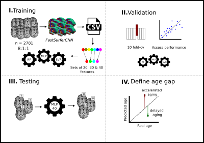 |
Increased T1w MRI-based brain age in chronic migraine patients
Rafael Navarro-González1,
David García-Azorín2,
Ángel L. Guerrero2,
Álvaro Planchuelo-Gómez1,3,
Santiago Aja-Fernández1,
and Rodrigo de Luis-García1
1Image Processing Laboratory, Universidad de Valladolid, Valladolid, Spain, 2Hospital Clínico Universitario de Valladolid, Valladolid, Spain, 3CUBRIC, Cardiff University, Cardiff, United Kingdom Keywords: Machine Learning/Artificial Intelligence, Aging, Migraine Brain-age is an emerging neuroimaging biomarker that represents the aging status of the brain using machine learning techniques from MRI data. It has been successfully applied to the study of different neurological and psychiatric conditions. We hypothesize that patients with migraine may show an increased brain age gap (difference between the age estimated from the MRI data and the chronological age). After building a brain age model from 2,781 healthy subjects, we tested this hypothesis on a dataset with 210 healthy controls and migraine patients. Results showed an increased brain age in chronic migraine patients with respect to healthy controls. |
||
5328.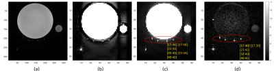 |
Enhancement of Pulse Simulation in Quantifying Procedure of Very
Slow CSF Flow Measurement with Shunt-FENSI
Mingxiao Zhang1,2,
Suguna Pappu3,4,
William C. Olivero3,4,
Jason M. Huston3,5,
and Bradley P. Sutton1,2,3
1Department of Bioengineering, University of Illinois at Urbana-Champaign, Champaign, IL, United States, 2Beckman Institute, University of Illinois at Urbana-Champaign, Urbana, IL, United States, 3Carle Illinois College of Medicine, University of Illinois at Urbana-Champaign, Urbana, IL, United States, 4Department of Neurosurgery, Carle Foundation Hospital, Urbana, IL, United States, 5Department of Radiology, Carle Foundation Hospital, Urbana, IL, United States Keywords: Neurofluids, Brain, Flow, Hydrocephalus, CSF, Quantitative Ventriculoperitoneal shunts drain excessive cerebrospinal fluid (CSF) for hydrocephalus patients, but shunts placed shortly after birth 50% fail within 2 years. Since rapid MRI is becoming the standard for imaging possible shunt malfunction, an MRI technique that assesses shunt flow at the same imaging setting, would be beneficial. Previously, the non-invasive quantitative method, Shunt Flow Enhancement of Signal Intensity (Shunt-FENSI), was proposed for diagnosing possible shunt failure. An improvement in its flow data simulation and quantification is included, more accurately reflecting the RF pulse application in the pulse sequence. Improvements are demonstrated on phantom, extraventricular drain (EVD), and shunt patients. |
||
5329.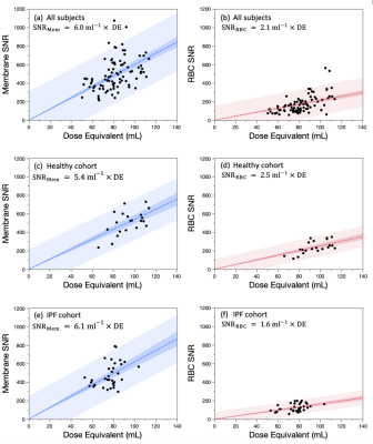 |
SNR and Dose Considerations for Hyperpolarized 129Xe Magnetic
Resonance Spectroscopy
Haoran Dai1,
Elianna Bier2,
David Mummy3,
Alexander Church3,
Aryil Bechtel3,
Anna Costelle1,
and Bastiaan Driehuys1,2,3
1Medical Physics Graduate Program, Duke University, Durham, NC, United States, 2Department of Biomedical Engineering, Duke University, Durham, NC, United States, 3Department of Radiology, Duke University, Durham, NC, United States Keywords: Hyperpolarized MR (Gas), Spectroscopy Hyperpolarized 129Xe MR spectroscopy is a powerful tool for evaluating gas exchange, blood oxygenation and hemodynamics. However, there is no established method for determining the HP 129Xe dose equivalent required to achieve scans of sufficient quality for analysis. Here, we establish a robust approach to calculate the 129Xe MRS SNR and assess its relationship to the 129Xe dose equivalent. |
||
5330.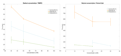 |
129Xe sublimation DNP towards hyperpolarized imaging standards
Emma Linnea Wiström1,
Jean-Noel Hyacinthe1,
Thanh Phong Lê1,
Rolf Gruetter1,
and Andrea Capozzi1,2
1Laboratory of Functional and Metabolic Imaging, EPFL, Lausanne, Switzerland, 2HYPERMAG, Technical University of Denmark, Copenhagen, Denmark Keywords: Hyperpolarized MR (Gas), Hybrid & Novel Systems Technology Sample preparation of 129Xe for dDNP with different glassing solvents, radical choice and concentrations affects the electron properties as well as the overall achieved 129Xe polarization. A short T1e and/or poor mixing is improved by ad hoc solutions that results in competitive polarization values in solid state measurements.
The sample preparation of 129Xe for DNP showed an improved incorporation of the gas atoms in the solvent when using ultrasonication. Applying microwave frequency modulation during the irradiation reached a higher 129Xe polarization. Extracted Xe was measured in a 1T NMR benchtop spectrometer to be 18s. |
||
5331.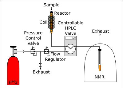 |
A highly automated experimental setup to perform PHIP via proton
exchange with high pressure and high reproducibility at various
magnetic fields
Jule Kuhn1,
Kolja Them1,
and Jan-Bernd Hövener1
1Section Biomedical Imaging, Department of Radiology and Neuroradiology, University Medical Center Schleswig - Holstein, Kiel University, Kiel, Germany Keywords: Hyperpolarized MR (Non-Gas), Contrast Agent The PHIP-X methodology is a novel approach to produce contrast agents for metabolic MRI by hyperpolarization and combines the high polarization resulting from pH2 addition with the versatility of proton exchange. However, the polarization yield and reproducibility have to be improved. The proposed setup provides reproducible and well controlled experimental conditions for PHIP-X, including a pH2 pressure of up to 100 bar and a magnetic field of up to 95 mT. Using this setup allowed to reach high 1H polarizations of the transfer agent and to transfer the polarization to 13C in the target molecule glucose successfully. |
||
5332.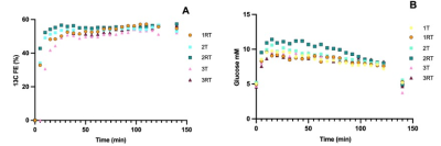 |
Reproducibility of 13C labeling of glutamate and glutamine in
human brain using selPOCE MRS at 7T upon [U-13C]-labeled glucose
infusion
Narjes Ahmadian1,2,
Sarah M Jacobs1,
Mark Gosselink1,
Wybe JM van der Kemp1,
Hans Hoogduin1,
Anastasia Coppoli3,
Graeme F Mason3,
Robin A de Graaf3,
Helia Norouzizadeh4,
Chantal Mahon5,
Sjoerd van Marle6,
Pieter van Eijsden2,
Dirk Cerneus6,
Corin O Miller5,
Inge De Lepeleire4,
Anthony S Basile5,
Dennis W Klomp1,
Jeanine J Prompers1,
and Evita C Wiegers1
1Radiology, University Medical Center Utrecht, Utrecht, Netherlands, 2Neurosurgery, University Medical Center Utrecht, Utrecht, Netherlands, 3Yale University School of Medicine, New Haven, CT, United States, 4MSD (Europe) Inc, Brussel, Belgium, 5Merck & Co. Inc, Kenilworth, NJ, United States, 6ICON plc, Groningen, Netherlands Keywords: Non-Proton, Metabolism, 13C The aim of this study is to assess the reproducibility of a selective proton-observed, carbon-edited (selPOCE) MRS sequences for the detection of glutamate/glutamine cycling at 7T in healthy participants. Participants were scanned twice while undergoing [U-13C] glucose infusion. The time course of Glu C45 and Gln C45 labeling were similar for test-retest measurements. This supports the application in future studies on measuring neuroenergetics and neurotransmitter cycling. |
||
5333.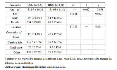 |
Prediction meningioma grade by constructing a cli-radiomics
model nomogram based on magnetic resonance imaging
Tao Han1,
Xianwang Liu1,
and Junlin Zhou1
1Lanzhou University Second Hospital, LanZhou, China Keywords: Tumors, Machine Learning/Artificial Intelligence This study investigate the feasibility of a cli-radiomics model in preoperative noninvasive prediction of meningioma grade. This study attempted to construct a radiomics model for predicting meningioma grade based on T1C and T2WI sequences using different classifiers, select the optimal radiomics model, and combine it with clinical labels to construct a nomogram. Decision curve analysis was used to verify the clinical validity of the nomogram, which provides a non-invasive and convenient alternative method for clinicians to predict meningioma grades. |
||
5334. |
The effect of elevated serum lactate on measures of apparent
cerebral metabolism and perfusion in the anesthetised rodent
brain.
Jordan McGing1,
Daniel Radford-Smith2,
Richard Healicon3,
Catriona H. E. Rooney3,
Ayaka Shinozaki1,
Surrin Deen3,
Tricia Seow3,
Vicky Ball3,
Fulvio Zaccagna 3,
Sean Smart4,
Daniel Anthony2,
Damian J Tyler1,3,
and James T Grist1,3,5,6
1Oxford Centre for Clinical Magnetic Resonance Research, University of Oxford, Oxford, United Kingdom, 2Department of Pharmacology, University of Oxford, Oxford, United Kingdom, 3Department of Physiology Anatomy and Genetics, University of Oxford, Oxford, United Kingdom, 4Nuffield Department of Clinical Neurosciences, University of Oxford, Oxford, United Kingdom, 5Department of Radiology, University of Oxford, Oxford, United Kingdom, 6Institute of Cancer and Genomic Sciences, University of Birmingham, Birmingham, United Kingdom Keywords: Neurodegeneration, Hyperpolarized MR (Non-Gas) It is unclear how serum lactate perturbations influence the quantification of cerebral blood flow (CBF) and metabolism using proton perfusion imaging and hyperpolarized 13C MRI. Low dosage L-lactate injection increased CBF and cerebral LDH flux, whilst high dosage lactate decreased CBF and maintained the increased LDH flux, relative to saline administration. Lactate administration did not alter apparent oxidative metabolism. This suggests the efficacy of probing oxidative metabolism with 13C MRI when alterations in serum lactate are present (e.g. in pathology). But highlights the requirement for serum lactate measurement prior to interpretation of 13C metabolic data. |
||
5335. |
7T MRI and CSF correlates of brain iron accumulation,
neurodegeneration and neuroinflammation in Huntington's Disease:
Study Protocol
Nadine van de Zande1,
Marjolein Bulk2,
Chloé Najac2,
Louise van der Weerd2,3,
Jeroen de Bresser2,
Jan Lewerenz4,
Itamar Ronen5,
and Susanne de Bot1
1Neurology, Leiden University Medical Centre, Leiden, Netherlands, 2Radiology, Leiden University Medical Centre, Leiden, Netherlands, 3Human Genetics, Leiden University Medical Centre, Leiden, Netherlands, 4Neurology, University of Ulm, Ulm, Germany, 5Clinical Imaging Sciences Centre, Brighton and Sussex Medical School, Brighton, United Kingdom Keywords: Neuroinflammation, Quantitative Susceptibility mapping, Huntington's Disease HD is a rare, autosomal dominant inherited, progressive, neurodegenerative disorder. Strong evidence suggests a significant role for iron accumulation and neuroinflammation in HD. Previous studies already showed iron accumulation in the brain of patients with HD, but no other study linked these results with well-accepted biofluid biomarkers for neuroinflammation, or with neuroimaging methods to assess neuroinflammation. This study will provide an important basis for the evaluation of brain iron levels and neuroinflammation metabolites as imaging biomarkers for disease state and progression in HD and their relationship with the salient pathophysiological mechanisms of the disease. |
||
5336. |
Brain atrophy and cognitive impairment in patients with
end-stage renal disease: A Voxel-Based Morphometry Study
Yuan Li1,
Yuhan Jiang1,
Bingbing Gao1,
Mingrui Qu1,
Qingwei Song1,
and Yanwei Miao1
1the First Affiliated Hospital of Dalian Medical University, Dalian, China Keywords: Nerves, Kidney End-stage renal disease is the final stage of chronic kidney disease, often complicated by abnormal brain structure and neurocognitive function. In this study, a voxel-based morphometry (VBM) measurement was used to observe the volume change of gray matter structure in ESRD patients. Patients with ESRD were found to have atrophy in the right Cerebelum_Crus1, bilateral Cerebelum_6, left Temporal_Pole_Sup, left Putamen, bilateral Calcarine (CAL), bilateral Lingual (LING), left Rolandic_Oper, left Heschl, right Cuneus, bilateral Postcentral. Furthermore, the volumes of the right Cerebelum_Crus1, bilateral CAL, left Cerebelum_6, bilateral LING were positively correlated with overall cognitive score. |
||
5337.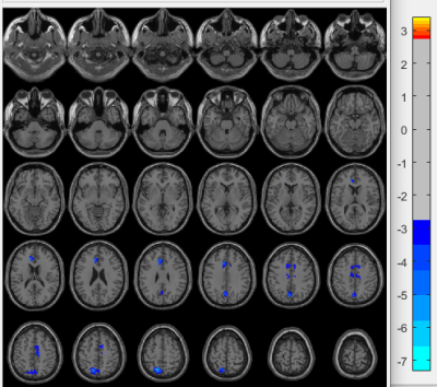 |
Neuropsychiatric systemic lupus erythematosus is associated with
a distinct type and cerebral structural changes
Huiyang Liu1,
Hu Liu1,
and Guoguang Fan1
1the First Hospital of China Medical University, shenyang, China Keywords: Gray Matter, Neuroinflammation, NPSLE We used the voxel-based morphometry (VBM) to investigate morphological alterations within gray matter (GM) in patients with neuropsychiatric systemic lupus (NPSLE). The two subgroups (inflammatory and ischemic phenotypes) have different pathological mechanisms and treatment methods. In the comparison of grey matter volumes within the two patient groups, we found brain atrophy was more severe in the inflammatory group and that the atrophied brain areas were consistent with previous studies of the blood-brain barrier in patients with autoimmune disease. Our findings could help to deeply understand the pathophysiology of NPSLE, and to treat lupus patients early to improve their prognosis. |
||
5338. |
Intra-plaque hemorrhage of the basilar artery is a risk factor
for recurrent pontine infarction
Chuanying Shi1,
Peng Wu2,
and Xiance Zhao2
1Liaocheng People's Hospital, Liaocheng, China, 2Philips Healthcare, Shang Hai, China Keywords: Blood vessels, Ischemia Compared with the first episode of pontine acute infarction, the risk factors of the basilar artery (BA) wall of recurrent pontine acute infarction have not been fully defined. In this study, high-resolution magnetic resonance vessel wall imaging (HRMR-VWI) was used to compare the changes of BA wall in patients with first-onset and recurrent pontine acute infarction, to find the risk factors of the BA wall in patients with recurrent pontine acute infarction. The result showed intra-plaque hemorrhage (IPH) of the BA wall identified on HRMR-VWI is associated with recurrent ischemic stroke. |
||
5339.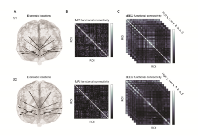 |
Spatial Correspondence Between fMRI and Frequency-Specific
Electrophysiological Networks in Treatment Resistant Depression
Niki Sabetfakhri1,
Joline Fan2,
Natalya Slepneva 1,
Julian Motzkin2,3,
Melanie Morrison4,
Leo Sugrue4,
Andrew Krystal1,
and A Moses Lee 1
1Department of Psychiatry and Behavioral Sciences, UCSF, San Francisco, CA, United States, 2Department of Neurology, UCSF, San Francisco, CA, United States, 3Department of Anesthesia and Perioperative Care, UCSF, San Francisco, CA, United States, 4Department of Radiology and Biomedical Imaging, UCSF, San Francisco, CA, United States Keywords: Psychiatric Disorders, fMRI (resting state), Stereoencephalography (sEEG) Here, we obtained pre-operative fMRI and intracranial recordings from two subjects with treatment-refractory depression undergoing stereoencephalography as part of a DBS study. Intracranial recordings were obtained from leads bilaterally implanted in the orbitofrontal cortex, subgenual cingulate, ventral striatum, hippocampus, and amygdala. fMRI-based resting-state functional connectivity (RSFC) was calculated for ROIs defined by the recording sites. We identified significant spatial correlations between power bandpassed in canonical frequency bands and fMRI RSFC. Correlations between both modalities were highest within the beta band. These data suggest that it is possible to map between networks defined by fMRI and intracranial electrophysiology. |
||
5340. |
Glymphatic system impairment in migraine: relation with chronic
pain
Xinying Wu 1,
Tong Fu1,
Yujia Gao1,
Hai Lin2,
Yongming Dai2,
Xiaobin Huang1,
Di Zhang1,
Xindao Yin1,
Lindong Liu1,
and Peng Wang1
1Department of Radiology, Nanjing first hospital,Nanjing Medical University, Nanjing, China, 2Central Research Institute, United Imaging Healthcare, Shanghai, China Keywords: Blood vessels, Diffusion Tensor Imaging The glymphatic system is a recently discovered waste drainage system in the brain that involves movement of the cerebrospinal fluid along the perivascular space. In this study, we conducted a non-invasive method, diffusion tensor image analysis along the perivascular space (DTI-ALPS), to investigate the glymphatic system’s function in migraine and its association with brain atrophy and clinical symptoms. Abnormalities were identified in the glymphatic system of patients with migraine, compared with normal controls. The associations between ALPS-index and neuropsychological performance suggested the potential of ALPS-index as a biomarker for brain dysfunction. |
||
5341.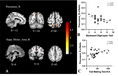 |
Dynamic Alterations of Spontaneous Neural Activity and Its
Association with Neurocognitive Function in Sport-related
Concussion
Wenjing Huang1,
Laiyang Ma1,
Wanjun Hu1,
Yu Zheng1,
Yuhui Xiong2,
and Jing Zhang1
1Lanzhou University Second Hospital, Lanzhou, China, 2GE Healthcare, Beijing, China Keywords: Traumatic brain injury, fMRI (resting state), sports-related concussion Sports-related concussion (SRC) is a complex and heterogeneous injury with psychological, cognitive, and functional consequences. Cognitive impairment is common following SRC, but the neural basis is unclear. In this study, the regional homogeneity (ReHo) and dynamic ReHo (d-ReHo) were obtained from rs-fMRI of both 31 SRC athletes and 7 non-SRC (NSRC) athletes. We found increased ReHo and d-ReHo in the SRC group, and the changes of d-ReHo in the right precuneus correlated with cognitive function. Our findings suggested that altered dynamics in the intrinsic brain activity might be a potential biomarker for explaining the cognitive function decline of SRC. |
||
5342.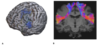 |
Exploring the Frontal Aslant Tract in Tourette syndrome: a
connectivity study
Domenico Aquino1,
Daniela Eldahaby2,
Emanuele La Corte3,
Vincenzo Levi3,
Elena Greco2,
Greta De Michelis1,
Riccardo Pascuzzo1,
Alessandro Gans2,
Malte Ottenhausen4,
Luigi Michele Romito5,
Maria Grazia Bruzzone1,
and Graziano Serrao2
1Neuroradiology Department, Fondazione IRCCS (Istituto di Ricovero e Cura a Carattere Scientifico) Istituto Neurologico Carlo Besta, Milan, Italy, 2Department of Health Sciences, Università degli Studi di Milano, Milan, Italy, 3Neurosurgery Department, Fondazione IRCCS (Istituto di Ricovero e Cura a Carattere Scientifico) Istituto Neurologico Carlo Besta, Milan, Italy, 4Department of Neurological Surgery, University Medical Center Mainz, Mainz, Germany, 5Parkinson's Disease and Movement Disorders Unit, Department of Clinical Neurosciences, Fondazione IRCCS (Istituto di Ricovero e Cura a Carattere Scientifico) Istituto Neurologico Carlo Besta, Milan, Italy Keywords: White Matter, Tractography & Fibre Modelling This study investigates for the first time microstructural alteration of the Frontal Aslant Tract (FAT) in nine subjects with TS compared to twelve healthy subjects. FAT was reconstructed trough Spherical Deconvolution (SD) and parameters derived from Diffusion Tensor Imaging (DTI) and Neuritis Orientation Dispersion and Density Imaging (NODDI) were extracted. We observed a Fractional Anisotropy (FA) reduction, indicating a greater disorganization of fibers, and accordingly an increased Orientation Dispersion Index (ODI), which indicates more dispersed fibers. We also highlighted a negative correlation between motor tic severity (quantified by the YGTSS) and neurite density (ND) and free water fraction (FISO). |
||
5343. |
The value of Synthetic MRI combined with histogram analysis in
predicting treatment response to chemoradiotherapy in
nasopharyngeal carcinoma
Fan Yang1,
Haoran Wei1,
Yujie Li1,
Xiaoduo Yu1,
Lizhi Xie2,
and Meng Lin1
1Department of Diagnostic Radiology, National Cancer Center/National Clinical Research Center for Cancer/Cancer Hospital, Chinese Academy of Medical Sciences and Peking Union Medical College, Beijing, 100021, China, Beijing, China, 2GE Healthcare, MR Research China, Beijing, Beijing, China Keywords: Head & Neck/ENT, MR Fingerprinting, Nasopharyngeal carcinoma; treatment response; Synthetic MRI Almost 80% nasopharyngeal carcinoma patients are diagnosed with locoregionally-advanced stage at the time of initial diagnosis. Early detection of treatment response is important for adjusting therapy regimens. SyMRI could not only generate multi-contrast images (including T1WI, T2WI and PDWI) in a single scan, but also generate quantitative T1, T2 and PD maps. Our study demonstrated that SyMRI combined with histogram analysis could non-invasive predict early treatment response. The predict performance of combined SyMRI and clinical factors was significantly higher than clinical only or TNM stage only. |
||
5344.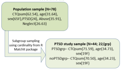 |
The association between adverse childhood experience, PTSD
diagnosis and White matter integrity.
Richard Okyere Nkrumah1,
Claudius von Schröder2,
Traute Demirakca 1,
Christian Schmahl2,
and Gabriele Ende1
1Department of Neuroimaging, Central Institute of Mental Health, Medical Faculty Mannheim, Heidelberg University, Mannheim, Germany, 2Clinic for Psychosomatics and Psychotherapeutic Medicine, Central Institute of Mental Health, Medical Faculty Mannheim, Heidelberg University, Mannheim, Germany Keywords: Psychiatric Disorders, Trauma, Adverse childhood experiences, PTSD, White matter integrity Adverse childhood experiences (ACE) are well-known risk factors for the development of PTSD later in life. The overall effect of ACE on WM integrity and the moderation effect of PTSD in this relation are still being researched. Using a total of 78 participants with any form of ACE, we show that ACE was negatively correlated with WM integrity in the genu and body of the corpus callosum and the bilateral uncinated fasciculus after controlling for overall psychological burden. The overall strength of this effect changed in corpus callosum and uncinated fasciculus when PTSD was added as a moderator variable. |
||
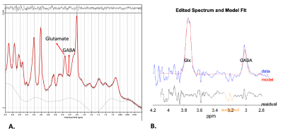 |
7-Tesla in-vivo 1H-magnetic resonance spectroscopy of glutamate
and GABA in 22q11.2 copy number variants.riants.
Chaira Serrarens1,
Desmond HY Tse2,
Esther Steijvers-Peeters2,
Kim Brouwers2,
David Linden1,
Claudia Vingerhoets1,
and Therese van Amelsvoort1
1Department of Psychiatry & Neuropsychology, School for Mental Health and Neuroscience, Maastricht University, Maastricht, Netherlands, 2Scannexus BV, Maastricht, Netherlands Keywords: Psychiatric Disorders, Spectroscopy, Genetic Diseases 22q11.2 copy number variants (22q11.2 CNVs) are associated with either an increased or a reduced risk of developing psychotic disorders and impaired cognitive functioning. Glutamatergic and GABAergic pathways are hypothesized to be disrupted in 22q11.2 CNV patients. Although a balance between glutamate and GABA is necessary for optimal brain functioning, to date, GABA has not been studied in 22q11.2 CNVs. Here, we investigated glutamate and GABA concentrations in the anterior cingulate cortex in patients with 22q11.2 CNVs using 7-Tesla 1H-MRS. Our results showed no significant differences in glutamate and GABA concentrations between 22q11.2 CNV patients and healthy controls. |
||
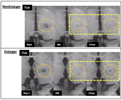 |
Differential lesion volume progression following MR-guided
focused ultrasound thalamotomy for Essential Tremor Tremor
Conrad P Rockel1,2,
Sarah Scott3,
Erin L Mazerolle4,
Samuel Pichardo1,2,
Davide Martino1,2,
Tejas Sankar5,
Zelma Kiss1,2,6,
and Bruce Pike1,2
1Radiology and Clinical Neurosciences, Cumming School of Medicine, University of Calgary, Calgary, AB, Canada, 2Hotchkiss Brain Institute, Cumming School of Medicine, University of Calgary, Calgary, AB, Canada, 3University of Calgary, Calgary, AB, Canada, 4Dept of Psychology, St Francis Xavier University, Antigonish, NS, Canada, 5Dept of Surgery, University of Alberta, Edmonton, AB, Canada, 6Dept of Surgery, University of Calgary, Calgary, AB, Canada Keywords: Neurodegeneration, Focused Ultrasound, Movement disorders; treatment Following MRgFUS thalamotomy for Essential Tremor, it was observed that lesions in some patients re-enlarged after the 3-month timepoint following surgery, based on T1-weighted MRI. After grouping patients as NonEnlargers and Enlargers based on this observation, we did not observe significant between-group differences in clinical measures of tremor beyond the 3-month timepoint. Patient demographic- and MRgFUS-related factors did not differ between groups. However, patients in the NonEnlarger group demonstrated significantly greater tremor severity prior to and in early timepoints following MRgFUS thalamotomy, suggesting that the relationship between tremor severity and longitudinal lesion progression may be worthy of further investigation. |
||
5347.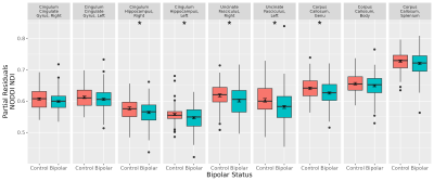 |
Reduced white matter axonal density in bipolar disorder using
Neurite Orientation Dispersion and Density Imaging (NODDI)Dispersion and Density Imaging (NODDI)
Gail I. S. Harmata1,
Hesam Abdolmotalleby1,
John E. Barsotti1,
Jess G. Fiedorowicz1,2,
Aislinn Williams1,
Gary Christensen1,
Jia Xu1,
Joseph J. Shaffer1,3,
Jeffrey D. Long1,
Jenny Gringer Richards1,
Leela Sathyaputri1,
Samantha L. Schmitz1,4,
John A. Wemmie1,
Vincent A. Magnotta1,
and Merry Mani1
1University of Iowa, Iowa City, IA, United States, 2University of Ottawa, Ottawa, ON, Canada, 3Kansas City University, Kansas City, MO, United States, 4Des Moines University, Des Moines, IA, United States Keywords: Psychiatric Disorders, White Matter, bipolar disorder Bipolar disorder is a serious psychiatric condition whose cause remains unknown. Previous diffusion MRI studies suggest white matter alterations may be involved, but standard diffusion tensor scalars provide limited information regarding potential pathophysiology. Here we used Neurite Orientation Dispersion and Density Imaging (NODDI) to examine select white matter bundles to provide additional information regarding changes in microstructure. We found that the uncinate fasciculus, cingulum hippocampus, and corpus callosum genu showed evidence of reduced axonal density, suggesting axonal loss or altered neurodevelopment. Additional work is necessary to determine how this pattern changes over time, and how it relates to mood lability. |
||
| 5348. |
Long-term cannabis use and brain structure: an MRI study of a
New Zealand longitudinal birth cohort
Rebecca M. Lee1,2,
James A. Foulds3,
Reza Shoorangiz2,
Mustafa M. Almuqbel4,
Campbell Le Heron1,2,5,
Lana Cleland3,
Ross J. Keenan4,
Roger Mulder3,
Richard J. Porter3,
Giles Newton-Howes6,
Katie M. Douglas3,
Anthony P. H. Butler7,
Joseph M. Boden3,
and Tracy R. Melzer1,2
1Department of Medicine, University of Otago, Christchurch, New Zealand, 2New Zealand Brain Research Institute, Christchurch, New Zealand, 3Department of Psychological Medicine, University of Otago, Christchurch, New Zealand, 4Pacific Radiology Group, Christchurch, New Zealand, 5Department of Neurology, Christchurch Hospital, Christchurch, New Zealand, 6Department of Psychological Medicine, University of Otago, Wellington, New Zealand, 7Department of Radiology, University of Otago, Christchurch, New Zealand Keywords: Gray Matter, Brain, Cannabis This study investigated whether cannabis use during adolescent and early adulthood was associated with long-term brain differences into middle-age, using structural T1-weighted, ASL, and diffusion MRI. Compared to non-using controls, users exhibited significantly less grey matter volume in the hippocampus and amygdala (p < 0.05). We observed no significant between-group differences in cerebral blood flow or white matter integrity. While cannabis may relate to long-term brain changes, prospective longitudinal MRI studies may help to elucidate causality. |
||
5349. |
Blood-brain barrier damage and new-onset refractory status
epilepticus: an explorative study using dynamic
contrast-enhanced MRI
Huiping Li1,
Xian Liu2,
Ruihong Wang1,
Aili Lu1,
Zhaohui Ma1,
Shibiao Wu1,
Hongji Lu1,
Yaming Du3,
Kan Deng4,
Lixin Wang1,
and Fang Yuan1
1Department of Neurocritical Care, The Second Affiliated Hospital of Guangzhou University of Chinese Medicine, Guangzhou, China, 2Department of Imaging, The Second Affiliated Hospital of Guangzhou University of Chinese Medicine, Guangzhou, China, 3Hubei University of Chinese Medicine, Wuhan, China, 4Philips Healthcare, Guangzhou, China Keywords: Epilepsy, DSC & DCE Perfusion, new-onset refractory status epilepticus; status epilepticus This is the first study investigating blood-brain barrier (BBB) dysfunction in new-onset refractory status epilepticus (NORSE) using dynamic contrast-enhanced MRI (DCE-MRI). BBBs of NORSE patients were impaired diffusely, and they had significantly higher BBB permeability in the basal ganglia and thalamus compared to encephalitis patients without status epilepticus (SE). Our preliminary findings demonstrate that BBB dysfunction in the basal ganglia and thalamus plays an important role in the pathophysiology of NORSE. |
||
5350.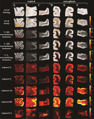 |
Mapping astrogliosis in the individual human brain using
multidimensional MRI
Dan Benjamini1,
David S Priemer2,
Daniel P Perl2,
David L Brody2,
and Peter J Basser3
1National Institute on Aging, Baltimore, MD, United States, 2Uniformed Services University of the Health Sciences, Bethesda, MD, United States, 3National Institute of Child Health and Human Development, Bethesda, MD, United States Keywords: Traumatic brain injury, Microstructure There are currently no noninvasive imaging methods available for astrogliosis mapping in the brain despite its essential role in the response to many disease states. In an ex vivo human brain study we used diffusion-relaxation MRI to derive a signature of astrogliosis and disentangle it from normative brain at the individual level using machine learning. We developed a within-subject anomaly detection procedure that generates MRI-based astrogliosis maps ex vivo, which were significantly and strongly correlated with co-registered histology. Our findings demonstrated spatial sensitivity and specificity in detecting reactive astrocytes, and could significantly impact the studying of injury, disease, and aging. |
||
 Back to Meeting Home
 Back to the Program-at-a-Glance
The International Society for Magnetic Resonance in Medicine is accredited by the Accreditation Council for Continuing Medical Education to provide continuing medical education for physicians.uing medical education for physicians.
 Back to Meeting Home
 Back to the Program-at-a-Glance
 View Presentation Video