Traditional Poster
Pediatrics
ISMRM & ISMRT Annual Meeting & Exhibition • 03-08 June 2023 • Toronto, ON, Canada

| Booth # | |||
|---|---|---|---|
5289.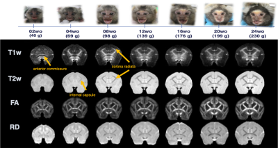 |
Heterogeneous regional growth process and speed in marmoset
brain: multi-modal longitudinal MRI study
Akiko Uematsu1,
Makoto Fukushima1,
Junich Hata2,
Ayako Murayama1,
Noriyuki Kishi3,
Takuya Hayashi1,
and Hideyuki Okano3,4
1RIKEN, Hyogo, Japan, 2Tokyo Metropolitan University, Tokyo, Japan, 3RIKEN, Saitama, Japan, 4Keio University School of Medicine, Tokyo, Japan Keywords: Normal development, Animals “The soul of a child of three is the same at 100” may be also true among all the primates. Here, we delineate the robust value changes of T1w, T2w, and DTI metrics for the first 2 to 3 months after birth in common marmoset brain. Different temporal changes across brain regions were found by Fixel-based analysis, suggesting that different neuronal contributors of age-related structural changes. Integrating multi-modal MRI images provided more detailed insight of tissue property changes especially through development as compared with a single-modal image analysis. |
||
5290.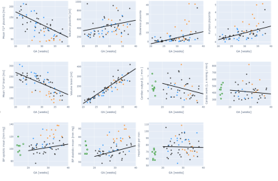 |
Characterisation of pregnancies affected by pre-eclampsia using
MRI
Megan Hall1,2,
Kathleen Colford1,
Daniel Cromb1,
Priya Jandu3,
Alison Ho1,2,
Anthony Price1,
Lucy Chappell2,
Mary Rutherford1,4,
Lisa Story1,2,
Pablo Lamata4,
and Jana Hutter1,4
1Centre for the Developing Brain, King's College London, London, United Kingdom, 2Department of Women and Children's Health, King's College London, London, United Kingdom, 3GKT School of Medicine, King's College London, London, United Kingdom, 4Centre for Medical Engineering, King's College London, London, United Kingdom Keywords: Fetal, Fetus, Placenta; hypertension; clinical translation Maternal cardiac, placental and fetal brain anatomical and functional MRI was obtained in 63 pregnancies, including 12 with pre-eclampsia. In pregnancies affected by pre-eclampsia, placental T2* was significantly reduced compared to normal pregnancy. All women with placental histopathological anomalies consistent with pre-eclampsia showed a reduced placental T2*. Fetal brain T2* was significantly lower in women with pre-eclampsia than those without, although brain volume was maintained. In women with pre-eclampsia, reduced fetal brain T2* was associated with middle cerebral artery Doppler abnormalities. Cardiac work was increased in women with pre-eclampsia. |
||
5291.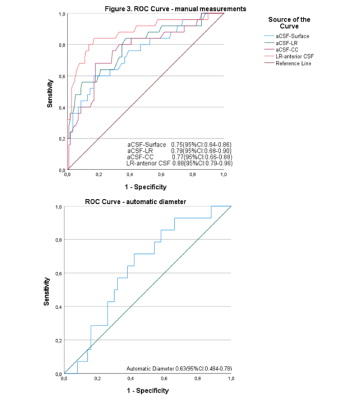 |
Optic nerve thickening on high-resolution MRI predicts
early-stage postlaminar optic nerve invasion of retinoblastoma.
Christiaan Marius de Bloeme1,
Sabien van Elst1,
Sophia Göricke2,
Pim de Graaf1,
and Marcus Christiaan de Jong1
1Radiology, Amsterdam UMC, Amsterdam, Netherlands, 2Diagnostic and Interventional Radiology and Neuroradiology, University Hospital Essen, Essen, Germany Keywords: Tumors, Tumor, optic nerve invasion; quantitative; pediatric; prediction; metastases; Two radiologists with different levels of experience measured the anterior optic nerve to predict postlaminar optic nerve invasion (PLONI) in retinoblastoma. In addition, quantitative measurements were performed using a deep-learning method 3D U-net. The results showed that measurements performed by the radiologists had an AUC of 0.88 for the detection of PLONI, while the preliminary results of the quantitative approach had an AUC of 0.63. Both methods show promising results to predict early-stage PLONI in retinoblastoma patients. |
||
5292.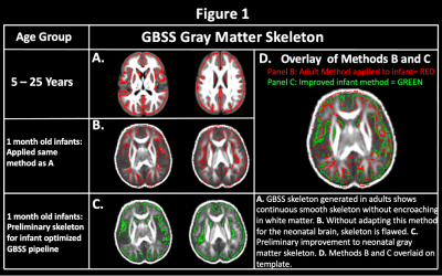 |
Adapting the NODDI-GBSS Framework for the 1-Month Infant Brain
Marissa DiPiero1,2,
Patrik Goncalves Rodrigues2,
Hassan Cordash2,
Jose Guerrero Gonzalez2,
Richard J. Davidson3,4,5,
Andrew Alexander2,3,6,
and Douglas C. Dean III2,6,7
1Neuroscience Training Program, University of Wisconsin-Madison, Madison, WI, United States, 2Waisman Center, University of Wisconsin-Madison, Madison, WI, United States, 3Department of Psychiatry, University of Wisconsin-Madison, Madison, WI, United States, 4Department of Psychology, University of Wisconsin-Madison, Madison, WI, United States, 5Center for Healthy Minds, University of Wisconsin-Madison, Madison, WI, United States, 6Department of Medical Physics, University of Wisconsin-Madison, Madison, WI, United States, 7Department of Pediatrics, University of Wisconsin-Madison, Madison, WI, United States Keywords: Neuro, Gray Matter The brain’s cytoarchitecture undergoes highly dynamic morphological changes during early life that establish the brain’s structural and functional framework and lays the foundation for future cognitive and behavioral skills. While differences in early cortical organization are thought to subserve future behavioral and psychiatric challenges, little is known regarding cortical organization in early life. For the first time, we adapt the NODDI-GBSS framework for the infant brain while keeping the data in its native diffusion space. We show feasibility for cortical skeletonization and show relationships with age across the GM microstructure within the first month of life. |
||
5293.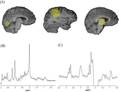 |
Glutamate in the visual cortex changes within the migraine cycle
in children and adolescents
Lydia Y Cho1,2,3,
Tiffany K Bell1,2,3,
Kate J Godfrey1,2,3,
Andrew D Hershey4,5,
Jonathan Kuziek2,3,6,
Mehak Stokoe1,2,3,
Kayla Millar1,2,3,
Serena L Orr2,3,6,
and Ashley D Harris1,2,3
1Department of Radiology, University of Calgary, Calgary, AB, Canada, 2Hotchkiss Brain Institute, University of Calgary, Calgary, AB, Canada, 3Alberta Children's Hospital Research Institute, University of Calgary, Calgary, AB, Canada, 4Division of Neurology, Cincinnati Children's Hospital Medical Center, Cincinnati, OH, United States, 5Department of Pediatrics, University of Cincinnati School of Medicine, Cincinnati, OH, United States, 6Departments of Pediatrics, Community Health Sciences, and Clinical Neurosciences, University of Calgary, Calgary, AB, Canada Keywords: Neuro, Spectroscopy, Migraine Migraine is a common neurological disorder in pediatrics, yet the pathophysiology remains unclear. Migraine is characterized by episodic attacks, suggesting a shifting excitation-inhibition imbalance that when tipped, will trigger an attack. We used magnetic resonance spectroscopy to measure glutamate and gamma-aminobutyric acid (GABA) concentrations to examine changes in these brain metabolites across different phases of the migraine attack in children and adolescents. We show fluctuations in glutamate with migraine cycle phases in the visual cortex. There were no observed changes in glutamate nor GABA in the sensorimotor cortex and the thalamus. |
||
5294.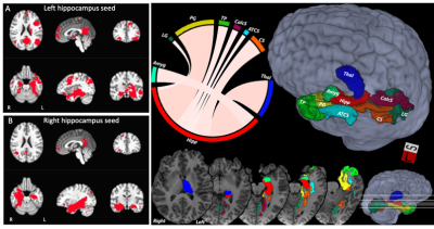 |
Memory and learning relationships with hippocampal morphometry
and multi-modal connectivity after pediatric severe TBI
Jose Guerrero-Gonzalez1,
Gregory Kirk2,
Rasmus Birn1,
Erin Bigler3,
Katherine Bowen4,
Aimee Broman5,
Bedda Rosario6,
Warwick Butt7,
Sue Beers8,
Michael Bell9,
Peter Ferrazzano10,
and Andrew Alexander1
1Medical Physics, University of Wisconsin - Madison, Madison, WI, United States, 2Waisman Center, University of Wisconsin - Madison, Madison, WI, United States, 3Department of Psychology and Neuroscience Center, Brigham Young University, Provo, UT, United States, 4Seattle Children’s Hospital, Seattle, WA, United States, 5Biostatistics, University of Wisconsin - Madison, Madison, WI, United States, 6Epidemiology, University of Pittsburgh, Pittsburgh, PA, United States, 7Critical Care, Faculty of Medicine, Melbourne University, Melbourne, Australia, 8Psychiatry, University of Pittsburgh, Pittsburgh, PA, United States, 9Pediatrics, Children’s National Medical Center, Washington, DC, United States, 10Pediatrics, University of Wisconsin - Madison, Madison, WI, United States Keywords: Neuro, Traumatic brain injury Traumatic brain injury in adolescents is a major public health concern, leading to tens of thousands of hospitalizations in the U.S. each year. Our study aims to identify multimodality imaging biomarkers of long-term neurocognitive outcome after severe adolescent TBI. For this investigation we explored memory performance using the California Verbal Learning Test (CVLT) in relation to structural and functional connectivity of the memory network, as well as hippocampal volume and fornix microstructure. |
||
5295.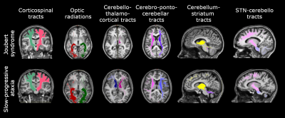 |
Whole-brain connectivity in patients with Pediatric Cerebellar
Ataxia
Silvia Maria Marchese1,
Fulvia Palesi2,
Mariagrazia Bruzzone3,
Anna Nigri3,
Chiara Maria Pantaleoni4,
Claudia AM Gandini Wheeler-Kingshott2,5,6,
Stefano D'Arrigo4,
Egidio D'Angelo2,6,
and Paolo Cavallari1
1Human Physiology Section of the DePT, Università degli Studi, Milano, Italy, 2Department of Brain and Behavioral Science, Università degli Studi, Pavia, Italy, 3Neuroradiology Department, Fondazione IRCCS Istituto Neurologico "C. Besta", Milano, Italy, 4Developmental Neurology Department, Fondazione IRCCS Istituto Neurologico "C. Besta", Milano, Italy, 5Department of Neuroinflammation, UCL Queen Square Institute of Neurology, Faculty of Brain Sciences, University College London, NMR Research Unit, Queen Square MS Centre, London, United Kingdom, 6Brain Connectivity Center, IRCCS Mondino Foundation, Pavia, Italy Keywords: Neuro, Brain Connectivity, Ataxia For the first time, brain networks of pediatric cerebellar ataxic patients were characterized in order to explain the different postural motor behavior of subjects suffering from non-progressive and slow-progressive illness. This work revealed volume differences in several cerebellar regions and specific alterations of white matter tracts. This result reinforces the hypothesis of the existence of a compensatory strategy which may involve cortical areas and basal ganglia to compensate for cerebellar deficits. |
||
5296.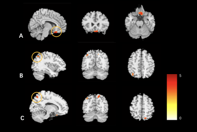 |
Structural and Functional Brain Alterations in Children with
Autism Spectrum Disorder
Di Zhou1,
Ting Hua1,
Xiance Zhao2,
and Guangyu Tang1
1Department of Radiology, Shanghai Tenth People’s Hospital, Tongji University School of Medicine, Shanghai, China, 2Philips Healthcare, Shanghai, China Keywords: Neuro, fMRI (resting state) MRI is an important tool available to detect brain functional and structural abnormalities of autism spectrum disorder (ASD) patients. In this study, we found that children with ASD have increased changes in structure and both increased and decreased changes in function in the brain, which suggests that there may be some underlying pathogenic mechanism in the brain of ASD. With the combination of brain structural and functional analysis can provide a new imaging perspective for understanding the neural mechanism of ASD. |
||
5297.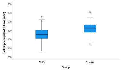 |
Hippocampal subfield alterations and working memory in children
with congenital heart disease
Ruth O'Gorman Tuura1,
Melanie Ehrler2,
Nadja Naef2,
Alenka Schmid2,
Felicitas Koch2,
Fraser Callaghan1,
Oliver Kretschmar3,
and Beatrice Latal2
1University Children's Hospital Zurich, Zurich, Switzerland, 2Child Development Center, University Children's Hospital Zurich, Zurich, Switzerland, 3Cardiology, University Children's Hospital Zurich, Zurich, Switzerland Keywords: Neuro, Segmentation Reduced hippocampal volumes are associated with working memory impairments in children with congenital heart disease (CHD). However, the volumes of individual hippocampal subfields are largely unexplored in the context of different working memory domains in CHD. In 57 children with complex CHD and 82 age-matched control children, the CHD group showed smaller hippocampal subvolumes, particularly for the hippocampal tail, and poorer verbal and spatial working memory scores, which were differentially related to the hippocampal subvolumes. In addition, hippocampal volumes in cyanotic CHD were significantly smaller than in acyanotic CHD, supporting the link between hypoxia, hippocampal damage, and memory impairment. |
||
5298.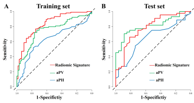 |
Noninvasive MRI Radiomics Features of adenohypophysis in
evaluation of HPG axis activation in children.
Dong Liu1,
Wenzhi Lv2,
Weiyin Vivian Liu3,
and Wenzhen Zhu1
1Department of Radiology, Tongji Hosptial ofTongji Medical College of Huazhong University of Science and Technology, Wuhan, China, 2Department of Artificial Intelligence,, Julei Technology Company, Wuhan 430000, Hubei, China, Wuhan, China, 3MR Research, GE Healthcare, Beijing 100176, China, Beijing, China Keywords: Adolescents, Pediatric, hypothalamic-pituitary-gonadal axis we report for a low-cost and rapid radiomics model based on MRI data in evaluation of hypothalamic-pituitary-gonadal (HPG) axis activation in children. In our study, radiomics model base on CUBE T1WI showed good prediction of HPG axis activation with the AUC of 0.84 in the training set and 0.81 in the test set. The AUC of the radiomics model was higher than that of aPV and aPH in the training set. In results of DCA analysis, radiomics signature showed higher net benefit than aPV and aPH models.The MRI radiomics model may serve as a noninvasive predictor of HPG axis activation. |
||
5299.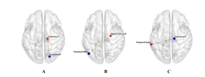 |
Abnormal spontaneous brain activity and functional connectivity
in children with new-onset type 1 diabetes mellitus
Kun Liu1,
Xiao-Yan Huang1,
Lu Han2,
and Zhi-Han Yan1
1Radiology, The Second Affiliated Hospital and Yuying Children’s Hospital of Wenzhou Medical University, Wenzhou, China, 2Philips Healthcare, Shanghai, China Keywords: Neuro, Diabetes, children, cognition, fMRI It is unclear whether brain activity and functional connectivity (FC) will change in type 1 diabetes mellitus (T1DM) children. We investigated brain activity and FC changes in new-onset T1DM children based on fractional amplitude of low-frequency fluctuation (fALFF) and seed-based FC analysis. We found that children with new-onset T1DM showed changed fALFF values and FCs in several brain regions. Changed fALFF values were correlated with intelligence quotient (IQ) and blood glucose level. These results suggest that altered brain spontaneous activity and FC in initial-stage T1DM patients may be the potential mechanisms of subsequent visual impairment and cognitive dysfunction. |
||
5300.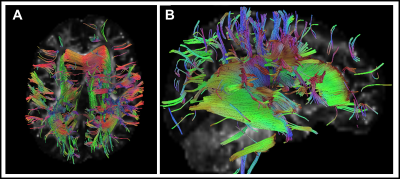 |
Longitudinal findings from differential tractography of patients
with type II GM1 gangliosidosis
Zeynep Vardar1,
Anna Kuhn1,
Jean M. Johnston2,
Precilla D'Souza2,
Maria T. Acosta2,
Cynthia J. Tifft2,
and Mohammed Salman Shazeeb1
1University of Massachusetts Chan Medical School, Worcester, MA, United States, 2National Institutes of Health, Bethesda, MD, United States Keywords: Rare disease, Rare disease GM1-gangliosidosis is a rare heritable lysosomal storage disorder caused by accumulation of GM1-ganglioside due to deficiency of the lysosomal enzyme b-galactosidase required for sphingolipid degradation. Progressive accumulation of GM1-ganglioside in the central nervous system induces hypomyelination that results in progressive neurodegeneration. This study used differential tractography in 11 type II GM1 patients to assess longitudinal white matter tract changes using fractional anisotropy (FA) in different regions of the brain in late-infantile and juvenile patients. FA decrease was observed predominantly in the corpus callosum, superior longitudinal fasciculus, and cingulum in supratentorial white matter structures, demonstrating the utility of differential tractography. |
||
5301.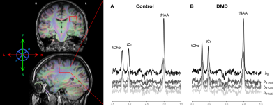 |
Probing the diffusion of water and intracellular metabolites to
assess white matter microstructure in Duchenne muscular
dystrophy
Rosanne Govaarts1,
Nathalie Doorenweerd1,2,3,
Emma M Broek1,
Maud E Tamsma1,
Itamar Ronen1,
Chloé F Najac1,
Kieren Hollingsworth3,
Erik H Niks1,
Volker Straub2,3,
and Hermien Kan1
1Leiden University Medical Center, Leiden, Netherlands, 2Newcastle Hospitals NHS Foundation Trust, Newcastle, United Kingdom, 3Newcastle University, Newcastle, United Kingdom Keywords: Neuro, Spectroscopy, Duchenne muscular dystrophy Besides motor impairment, Duchenne muscular dystrophy (DMD) patients experience cognitive and behavioural symptoms. Altered white matter microstructure has been shown with diffusion tensor imaging (DTI) in this population. Here, we combined single volume 1H diffusion-weighted spectroscopy (DWS) and DTI to disentangle intra- and extracellular contributions. Mean apparent diffusion coefficients of N-acetyl aspartate, choline, and creatine were comparable between DMD patients and healthy controls. In the same volume, DMD patients showed increased mean water diffusivity. This suggests that altered white matter microstructure is likely due to extracellular, rather than intracellular, changes. |
||
5302.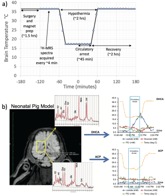 |
The Circulatory Arrest Recovery Ammonia Problem (CARAP)
Hypothesis
Daniel Spielman1,
Meng Gu2,
Hunter Liu1,
Shie-Chau Liu2,
Ralph Hurd2,
Kirk Riemer3,
Kenichi Okamura3,
and Frank Hanley3
1Stanford University, Stanford, CA, United States, 2Radiology, Stanford University, Stanford, CA, United States, 3Cardiothoracic Surgery, Stanford University, Stanford, CA, United States Keywords: Neonatal, Spectroscopy, brain, cardiopulmonary bypass, neuronal injury, surgery Brain injury remains an ongoing concern for patients requiring cardiopulmonary bypass (CPB) surgery. Observations in a neonatal pig model of build-up of blood ammonia levels may be a significant unrecognized source of metabolic stress in these surgeries. Ammonia entering the brain upon restarting the CPB pump appears highly correlated with the level of hypothermia and glutamine/glutamate appears highly correlated with brain lactate levels. These changes are also strongly dependent on the choice of surgical parameters such the use of deep hypothermia cardiac arrest (whereby all blood flow is stopped) versus antegrade cerebral perfusion (whereby brain blood flow is maintained). |
||
5303. |
Functional Brain Alterations in Children with Autism Spectrum
Disorder after Fecal Microbiota Transplantation
Di Zhou1,
Ting Hua1,
Xiance Zhao2,
and Guangyu Tang1
1Department of Radiology, Shanghai Tenth People’s Hospital, Tongji University School of Medicine, Shanghai, China, 2Philips Healthcare, Shanghai, China Keywords: Neuro, fMRI (resting state) MRI is an important tool available to detect brain functional and structural abnormalities of autism spectrum disorder (ASD) patients. In this study, we combined fecal microbiota transplantation (FMT) and neuroimages to follow-up and explore the change in functional activity in children with ASD. The combination of FMT and MRI can provide a new imaging perspective for understanding the neural mechanism of gut microbiota in ASD. |
||
5304.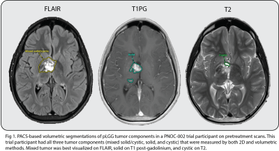 |
Comparing Volumetric to 2D Methods in Assessing Post-treatment
Response of Different Pediatric Low-Grade Glioma (pLGG) Tumor
Components
Divya Ramakrishnan1,
Marc von Reppert2,
Sarah C Bruningk3,
Fatima Memon1,
Mark Krycia1,
Matthew Sala1,
Ryan Bahar1,
Anahita Fathi Kazerooni4,
Ali Nabavizadeh4,
Annette Molinaro5,
Daphne Haas-Kogan6,
Theodore Nicolaides7,
Sabine Mueller5,
Michael Prados5,
and Mariam Aboian1
1Department of Radiology, Yale School of Medicine, New Haven, CT, United States, 2University of Leipzig, Leipzig, Germany, 3ETH Zurich, Zurich, Switzerland, 4Department of Radiology, University of Pennsylvania, Philadelphia, PA, United States, 5Department of Neurology, Neurosurgery, and Pediatrics, UCSF, San Francisco, CA, United States, 6Harvard Medical School, Boston, MA, United States, 7NYU Langone Medical Center, New York, NY, United States Keywords: Neuro, Tumor, Volumetrics Accurate response assessment is critical in pediatric low-grade glioma (pLGG) as there is significant morbidity associated with continued treatment. Current standards for assessment rely on 2D measurements. Given the heterogenous nature of pLGG, volumetric assessment of treatment response may prove more effective. In this study, we compared 2D and volumetric methods of assessing treatment response in patients from the PNOC-002 clinical trial of pLGG. We analyzed the quantitative differences between 2D and volumetric measurements of tumor volume percent change post-treatment. We also compared classification of treatment response based on application of RANO criteria to various pLGG tumor components. |
||
5305.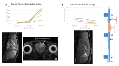 |
Changes in Cellular and Vascular Phenotype in Pediatric
Ependymoma Models by Multi-Parametric MRI: Effects of Tumor Size
and Radiation Treatment
Jane Manalo1,
Andrea M Griesinger2,
Jenna Steiner1,
Angela Pierce2,
Nicholas K Foreman3,
and Natalie Julie Serkova4
1Radiology, University of Colorado Anschutz, Aurora, CO, United States, 2Pediatric Neurooncology, University of Colorado Anschutz, Aurora, CO, United States, 3Pediatric Hematology Oncology, The Children's Hospital of Colorado, Aurora, CO, United States, 4Radiology, University of Colorado Anschutz, Denver, CO, United States Keywords: Cancer, Animals, cell size imaging At 2022 ISMRM, we presented on simulation results of the selective size imaging using filters via diffusion times (SSIFT) in perfused irradiated cells and flank xenograft models. Here, using SSIFT and iron-oxide vessel-size imaging, we report on ependymoma (EPN) phenotypes, comparing small and large intracranial lesions: variable cell sizes fitted into SSIFT iAUC (S=14 microns small EPN, S=19 medium and S=12 large), increased vessel density index (Q=0.54 small, Q=0.62 large EPN), and low ADC (0.63x10⁻³ small, 0.58x10⁻³ mm2/s large EPN). Chemo-radiation treatment led to decreased gross tumor volumes, necrosis with decreased cell sizes and increased ADCs. |
||
5306.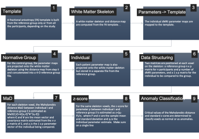 |
Univariate and multivariate individual-specific tract-based
spatial statistics (iTBSS) for white matter microstructure
anomaly detection
Jose Guerrero-Gonzalez1,
Jothi Venkatesh2,
Peter Ferrazzano3,
and Andrew Alexander1
1Medical Physics, University of Wisconsin - Madison, Madison, WI, United States, 2Department of Physics, The Clatterbridge Cancer Centre NHS Foundation Trust, University of Liverpool, Liverpool, United Kingdom, 3Pediatrics, University of Wisconsin - Madison, Madison, WI, United States Keywords: Neuro, Traumatic brain injury, Precision Medicine Tract-based spatial statistics (TBSS) is a voxel-based analysis (VBA) method for diffusion MRI (dMRI) data along a skelentonized representation of the brain white matter. When applied to group-level analyses of dMRI data, TBSS has been shown to improve sensitivity, objectivity, and interpretability. In this work, we introduce individual-specific tract-based spatial statistics for anomaly detection in white matter micro-structure. Results from implementation on severe pediatric TBI brains reveal heterogenous patterns of atypical white matter. |
||
5307.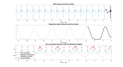 |
Free breathing, multi-shot real-time imaging using adaptive
k-space sampling (ARKS) for iCMR
Nuri Chung1,
Ana Rodriguez-Soto2,
Sanjeet Hegde3,
Eleanor L. Schuchardt3,
Brent M. Gordon3,
Ileen Cronin3,
and Francisco Contijoch1,2
1Bioengineering, University of California San Diego, La Jolla, CA, United States, 2Radiology, University of California San Diego, La Jolla, CA, United States, 3Pediatrics, Rady Children's Hospital-San Diego, Heart Institute, San Diego, CA, United States Keywords: Cardiovascular, Heart Adaptive k-space sampling (ARKS) enables real-time multi-shot imaging by quickly finding data acquired during similar cardiac phases1. In this study, we used pediatric physiologic signals to simulate ARKS and extended the approach to incorporate respiratory gating and enable free breathing acquisition. Respiratory gating was performed by cross-correlating the recent respiratory signal with previously acquired values. A correlation cutoff was used to select similar periods. As expected, higher correlation cutoffs led to lower acceptance rates. For a search period of 32 beats, respiratory gating with a correlation cutoff of 0.5 identified 6 heartbeats for multi-beat imaging. |
||
5308.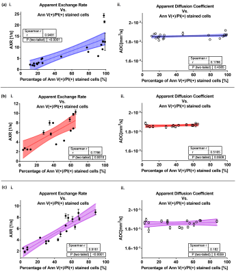 |
Monitoring in vitro and in vivo cell death using AXR, ADC and
hyperpolarized [1,4-13C2]fumarate
Athanasia Kaika1,
Luca Nagel1,
Ulrike Höckendorf2,
Geoffrey J. Topping1,
Irina Beer3,
Frits H. A. van Heijster1,
Philipp J. Jost4,5,
Natalia P. Ivleva3,
and Franz Schilling1
1School of Medicine, Klinikum rechts der Isar, Department of Nuclear Medicine, Technical University of Munich (TUM), Munich, Germany, 2School of Medicine, Klinikum rechts der Isar, Department of Medicine III, Technical University of Munich (TUM), Munich, Germany, 3Chair of Analytical Chemistry and Water Chemistry, Institute of Water Chemistry, Technical University of Munich (TUM), Garching, Germany, 4Chair of Clinical Division of Oncology Department of Internal Medicine, Medical University of Graz, Graz, Austria, 5Chair of University Palliative Care Unit, Medical University of Graz, Graz, Austria Keywords: Diffusion/other diffusion imaging techniques, Microstructure, FEXSY, FEXI, exchange, water transmembrane permeability, cell death Filter exchange spectroscopy (FEXSY) and imaging (FEXI) were used to measure apparent exchange rate (AXR) in acute myeloid leukemia cells undergoing apoptosis, necroptosis, or necrosis. Sensitivity of AXR to membrane permeabilization in vitro, while ADC was stable, were confirmed by Annexin V/PI staining and by scanning electron microscopy of microstructural leaks upon necrosis. AXR of murine EL4 lymphoma showed negative and positive correlation with ADC and malate/fumarate ratio, respectively. Tumor H&E histological analyses show clusters of diffuse necrosis in the solid tumor region, which likely contribute to high AXR and MFR variation while ADC is still low. |
||
5309.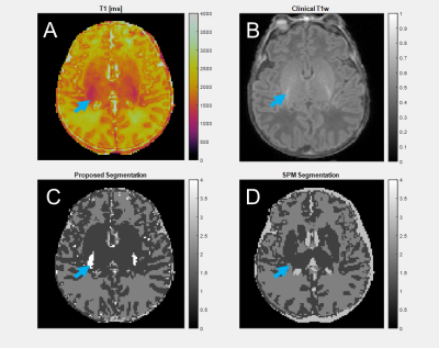 |
Age-Agnostic, Unsupervised Segmentation of Infant Brains using
Magnetic Resonance Fingerprinting
Richard James Adams1,
Pew-Thian Yap2,
and Dan Ma1
1Biomedical Engineering, Case Western Reserve University, Cleveland, OH, United States, 2Radiology, University of North Carolina, Chapel Hill, NC, United States Keywords: Neuro, Segmentation, Atlas-Free Brain segmentation is challenging in infants, as rapid changes to tissue properties and shapes during developmental growth make atlas-based modeling difficult. We use MRF-derived image features and density-based clustering to segment 2D brain slices from subjects without assumptions about subject age, brain shape, or image intensity. Segmentations from the proposed method closely match SPM without needing different atlases for subjects of varying ages. With flexible assumptions about the number of tissues present in an image, the proposed method identifies additional tissues such as CSF partial volume voxels and neonatal myelination that are not segmented by atlas-based approaches. |
||
5310.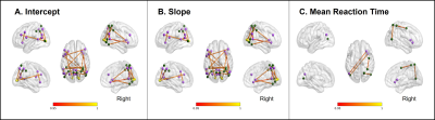 |
Attention network dysconnectivity and impaired visual search in
individuals with early developmental brain injury
Marie Drottar1,
Claire E Manley2,
Lotfi B Merabet2,3,
and Corinna Bauer1,3
1Lab for Neuroimaging and Vision Science, Gordon Center for Medical Imaging at Mass General Hospital, Boston, MA, United States, 2Schepens Eye Research Institute,Massachusetts Eye and Ear Infirmary, Boston, MA, United States, 3Department of Ophthalmology, Harvard Medical School, Boston, MA, United States Keywords: Neuro, Brain Connectivity, multimodal This study investigated the relationship between structural and functional disconnectivity of the dorsal and ventral attention networks and performance on a visual conjunction search task in individuals with early developmental brain injury (EDBI). Individuals with EDBI performed significantly worse than controls on the conjunction search task, as indicated by increased mean search time, higher y-intercept, and increased search time as a function of task difficulty (i.e. steeper slope). Each of these behavioural outcomes was positively correlated with connectivity measures in the dorsal and ventral attention networks, suggesting that aberrant functional and structural connectivity may underlie the observed visual search impairments. |
||
5311.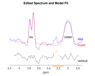 |
Changes in excitation/inhibition balance in infants with brain
injury evaluated by MEGA-edited MRS
Hui LIhong1,
Lin Liangjie2,
Cui Linfei2,
Yan Chenyu1,
Wei Wenjin1,
Zhang Yong1,
and Meng Yun1
1The First Affiliated Hospital of Zhengzhou University, Zhengzhou, China, 2Clinical and Technical Support, Philips Healthcare, Beijing, China Keywords: Prenatal, Pediatric In this study, Infants with a history of encephalopathy at born and normal infants were performed the advanced J-edited 1H MR spectroscopy technique (MEGA-PRESS) for observing the changing of excitation/inhibition (E/I) balance of the infants. The results showed significantly difference in Glx [Cr] concentrations between the two groups, and loss of excitation/inhibition (E/I) balance in infants with a history of encephalopathy. The changing of excitation/inhibition (E/I) balance can partial explain the underlying metabolic mechanism. |
||
5312.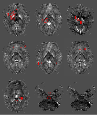 |
QSM Based Decreased Iron Deposition Correlates with Reduced
Neurodevelopmental Status in Children with Autism Spectrum
Disorder
Lei Du1,
Fang Ye2,
and Bing Liu1
1Radiology, China-Japan Friendship Hospital, Beijing, China, 2Pediatrics, China-Japan Friendship Hospital, Beijing, China Keywords: Normal development, Neuro, autism spectrum disorder, Gesell Developmental Schedules, Autism Behavior Checklist, iron, child To investigate potential correlations between the susceptibility values of certain brain regions and the severity of disease or neurodevelopmental status in children with autism spectrum disorder (ASD). 18 ASD children and 15 healthy controls (HC) were recruited. 11 brain regions as regions of interest. Pearson and Spearman partial correlation analysis was used to depict the correlations between the susceptibility values, the ABC scores, and the GDS scores in ASD group. We found that the susceptibility value of the right globus pallidus was positively correlated with the GDS-fine motor scale score. |
||
| 5313. |
Myelin water and T1 mapping of lesions and normal appearing
white matter in young adults with neonatal brain injury
Marie Drottar1,
Yansong Zhao2,
and Corinna Bauer1
1Gordon Center for Medical Imaging, Department of Radiology, Massachusetts General Hospital, Boston, MA, United States, 2Philips Healthcare, Cambridge, MA, United States Keywords: Neuro, White Matter, relaxometry This study used myelin water fraction (MWF) and geometric mean T2 time of the intra- and extra-cellular water fraction (IET2) derived from a multi-spin echo sequence with compressed sensing (METRICS)2, as well as T1 mapping derived from MP1RAGE to evaluate the long-term changes in myelination and white matter integrity in youths with neonatal brain injury. We observed significant differences in T1 values and IET2 of the normal appearing white matter, but not in the white matter lesions in a population of young adults with neonatal brain injury compared to controls. |
||
5314.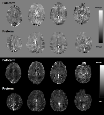 |
Comparison of brain quantitative susceptibility mapping between
preterm and full-term newborns
Shin-Eui Park1,
Eric J. Mallack1,
Yi Wang1,
Thanh D. Nguyen1,
and Zungho Zun1
1Weill Cornell Medicine, New York, NY, United States Keywords: Neonatal, Quantitative Susceptibility mapping Quantitative susceptibility mapping (QSM) is an emerging technique and may be utilized to assess neurodevelopmental disabilities characterized by insufficient iron content and myelination. In this study, a total of 23 full-term and 12 preterm newborns were studied using QSM. Compared to full-term, mean regional susceptibility of preterm newborns was significantly higher in the parietal white matter and was significantly lower in the frontal gray matter. This may be indicative of a regional deficit in iron deposition and myelination of the preterm newborn brain, and suggest that QSM may be used to identify early evidence of impaired neurodevelopment. |
||
5315.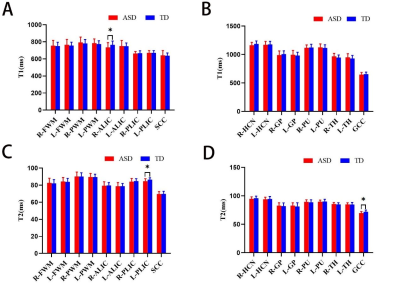 |
Quantitative relaxometry assessment of brain microstructural
abnormality of preschool children with autism spectrum disorder
by Synthetic MRI
Shuangyu Li1,
Xin Zhao1,
Lin Lu1,
Jinxia Guo2,
Qingna Xing1,
Yongbing Sun3,
Desheng Xuan1,
and Xiaoan Zhang1
1Department of Radiology, The Third Affiliated Hospital of Zhengzhou University, Zhengzhou, China, 2GE Healthcare, MR Research China, Beijing, China, 3Zhengzhou University People’s Hospital, Zhengzhou, China Keywords: Neuro, Neuroscience Quantitative T1/T2 relaxometry can be simultaneously generated from Synthetic MRI (SyMRI) in one single scanning and short time. This study aims to use SyMRI to evaluate the brain structural changes in preschool children with autism spectrum disorder (ASD), and the correlation between T1/T2 and ASD scores, as well as the diagnostic efficacy. We found that Synthetic T1 and T2 could be helpful for brain abnormity assessing, pathophysiological mechanism understanding and diagnosis of ASD. |
||
5316.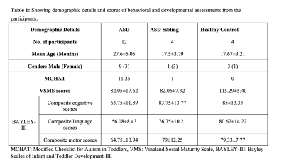 |
Altered cerebellar lobular volumes correlate with
neurodevelopment and cognitive scores in children with ASD and
ASD-siblings at an early age
Manoj Kumar1,
Chandrakanta Hiremath2,
Eshita Bansal2,
Sunil Kumar Khokhar2,
Kommu Johnvijay Sagar3,
Swetha Narayanan4,
Akhila S. Girimaji5,
BK Yamini5,
Rose Dawn Bharath2,
Jitender Saini2,
and M. Thamos Kishore4
1Neuroimaging and Interventional Radiology, National Institute of Mental Health and Neurosciences, Bangalore, India, 2Neuroimaging and Interventional Radiology, National Institute of Mental Health and Neurosciences, Bengaluru, India, 3Child and Adolescents Psychiatry, National Institute of Mental Health and Neurosciences, Bengaluru, India, 4Clinical Psychology, National Institute of Mental Health and Neurosciences, Bengaluru, India, 5Speech pathology and Audiology, National Institute of Mental Health and Neurosciences, Bengaluru, India Keywords: Neonatal, Neonatal, Neurodevelopmental disorders, ASD Neuroimaging methods comparing individuals with ASD and healthy-controls posited a distinction in various cerebellar regions; including arrested growth of posterior vermis and reduced gray matter in right Crus I, and lobule VIII and IX. Younger cohorts of ASD, ASD-siblings, and healthy-children demonstrates volumetric differences in cerebellar lobules and look for correlation with neurodevelopmental measures. Multiple cerebellar lobules demonstrate abnormal volumes in ASD compared to ASD-sibling and healthy controls, including Vermis, dentate, and lobule I-V. Lobular volumes were also significantly correlates with social quotient, language, and cognition measures in ASD. |
||
5317.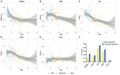 |
Characterising the lifespan trajectory of six essential
neurometabolites in a cohort of 100 participants.
Alice Thomson1,2,
Duanghathai Pasanta1,
Hannah Hwa1,
Tomoki Arichi2,3,
Richard Edden4,
Xiaoqian Chai5,
and Nicolaas Puts1,2
1Department of Forensic and Neurodevelopmental Sciences, Institute of Psychiatry, King's College London, London, United Kingdom, 2MRC Centre for Developmental Neurobiology, King's College London, London, United Kingdom, 3Centre for the Developing Brain, King's College London, London, United Kingdom, 4Department of Radiology and Radiological Science, The Johns Hopkins University School of Medicine, Baltimore, MD, United States, 5Department of Neurology and Neurosurgery, McGill University, Montreal, QC, Canada Keywords: Normal development, Spectroscopy, GABA, Normative, metabolites, childhood, adolescence, adult We characterised the normative lifespan trajectories of brain neurometabolites, as measured by 1H-Magnetic Resonance spectroscopy (1H-MRS) from the posterior parietal cortex across 100 individuals (aged 5-40 yrs). Glutamate + glutamine (Glx), N-acetylaspartate (NAA), gamma-aminobutyric acid (GABA+) and glutathione (GSH) showed non-linear trajectories, decreasing steeply in childhood/adolescence before a gradual, significant, decline across early adulthood. Results suggest age associated changes in brain composition may contribute to the observed trajectories. Importantly, a non-linear regression modelling approach was found to be more appropriate for neurometabolite trajectories than a linear regression model and should be considered in future to prevent simplification of data trends. |
||
5318.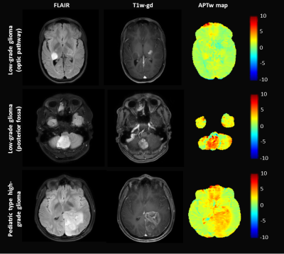 |
Initial experience with Amide Proton Transfer weighted imaging
in pediatric neuro-oncology
Iris Obdeijn1,
Evita Wiegers1,
Sabine Plasschaert2,
Kim van de Ven3,
Dennis Klomp1,
Hans Hoogduin1,
Maarten Lequin1,2,
and Jannie Wijnen1
1Department of Radiology and Nuclear Medicine, University Medical Center Utrecht, Utrecht, Netherlands, 2Department of Pediatric Neuro-Oncology, Princess Maxima Center for pediatric oncology, Utrecht, Netherlands, 3BIU MR, Philips Healthcare, Best, Netherlands Keywords: Neuro, CEST & MT APTw imaging has shown promising applications in diagnosing adult brain tumors. However, pediatric brain tumors differ from their adult counterparts in terms of clinical, biological, and radiological appearance. Therefore, we investigated if and how APTw imaging can be used in clinical-decision making in pediatric neuro-oncology. First, we showed the repeatability of APTw maps, allowing longitudinal assessment. Then APTw imaging was evaluated in eleven children with a brain tumor. APTw values are higher in pediatric brain tumors compared to normal-appearing white matter. Unlike studies in adults, APTw values were not significant different between low-grade glioma and high-grade glioma in these children. |
||
5319. |
Evaluation of Deep Gray Matter Iron and Myelin Changes in
children with Attention-deficit/hyperactivity disorder
shu su1,
Yingqian Chen 1,
Long Qian 2,
Yan Dai 1,
Liping Lin1,
and Zhiyun Yang1
1First Affiliated Hospital, Sun Yat-sen University, guangzhou, China, 2GE Healthcare, guangzhou, China Keywords: Neuro, Quantitative Susceptibility mapping Application of magnetic susceptibility have been consistently demonstrated in the subcortical gray matter of ADHD children, but some uncertainties remain concerning the underlying neurobiological processes. We applied quantitative susceptibility mapping and synthetic magnetic resonance imaging (SyMRI) to clarify the relative contribution of iron and myelin changes to deep gray matter changes in ADHD. We found that the iron and myelin concentration of these subcortical structures in ADHD were delayed in the developmental trajectory, which suggested that ADHD may be characterized by a delay in subcortical maturation and dysfunction in dopaminergic transmission. |
||
 Back to Meeting Home
 Back to the Program-at-a-Glance
The International Society for Magnetic Resonance in Medicine is accredited by the Accreditation Council for Continuing Medical Education to provide continuing medical education for physicians.
 Back to Meeting Home
 Back to the Program-at-a-Glance
 View Presentation Video