Traditional Poster
Preclinical
ISMRM & ISMRT Annual Meeting & Exhibition • 03-08 June 2023 • Toronto, ON, Canada

| Booth # | |||
|---|---|---|---|
5369.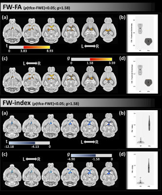 |
Using FW-DTI and VBM to identify white matter microstructural
integrity and grey matter density in a preclinical model of
Alzheimer’s disease
Maurizio Bergamino1,
Megan R Nelson1,2,
Asfia Numani1,
Matthew L Scarpelli3,
Deborah Healey4,
Alberto Fuentes1,
Gregory Turner1,
and Ashley M Stokes1
1Barrow Neurological Institute, Phoenix, AZ, United States, 2Arizona State University, Tempe, AZ, United States, 3Purdue University, West Lafayette, IN, United States, 4MD Anderson Cancer center, Houston, TX, United States Keywords: Data Analysis, Diffusion/other diffusion imaging techniques The objective of this study was to assess white matter integrity and grey matter volume density in a mouse model of Alzheimer’s disease (3xTg-AD) using free-water (FW) diffusion tensor imaging (FW-DTI) and voxel-based morphometry (VBM). Additionally, histological analyses of amyloid and tau protein in different regions across the brain were examined. |
||
| 5370. |
Quantitative analysis of metabolites in mouse optical track
after severe traumatic brain injury using magnetic resonance
spectroscopy
Roxan Ara1,
Fengchong Kong1,
Manish Kumar2,
Hannah MCMICHAEL2,
Mario ESPINOSA PALACIO2,
Meenakshi AHLUWALIA2,
Krishnan M DHANDAPANI2,
Kumar VAIBHAV2,3,
and Asamoah Bosomtwi4
1Georgia Cancer Center, Augusta University, Augusta, GA, United States, 2Neurosurgery, Augusta University, Augusta, GA, United States, 3Oral Biology and Diagnostic, Augusta University, Augusta, GA, United States, 4Georgia Cancer Center, Georgia medical College, Augusta, GA, United States Keywords: Data Acquisition, Metabolism, Spectroscopy Traumatic brain injury (TBI) is associated with an increased risk of cognitive and neurodegenerative complications that may develop and persist years after injury. Altered metabolism is considered to be the earliest possible sign of tissue injury and will be present before any structural changes can be detected. Magnetic resonance spectroscopy is a useful technology for longitudinal assessments, which are critical for understanding altered neurochemical concentrations following TBI, suggesting impaired neurotransmission and energy generation, neuronal injury/death, and oxidative stress |
||
5371.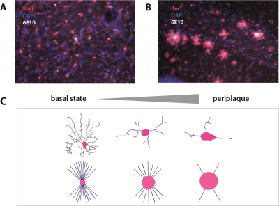 |
Developing novel MRI biomarkers of amyloid beta aggregation and
inflammation in APPswe/PS1dEd mouse model of Alzheimer’s Disease
Patricia Martínez Tazo1,
Mohamed Kotb Selim1,
Santiago Canals2,
Jose Vicente Sanchez-Mut1,
and Silvia De Santis1
1Molecular Neurobiology and Neuropathology, Institute of Neuroscience of Alicante, Sant Joan d'Alacant, Alicante, Spain, 2Cellular and Systems Neurobiology, Institute of Neuroscience of Alicante, Sant Joan d'Alacant, Alicante, Spain Keywords: Alzheimer's Disease, Microstructure We present a framework to extract novel MRI biomarkers, based on diffusion-weighted contrast, capable of capturing microstructural alterations in the APPswe/PS1dE9 mouse model of Alzheimer’s disease. To validate the MRI framework, imaging was complemented with histology, with the aim of elucidating the cell-scale biological basis. We demonstrate that MRI signal carries the fingerprint of Alzheimer’s disease characteristic plaque aggregation and associated neuroinflammation patterns in a cohort of 18 months old APPswe/PS1dE9 mice as compared to healthy age-matched controls. This framework sets the basis for an in-vivo multimodal imaging protocol, translatable to humans, for early detection of Alzheimer-specific microstructural alterations. |
||
5372.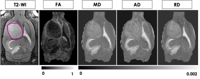 |
Diffusion Tensor MRI in a Rat C6 Glioma Model at 9.4T
Maryam Mozaffari1,2,
Naila Rahman1,2,
Patrick McCunn1,2,
Nivin Nystrom1,2,
Alex Li2,
Miranda Bellyou2,
Corey Baron1,2,
Timothy Scholl1,2,
and Robert Bartha1,2
1Medical Biophysics, Western University, London, ON, Canada, 2Centre for Functional and Metabolic Mapping, Robarts Research Institute, London, ON, Canada Keywords: Diffusion/other diffusion imaging techniques, Brain, Animal model The objective of this study was to characterize the diffusion properties of water in and around C6 glioma tumors in rats at 9.4T. DTI showed differences in some diffusion metrics between tumor tissue and surrounding tissues. Significantly higher FA, MD, and AD values were found in the tumor region compared to the contralateral side. DTI measurement of tumor microstructure and its surroundings could help to enhance the efficacy of diagnosis in different human malignancies. |
||
3728.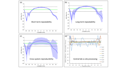 |
ADC Repeatability and Reproducibility of NCI CIRP Network
Pre-Clinical MRIs
Dariya Malyarenko1,
Ghoncheh Amouzandeh1,2,
Stephen Pickup3,
Rong Zhou3,
Henry Charles Manning4,
Seth T Gamon4,
Kooresh I Shoghi5,
James D Quirk5,
Renuka Sriram6,
Peder Larson6,
Michael T Lewis7,
Robia G Pautler7,
Paul E Kinahan8,
Mark Muzi8,
and Thomas L. Chenevert1
1Radiology, University of Michigan, Ann Arbor, MI, United States, 2neuro42, San Francisco, CA, United States, 3Radiology, University of Pennsylvania, Philadelphia, PA, United States, 4Cancer Systems Imaging, The University of Texas MDACC, Houston, TX, United States, 5Mallinckrodt Institute of Radiology, Washington University School of Medicine, St. Louis, MO, United States, 6Department of Radiology & Biomedical Imaging, University of California San Francisco, San Francisco, CA, United States, 7Baylor College of Medicine, Houston, TX, United States, 8Radiology, University of Washington, Seattle, WA, United States Keywords: System Imperfections: Measurement & Correction, Precision & Accuracy The goal of this work was to assess ADC repeatability, reproducibility, and bias of Co-Clinical Imaging Research Resource Program (CIRP) network MRIs using standardized procedures for comparison to corresponding performance of clinical MRIs. A temperature-controlled phantom provided an absolute reference standard and means to assess spatial uniformity of these metrics. Seven institutions participated in the study where DWI were acquired over multiple days on 10 pre-clinical scanners, from 3 vendors at 6 field strengths. Technical level repeatability and reproducibility metrics, and spatial uniformity patterns are comparable to that observed on human systems using similar phantoms and test procedures. |
||
 Back to Meeting Home
 Back to the Program-at-a-Glance
The International Society for Magnetic Resonance in Medicine is accredited by the Accreditation Council for Continuing Medical Education to provide continuing medical education for physicians.
 Back to Meeting Home
 Back to the Program-at-a-Glance
 View Presentation Video