Traditional Poster
Physics & Engineering
ISMRM & ISMRT Annual Meeting & Exhibition • 03-08 June 2023 • Toronto, ON, Canada

| Booth # | |||
|---|---|---|---|
5276.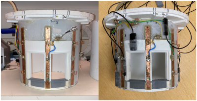 |
Workflow and performance measures for integrating magnetic field
monitoring with an 8-channel pediatric head coil for 7T MRI
Christian Sprang1,2,
Pedram Yazdanbakhsh1,3,
Marcus Couch4,
Sajjad Feizollah1,3,
Christine Tardif1,2,3,
and David A Rudko1,2,3
1McConnell Brain Imaging Centre, Montreal Neurological Institute and Hospital, Montreal, QC, Canada, 2Department of Biomedical Engineering, McGill University, Montreal, QC, Canada, 3Department of Neurology and Neurosurgery, McGill University, Montreal, QC, Canada, 4Siemens Healthcare Limited, Montreal, QC, Canada Keywords: System Imperfections: Measurement & Correction, RF Arrays & Systems Ultra-high field MRI is particularly susceptible to dynamic magnetic field fluctuations. To address this, commercial field probes were integrated into an 8-channel dipole Tx/Rx 7 T pediatric head coil. Coil performance was assessed before and after probe integration. Probe performance was compared on and off the coil. After probe integration, reflection coefficients were maintained below -10dB in all channels, noise correlation was shown to improve, and maximum SNR was shown to decrease. Probe performance did not change substantially once mounted on the coil. Future work will aim to address the reduction in SNR observed after probe integration. |
||
5277.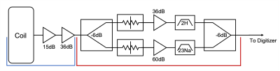 |
A Channelized Front-End for Single Port Multi-Tuned RF Coils
Courtney Bauer1,
Jue Hou1,
Chenhao Sun1,
and Steven M. Wright1
1Department of Electrical and Computer Engineering, Texas A&M University, College Station, TX, United States Keywords: RF Arrays & Systems, RF Arrays & Systems Increased interest in simultaneous and interleaved MR imaging and spectroscopy has led to the increased interest in multi-tuned coils and coil arrays. The introduction of single port, multinuclear coils resolves the complexity resulting from nested coils, but introduces new challenges in optimization of signal conditioning. Presented here is a channelization approach that enables frequency specific gain and filtering of signals from single port multinuclear coils. |
||
5278.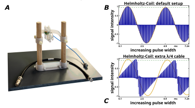 |
A 3T Helmholtz coil with either reduced or maximized radiation
damping effects
Roland Müller1,
Niklas Wallstein1,
André Pampel1,
and Harald E. Möller1
1Max Planck Institute for Human Cognitive and Brain Sciences, Leipzig, Germany Keywords: Non-Array RF Coils, Antennas & Waveguides, Ex-Vivo Applications, Tiltable RF Coil, Radiation Damping Radiation damping (RD) effects may confound experiments targeted at the quantification of MR contrast parameters, in particular, in experiments in small ex-vivo specimens performed with dedicated coils supporting a high filling factor. To address this problem, a long-known principle during reception with coil arrays (preamplifier decoupling) was also applied in the transmit branch of the TxRx switch. Furthermore, adding an extra λ/4 cable between coil and TxRx switch allows to switch between minimum and maximum RD with otherwise almost equal coil characteristics. This may be further exploited for for testing pulse sequences with regards to potential RD effects. |
||
5279.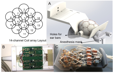 |
Design and construction of a 14-channel receive-only array for
high resolution MRI of marmosets’ brain at 9.4T
Daniel Papoti1,2,
Diego Szczupak1,
David Schaeffer1,
and Afonso Silva1
1Department of Neurobiology, University of Pittsburgh, Pittsburgh, PA, United States, 2Center for Engineering, Modeling and Applied Social Sciences, Federal University of ABC, Sao Bernardo do Campos, Brazil Keywords: RF Arrays & Systems, RF Arrays & Systems, Receive Arrays The present work describes the design characterization and tests of a 14-channel receive-only array for marmoset brain MRI at 9.4T. The coil was designed to maximize the SNR over the entire head of a common marmoset, considering the anesthesia apparatus, such as ear bars and anesthesia mask. |
||
5280.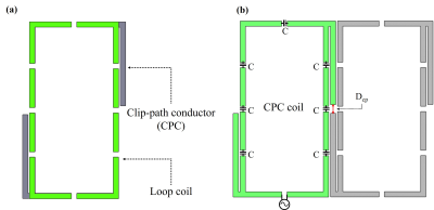 |
Geometrical Decoupling using Clip-path Conductor for 7T
Transceiver Coil Array
Taewoo Nam1,
Yonghwa Jeong1,
Minyeong Seo2,
Eunwoo Lee3,
Junseong Son3,
Taekwan Lee4,
Young Noh5,
Donghyuk Kim2,
Daniel Hernandez2,
and Kyoung-Nam Kim3
1Department of Health Sciences and Technology, GAIHST, Gachon University, Incheon, Korea, Republic of, 2Neuroscience Research Institute, Gachon University, Incheon, Korea, Republic of, 3Department of Biomedical Engineering, Gachon University, Incheon, Korea, Republic of, 4Brain Core Research Facility, Korea Brain Research Institute, Daegu, Korea, Republic of, 5Department of Neurology, Gill Medical Center, Gachon University College of Medicine, Incheon, Korea, Republic of Keywords: RF Arrays & Systems, RF Arrays & Systems, Clip-path, Transceiver Array, Decoupling, MRI We propose new geometrical decoupling method using clip-path conductor (CPC) to reduce the mutual coupling between individual elements in RF coil array. The 8-element CPC transceiver array was constructed based on electromagnetic (EM) simulation and applied to whole brain imaging at 7T. |
||
5281.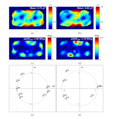 |
An 8-channel Transmit Array for Prostate MRI at 7T Using Six
Loops and Two Dipoles: A Simulation Study.
Aleksej Polpudenko1,
David Andrew Porter1,
and Shajan Gunamony1,2
1Imaging Centre of Excellence, University of Glasgow, Glasgow, United Kingdom, 2MR CoilTech Limited, Glasgow, United Kingdom Keywords: RF Arrays & Systems, Prostate Prospective 8 channel (6Tx loop + 2Tx dipole) body array intended for prostate MRI at 7T was simulated and its B1+ and SAR performance evaluated. The study demonstrated the capability of loop-based arrays to offer competitive levels of prostate imaging performance compared to current state-of-the-art hybrid and dipole arrays reported in the literature. |
||
5282.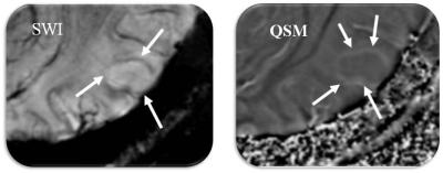 |
Demonstration of Distinct Iron Deposition Patterns in Gliomas
using 7T Quantitative Susceptibility Mapping
Demetrius Eugene Lee, B.S.1,2,
Kristina Vineis, B.S.1,2,
Laiz Godoy, M.D.1,2,
Lisa Desiderio, RT (R)(MR)1,2,
Suyash Mohan, M.D.1,2,
and Sanjeev Chawla, Ph.D1,2
1Radiology, Perelman School of Medicine, Philadelphia, PA, United States, 2University of Pennsylvania, Philadelphia, PA, United States Keywords: Tumors, Blood vessels, Glioma Evidence has shown that abnormal iron levels and gliomas are heavily correlated. Due to this, iron presents a possibility to serve as a glioma biomarker. This study will utilize susceptibility-weighted imaging (SWI) to identify the glioma, and quantitative susceptibility mapping (QSM) to explicitly classify iron deposits in the glioma on an ultra-high field (7T) scanner. A total of 6 untreated glioma patients with high- and low-grade gliomas underwent 7T QSM. Patients with high-grade gliomas showed distinct, geometric iron deposits that low-grade gliomas did not. These results illustrate that 7T QSM may be helpful in classifying low- and high-grade gliomas. |
||
5283.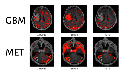 |
Differences in vascularity between recurrent glioblastoma and
brain metastasis using dynamic contrast enhanced MRI
Sapir Fajerzstein1,2,3,
Moran Artzi1,3,4,
Deborah T. Blumenthal4,5,
Dror Limon4,6,
Orna Aizenstein7,
Felix Bokestein4,5,
Netanell Avisdris1,
and Dafna Ben, Bashat1,3,4
1Sagol Brain Institute, Tel Aviv Sourasky Medical Center (TASMC), Tel Aviv, Israel, 2The Iby and Aladar Fleischman Faculty of Engineering TAU, Tel Aviv, Israel, 3Sagol School of Neuroscience, Tel Aviv University, Tel Aviv, Israel, 4Sackler Faculty of Medicine, Tel Aviv University (TAU), Tel Aviv, Israel, 5Division of Oncology, TASMC, Tel Aviv, Israel, 6Neuro-Oncology Service, TASMC, Tel Aviv, Israel, 7Division of Radiology, TASMC, Tel Aviv, Israel Keywords: Tumors, DSC & DCE Perfusion High grade glioma (HGG) and brain-metastasis are known to have different vascularity. Dynamic contrast-enhanced (DCE) was suggested to assess disease progression, and to differentiate between tumor recurrence and radiation necrosis. However, previous methods did not relate to the differences in vascularity of the two tumor types and often offer the same method and same threshold values. We show differences between groups with: higher vascularity, slightly increased permeability, higher percent of arteries overlapping of the tumor, and differences in several radiomics features, in recurrent HGG compared with recurrent brain-metastasis. Future studies should relate to these differences between the two tumor types. |
||
5284.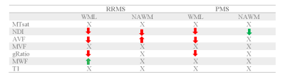 |
Longitudinal MRI study of white matter in multiple sclerosis
using surrogates measures of myelin and axonal damage.
Gretel Sanabria Sanabria Diaz1,2,3,
Lester Melie-Garcia1,2,3,
Po-Jui Lu 1,2,3,
Muhamed Barakovic1,2,3,
Mario Alberto Ocampo Pineda1,2,3,
Xinjie Chen1,2,3,
Matthias Weigel1,3,4,
Nina Siebenborn1,2,3,
Esther Ruberte Jiménez1,2,3,
Alessandro Cagol1,2,3,
Riccardo Galbusera1,2,3,
Antoine Lutti5,
Jens Kuhle2,3,
Ludwig Kappos1,2,3,
and Cristina Granziera 1,2,3
1Translational Imaging in Neurology (ThINk) Basel, Department of Biomedical Engineering, University Hospital Basel and University of Basel, Basel, Switzerland, 2Department of Neurology, University Hospital Basel, Basel, Switzerland, 3Research Center for Clinical Neuroimmunology and Neuroscience Basel (RC2NB), University Hospital Basel and University of Basel, Basel, Switzerland, 4Division of Radiological Physics, Department of Radiology, University Hospital Basel, Basel, Switzerland, 5Laboratory for Research in Neuroimaging, Department of Clinical Neuroscience, Lausanne University Hospital and University of Lausanne, Lausanne, Switzerland Keywords: Multiple Sclerosis, Multiple Sclerosis Damage to the myelin sheath and the neuroaxonal unit are features of multiple sclerosis, as well as reparative processes for both. However, a detailed characterization of the dynamics of those in vivo is challenging. In this longitudinal study, we applied a multi-contrast quantitative MRI approach to disentangle lesion progression in vivo in patients with MS. The microstructural measures were compared between multiple sclerosis groups (55 relapsing-remitting, 24 progressive) and 34 healthy controls. Our results indicate changes in microstructural MRI measures in white matter lesions and normal appearing tissue related to myelin and axonal integrity in RRMS and PMS. |
||
5285. |
In Vivo Imaging of LC-NE Integrity: Mechanisms Underlying Heath
Disparity for Alzheimer’s Disease
Yu-Shin Ding1,
Jiacheng Wang1,
Artem Mikheev1,
jingyun chen1,
and James Babb1
1NYUSoM, New York, NY, United States Keywords: Alzheimer's Disease, PET/MR Although blacks are at 2-3 times higher prevalence rate of developing AD, blacks have been under-included in many prominent AD clinical trials. The current biomarker classification system (ATN) can’t explain the increased prevalence in blacks of both AD and vascular risk factors for AD such as diabetes and hypertension when compared to whites. Our decade-long PET/MR studies have demonstrated a special vulnerability of locus coeruleus (LC) to aging and stress. Our recent study showed that a faster decline of LC function occurs in blacks. Thus, imaging LC represents a novel biomarker approach to mechanisms underlying health disparity for AD. |
||
5286.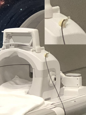 |
Measurement of Sound Pressure Level (SPL) at 3T and 7T for
optimization of Quiet Dynamic Zero Echo Time (ZTE) MRI
Joshua T. Hanson1,
James H. Holmes2,
Vincent A. Magnotta2,
and Curtis A. Corum3
1Champaign Imaging LLC, Shoreview, MN, United States, 2Radiology, University of Iowa, Iowa City, IA, United States, 3Champaign imaging LLC, Shoreview, MN, United States Keywords: Safety, Gradients, Acoustic Measurement, ZTE, 3D Radial, Silent This abstract aims to provide a general method of measuring the acoustic noise produced by a scanning protocol and to present the acoustic differences in absolute sound pressure level. To demonstrate these methods we show results using conventional Cartesian and acoustically quieter ZTE based protocols on 3T and 7T systems. |
||
5287.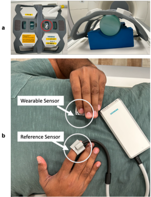 |
Evaluation of a Wearable Bluetooth Sensor at 0.55T
Felix Munoz1,
Krishna Nayak2,
and Yasser Khan2
1Biomedical Engineering, University of Southern California, Los Angeles, CA, United States, 2Ming Hsieh Department of Electrical and Computer Engineering, University of Southern California, Los Angeles, CA, United States Keywords: New Devices, Low-Field MRI, Wearable Devices Compatibility of Bluetooth low energy (BLE) wearable sensors in the MRI environment will enable the creative use of wearable devices to monitor vital signs such as heart rate, respiration rate, blood pressure, temperature, and biochemical markers during a scan. In this work, we demonstrate efficacy of BLE sensors at the novel 0.55T MRI field strength and evaluate the noise in a wearable caused by rapidly switching MRI gradients, as well as MRI noise/artifacts introduced by a BLE wearable. |
||
5288.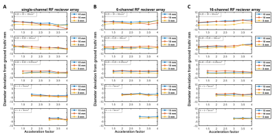 |
Impacts of compressed sense-parallel imaging on lung 19F-MRI
ventilation imaging measured with tissue-mimicking 1H phantoms.
Dominic Harrison1,2,
Mary Neal1,2,
Kieren Hollingsworth1,2,
and Pete Thelwall1,2
1Newcastle Magnetic Resonance Centre, Newcastle University, Newcastle upon Tyne, United Kingdom, 2Translational and Clinical Research Institute, Newcastle University, Newcastle upon Tyne, United Kingdom Keywords: Lung, Non-Proton, 19F-MRI We used 1H test objects to assess the ability to detect and quantify lung ventilation defects using accelerated 19F-MRI ventilation scan protocols. The test objects replicated the spin density and relaxation properties of inhaled perfluoropropane gas and are constructed with signal voids of known dimensions to simulate ventilation defects. Scans were acquired on single- and multi-channel RF receiver arrays, with multiple compressed sensing and parallel imaging (CS/CS-PI) accelerations and scan resolutions at fixed scan duration and field-of-view (FOV). A negative relationship between acceleration and measured ventilation defect diameter was observed with increasing CS acceleration factor, but not for CS-PI scans. |
||
 Back to Meeting Home
 Back to the Program-at-a-Glance
The International Society for Magnetic Resonance in Medicine is accredited by the Accreditation Council for Continuing Medical Education to provide continuing medical education for physicians.
 Back to Meeting Home
 Back to the Program-at-a-Glance
 View Presentation Video