Weekend Course
Tissue Oxygenation: MRI Measurement & Clinical Implications
ISMRM & ISMRT Annual Meeting & Exhibition • 03-08 June 2023 • Toronto, ON, Canada

Weekend Course
Tissue Oxygenation: MRI Measurement & Clinical Implications
| 13:00 | Technical Aspects of MRI Oxygenation Measurement Thoralf Niendorf | |
| 13:35 |
 |
Assessing tumor oxygenation as a predictive imaging biomarker
Ralph Mason
Keywords: Cross-organ: Cancer, Cross-organ: Oxygenation, Contrast mechanisms: Relaxometry Hypoxia is associated with tumor growth and development, and strongly influences some treatment response. Various MRI techniques have been developed to assess tumor hypoxia and dynamic response to interventions non-invasively. 19F, 1H or ESR provide quantitative estimates of pO2 revealing intra tumoral heterogeneity and differential response to interventions, but require exogenous reporter agents. Recently, oxygen-sensitive approaches based on direct interrogation of tissue water (R2* and R1) have been demonstrated in both pre-clinical studies and translational trials in humans, as potential predictive imaging biomarkers. The review will consider state of the art and future opportunities for development and applications. |
| 14:10 |
MRI of Cerebral Oxygenation: a quest to quantify the BOLD effect
Thomas Christen
Keywords: Neuro: Brain, Image acquisition: Quantification, Image acquisition: MR Fingerprinting In this presentation, we will follow a line of research that aims to estimate brain oxygen extraction fraction by quantifying the famous Blood Oxygen Level Dependent (BOLD) effect. We will see how the models and data acquisitions patterns have been refined over the years, and how these “quantitative BOLD” methods have been eventually fused with other MR approaches such as quantitative susceptibility mapping (QSM) or MR fingerprinting (MRF). Clinical and preclinical results will be presented in healthy brains as well as in pathologies such as stroke or neurodegenerative diseases. |
|
| 14:45 |
Break & Meet the Teachers |
|
| 15:15 |
 |
MRI of Placental Oxygenation
Esra Abaci Turk
Keywords: Cross-organ: Oxygenation The placenta is an important organ that serves as a critical interface between a mother and her fetus. Deterioration in placental function throughout pregnancy is a leading cause of perinatal morbidity and mortality. Monitoring placental function throughout pregnancy is therefore a critical component of antenatal care, but few tools currently exist for direct assessment of placental function. MRI provides a tremendous potential for monitoring placental function. Development of quantitative MRI measures of placental oxygen delivery and transport between the mother and the fetus would increase our ability to detect placental insufficiency and could motivate and evaluate potential therapeutic interventions. |
| 15:50 |
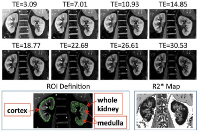 |
MRI of Renal Oxygen Availability
Pottumarthi Prasad
Keywords: Body: Kidney, Contrast mechanisms: fMRI, Cross-organ: Oxygenation Unlike most organs, in the kidneys, oxygen consumption changes with blood flow and increase in blood flow doesn't necessary lead to increased oxygen delivery. Further, there is a regional variation in blood and oxygen supply within the kidneys necessitating imaging based approach. BOLD MRI is the only non-invasive method to-date to evaluate renal oxygen availability. It is most useful for detecting acute changes following pharmacologic maneuvers. Limitations in conventional ROI analysis have been identified, creating an interest in alternative methods, including whole kidney analysis such as twelve layer concentric objects (TLCO). |
| 16:25 |
MRI of Cardiac Oxygenation
Lian-Ming Wu
|
 Back to Meeting Home
 Back to the Program-at-a-Glance
The International Society for Magnetic Resonance in Medicine is accredited by the Accreditation Council for Continuing Medical Education to provide continuing medical education for physicians.
 Back to Meeting Home
 Back to the Program-at-a-Glance
 View Presentation Video