Weekend Course
Validation of Microstructure Mapping with Diffusion MRI
ISMRM & ISMRT Annual Meeting & Exhibition • 03-08 June 2023 • Toronto, ON, Canada

Weekend Course
Validation of Microstructure Mapping with Diffusion MRI
| 13:00 |
 |
Diffusion Microstructure Models in the Brain
Santiago Coelho
Keywords: Contrast mechanisms: Diffusion, Contrast mechanisms: Microstructure, Neuro: Brain This talk examines current diffusion MRI modeling approaches for brain tissue. Biophysical models and signal representations are contrasted. How diffusion coarse-graining facilitates modeling is explained. Emphasis is put on the identification of common modeling assumptions and examples on how to validate them. High diffusion weightings, long and short diffusion times, and variable tensor encodings are discussed together with state-of-the-art modeling techniques on each. Finally, present and future directions for modeling white and gray matter are discussed. |
| 13:25 |
Diffusion Microstructure Models in the Body and Tumor
Sungheon Gene Kim
Keywords: Contrast mechanisms: Diffusion, Body: Body, Cross-organ: Cancer This lecture provides an overview of how diffusion MRI has been used to measure tissue microstructural properties in various tissues and organs outside the brain. We will focus on (i) diffusion microstructure models in the body that have been used to measure the tissue microstructural properties in specific examples including muscle, breast, and prostate; and (ii) diffusion microstructure models for tumor that can be used to measure cancer cell properties, such as cell size and transcytolemmal water exchange rate. The need and challenges for validation will also be discussed. |
|
| 13:50 |
Diffusion Hardware Phantoms
Penny Hubbard Cristinacce
Keywords: Contrast mechanisms: Diffusion This lecture will describe the array of hardware diffusion phantoms developed to help validate advanced diffusion MR methods. These physical phantoms can include liquids, solutions, liquid crystals, gels, beads, capillaries, textiles, polymer fibers, yeast cells and plants, as well as more elaborate physical assemblies and combinations of structures. Phantoms designed to validate tractography, microscopic anisotropy, restricted diffusion, inter-compartmental exchange, pseudo-diffusion/flow, and multi-parametric methods, such as relaxation-diffusion correlations, will be described and their advantages and disadvantages discussed. |
|
| 14:15 |
Diffusion Numerical Phantoms
Hong-Hsi Lee
Keywords: Contrast mechanisms: Microstructure, Contrast mechanisms: Diffusion Performing diffusion simulations in numerical phantom mimicking realistic tissue microstructure helps to test the sensitivity of diffusion MRI to tissue features and validate the biophysical models. Realistic cell geometries for simulations have been directly reconstructed from the microscopy data of biological tissues in 3-dimension. In addition to the diffusion within intra-cellular space, it is possible to perform simulations of diffusion in the extra-cellular space, as well as of the exchange between intra- and extra-cellular spaces. The tissue preparation preserving extra-cellular space in histology is non-trivial, prompting the development of pipelines to generate semi-realistic tissue microstructure by packing multiple artificial “cells”. |
|
| 14:40 |
Break & Meet the Teachers |
|
| 15:05 |
 |
Preclinical and Histological Validation of Brain Microstructure
Mapping
Alejandra Sierra
Keywords: Neuro: Brain, Contrast mechanisms: Diffusion, : Preclinical/Animal The brain tissue has a complex microstructural environment, which is averaged in the coarse resolution of MRI. In this talk, we will explore the potential of conventional and advanced histological methods, e.g., light or electron microscopy, offering a variety of tissue information at higher resolution than diffusion MRI. We will talk about extracting quantitative information or 3D modeling of brain tissue microstructure relevant to diffusion MRI. We will discuss how to develop strategies for validation using a multimodality approach and what are the benefits and challenges of combining histological information with greater sensitivity and the coarse information of diffusion MRI. |
| 15:30 |
 |
Preclinical & Histological Validation of Body/Tumor
Microstructure Mapping
Yuko Someya
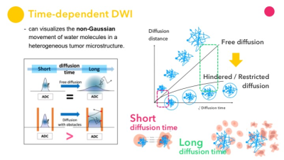
Keywords: : Preclinical/Animal, Cross-organ: Cancer Diffusion MRI, which visualizes the non-Gaussian movement of water molecules in a heterogeneous tumor microenvironment, has the potential to add new information about tissue microstructure non-invasively. This talks Introduce diffusion MRI methods for studying tumor microstructure, and then describe their sensitivity to tumor microstructural differences (cell size, shapes and configurations, cellularity, amount of extracellular space, intra-tumor heterogeneity, etc.), considering how the submicron resolution of tissue microscopy and the millimeter resolution of diffusion MRI can be linked to. Since the assessment of tumor angiogenesis is essential in oncology, the assessment of tissue microvasculature through the IVIM approach will also be discussed. |
| 15:55 |
Preclinical Validation of Fiber Orientations and Tractography
Marios Georgiadis
Keywords: Neuro: Brain connectivity, Neuro: White matter, Contrast mechanisms: Diffusion Myelinated axons enable fast and energy-efficient signal transmission. However, axons can have micron-level diameter and multiple centimeters length, so axon trajectories are hard to follow. Multiple modalities can reveal axon paths, typically by providing axonal orientations followed by tractography algorithms. Each technology has limitations preventing it from being considered a “gold standard”. Diffusion MRI (dMRI) provides beautiful tractograms, however its readouts are not specific to myelinated axons, and need validation. This talk will present the 2D and 3D methods used to image axonal orientations and trajectories, and review results from each method as well as past and ongoing validation efforts. |
|
| 16:20 |
Validation in Clinic & Population Studies
Catherine Lebel
Keywords: Contrast mechanisms: Diffusion Validations in phantoms, preclinical, and histological studies have greatly enhanced our understanding of what in vivo diffusion measures reflect. However, there remain complexities in human imaging that can confound interpretations and mean that phantom, animal, and histology studies do not always perfectly translate. Validation in clinical and population studies remains important to better inform interpretation of diffusion metrics in human studies. I will cover diffusion validation studies in clinical and population studies in living humans. In particular, studies relating various diffusion and other metrics to one another provide context for interpretation and help evaluate the specificity of various diffusion metrics. |
 Back to Meeting Home
 Back to the Program-at-a-Glance
The International Society for Magnetic Resonance in Medicine is accredited by the Accreditation Council for Continuing Medical Education to provide continuing medical education for physicians.
 Back to Meeting Home
 Back to the Program-at-a-Glance
 View Presentation Video