Weekend Course
Advances in Image Analysis: How to Get the Most from Your Images
ISMRM & ISMRT Annual Meeting & Exhibition • 03-08 June 2023 • Toronto, ON, Canada

Weekend Course
Advances in Image Analysis: How to Get the Most from Your Images
| Core principles in image analysis | |||
| 07:45 |
Methods for Improving Image Quality: A Diffusion MRI Perspective
M. Okan Irfanoglu
Keywords: Image acquisition: Image processing, Image acquisition: Artefacts, Image acquisition: Motion correction MR images suffer from several imperfections including (but not limited to) low SNR, artifacts and image distortions. In this seminar, we will first discuss whether manipulating images with image processing or machine learning techniques is beneficial or damaging, and then we will go over techniques (and tools) to improve image quality for each of these issues. Our examples will mostly focus on diffusion MRI but what can be done for other modalities such as anatomical or functional images will also be discussed and illustrated |
||
| 08:15 |
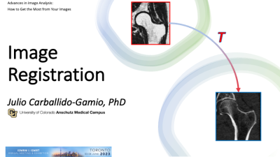 |
Image Registration
Julio Carballido-Gamio
Keywords: Image acquisition: Image processing The applications of Image Registration in medical imaging are numerous, ranging from the alignment of longitudinal scans to segmentation of anatomical structures. In this session, I will cover material that will help you understand the different components of the intensity-based image registration framework. |
|
| 08:45 |
Basic Principles of Model-Based Fitting for MRI
Dong-Hyun Kim
Keywords: Image acquisition: Modelling Model-based fitting is a powerful technique for extracting quantitative information from MRI data. We will discuss the challenges associated with model-based fitting, including model selection, noise and artifacts, parameter estimation, validation and reproducibility, and computation time. |
||
| 09:15 |
Voxel vs. ROI-Based Statistical Analyses: From Histograms to
Patients
Kyrre Emblem
Keywords: Image acquisition: Image processing, Transferable skills: Reproducible research, Image acquisition: Visualization Making good use of MRI data from a clinical study can be a challenge, especially when faced with the task of analyzing data from advanced imaging techniques in small patient cohorts. This talk will address some of the current challenges with image analyses in a clinical setting. Using neuroimaging and cancer as examples, the talk will discuss potential strategies to help produce and evaluate robust, repeatable, and clinically meaningful image parameters in patient studies with the typical low sample size. Different approaches for assessing and analyzing resulting parametric maps will be presented, including use of dynamic and longitudinal imaging data. |
||
| 09:45 |
Break & Meet the Teachers |
||
| Advanced image analysis methods | |||
| 10:00 |
Machine Learning: What to Use & When
Peter LaViolette
Keywords: Image acquisition: Machine learning In this seminar I will discuss many ways that machine learning (ML) can be used for image processing, from model fitting to image segmentation. Many approaches vary depending on the application. A broad overview using radio-pathomics as an example will be discussed. |
||
| 10:30 |
 |
Network-Based Analyses: Graph Theory for Evaluating Brain
Connectivity
Olga Tymofiyeva
Keywords: Neuro: Brain connectivity I will start by sharing my dream of a clinical application of network-based analyses: a short MRI scan for adolescents in primary care that would enable 1) brain network-based diagnosis of psychiatric disorders, 2) brain network-based treatment of psychiatric disorders, and 3) brain network-based prevention of psychiatric disorders. I will then talk about the challenges of the two main steps in constructing a brain network: i) choosing connectivity measures that will serve as the network "edges" and ii) dividing the brain into regions that will serve as the network "nodes." Various local and global network properties will also be discussed. |
|
| 11:00 |
 |
Tutorial: Practical Considerations When Performing Radiomics
Masoom Haider
Keywords: Body: Body, Cross-organ: Cancer, Image acquisition: Machine learning Radiomics has not yet made it into clinical care. For this to happen high quality biomarker technical validation is required. In this presentation, we go through key items to consider for meaningful radiomics research. This includes the availability of high-quality source data, proper technical validation methodology, good-quality analysis, external validation, a clear understanding of the use case. |
|
 Back to Meeting Home
 Back to the Program-at-a-Glance
The International Society for Magnetic Resonance in Medicine is accredited by the Accreditation Council for Continuing Medical Education to provide continuing medical education for physicians.
 Back to Meeting Home
 Back to the Program-at-a-Glance
 View Presentation Video