Weekend Course
Myelin Imaging: Strategies & Applications
ISMRM & ISMRT Annual Meeting & Exhibition • 03-08 June 2023 • Toronto, ON, Canada

Weekend Course
Myelin Imaging: Strategies & Applications
| 07:45 |
Introduction to Myelin Water Imaging: Overview, Motivation,
Historical Perspective & Classical Solutions
Cornelia Laule
Keywords: Neuro: White matter, Neuro: Brain, Neuro: Spinal cord This presentation will provide an introduction to myelin water imaging (MWI), a quantitative MRI technique that can detect myelin in vivo. MWI was first proposed in the early 1990s as a method to measure the water trapped between the myelin bilayers in brain tissue. They used a multi-echo T2 relaxation sequence fit to a multi-exponential model to quantify signal arising from myelin water, as well as intra- and extra-cellular water, and subsequently applied MWI to study multiple sclerosis. Since then, MWI has been improved, validated and applied to various neurological disorders and healthy populations by many researchers around the world. |
|
| 08:10 |
 |
Other views on Myelin Water Imaging: Exploring the short
apparent T1 and T2* of Myelin Water
Jose Marques
Keywords: Neuro: Brain, Contrast mechanisms: Electromagnetic Tissue Properties, Contrast mechanisms: Relaxometry Traditional MWI relies on the observation of a short T2 component (Mackay et al., 1994), yet there are other ways in which myelin affects the observed MR signal: by affecting the longitudinal magnetization recovery thanks to magnetization and chemical exchange mechanisms and by changing the free induction decay of the visible signal due to the myelin diamagnetic properties. This talk will focus on three recently proposed methods that explore these properties: ViSTa (Oh et al Neuroimage, 2013), gradient echo based myelin water imaging (Nam et al, Neuroimage 2015) and multi-compartment relaxometry myelin water imaging (Chan et al, Neuroimage, 2020). |
| 08:35 |
Applications: Multiple Sclerosis
Shannon Kolind
Keywords: Neuro: Brain, Neuro: Neurodegeneration, Neuro: Spinal cord This section will introduce the use of myelin imaging for better understanding and treatment of multiple sclerosis. It will focus on current multi-centre research studies, clinical trials, unmet needs and future directions. |
|
| 09:00 | Applications: Normal Development & Aging Ling Ling Chan | |
| 09:25 |
Break & Meet the Teachers |
|
| 09:50 |
Applications: Neurodegenerative diseases
Cristina Granziera
Keywords: Neuro: Brain, Neuro: Neurodegeneration This presentation will provide an overview of the application of myelin-sensitive MRI techniques to degenerative disorders Alzheimer's disease, Huntington's disease, traumatic brain injury, Parkinson's disease, and cerebral small vessel disease. We will discuss the findings but also critically consider the approach used in paradigmatic studies, which investigated myelin damage in patients affected by neurodegenerative pathologies. |
|
| 10:15 |
Myelin Imaging in Preclinical Animal Models: Techniques &
Applications
Ella Wilczynski
Keywords: Neuro: Brain, Neuro: White matter, : Preclinical/Animal Preclinical studies are essential for understanding myelin-related disorders, developing innovative methodologies, and comprehending their diverse manifestations. In this talk, we will explore the reasons for conducting preclinical experiments, discuss their advantages and disadvantages, and outline key adaptations for transitioning between preclinical and clinical sequences. Examples of histology validations, diverse animal models, and disparities with the human context will be presented. Valuable tips for researchers entering the field of myelin preclinical studies will also be provided. Join us to gain valuable insights into the crucial role of preclinical research in unraveling myelin pathologies. |
|
| 10:40 |
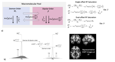 |
MT & ihMT
Guillaume Duhamel
Keywords: Neuro: White matter, Contrast mechanisms: Microstructure, Contrast mechanisms: CEST & MT This course provides an outline of the potential of magnetization transfer techniques for myelin imaging. In particular, this presentation will show how inhomogeneous magnetization transfer (ihMT) can overcome limitations of the basic MT contrasts (MTR, MPF) to provide more specific information related to myelin. The potential and scientific/technical challenges of these approaches for clinical applications are discussed. |
| 11:05 |
 |
Direct UTE MR Imaging of Myelin
Yajun Ma
Keywords: Neuro: Brain, Neuro: White matter Myelin is a lipid-protein bilayer, which plays an essential role for brain function in facilitating the rapid conduction of action potentials in the axon. Loss of myelin is the hallmark of numerous inflammatory andneurodegenerative disorders. UTE or ZTE MRI sequences with echo times shorter than 100 us make it possible to direct image and quantify myelin content in brain. This lecture summarizes the recent development and applications of UTE and ZTE techniques for evaluation of myelin changes ex vivo and in vivo. |
 Back to Meeting Home
 Back to the Program-at-a-Glance
The International Society for Magnetic Resonance in Medicine is accredited by the Accreditation Council for Continuing Medical Education to provide continuing medical education for physicians.
 Back to Meeting Home
 Back to the Program-at-a-Glance
 View Presentation Video