Weekend Course
Key Contrast Mechanisms for Imaging Neuroinflammation
ISMRM & ISMRT Annual Meeting & Exhibition • 03-08 June 2023 • Toronto, ON, Canada

Weekend Course
Key Contrast Mechanisms for Imaging Neuroinflammation
| Contrast Agents | |||
| 13:15 |
Using Iron Oxides to Track Neuroinflammation
Vanessa Wiggermann
Keywords: Contrast mechanisms: Contrast agents, Cross-organ: Inflammation, Image acquisition: Quantification Iron oxide nanoparticles are a promising approach to potentially track inflammation in vivo, and could additionally aid in targeted drug delivery and other mechanisms. Their exact properties are governed by their size and shell, providing great versatility in tagging and tracking different cells. Due to their iron-loading, MRI techniques that are sensitive to magnetic susceptibility changes allow to track their location. In this talk, we will provide an introduction to these nanoparticles as contrast agents and discuss MRI-based tracking as well as magnetic particle imaging, a novel approach to quantify cell numbers in vivo. |
||
| 13:40 | Imaging Blood-Brain Barrier Permeability with Gd Audrey Chagnot, Axel Montagne | ||
| Endogenous Contrast: Iron | |||
| 14:05 |
 |
Iron Imaging Using QSM, R2* & SWI
Sina Straub
Keywords: Contrast mechanisms: Susceptibility, Contrast mechanisms: Relaxometry, Neuro: Brain The basics of SWI, QSM and R2* will be introduced along with the contrast mechanisms for iron imaging. Sensitivity, specificity and limitations will be illustrated as well as examples for iron imaging in neurologic disorders. Susceptibility is a measure of how much a material gets magnetized in an external magnetic field. Paramagnetic materials have unpaired electrons and paramagnetism is temperature dependent. QSM differentiates between para- and diamagnetic materials. R2* relaxometry is sensitive to both iron and myelin. QSM and SWI can differentiate MS lesion types. QSM and R2* quantify age- and pathology related iron accumulation in brain nuclei and cortices. |
|
| 14:30 |
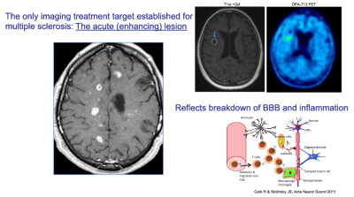 |
Case Study: Active Lesions in MS
Susan Gauthier
Keywords: Neuro: Brain Chronic CNS inflammation in the MS lesion is maintained with pro-inflammatory microglia at the rim of chronic active MS lesions. The presence of iron-laden microglia found at the rim of a subset of chronic active MS lesions affords a substrate to detect these lesions with gradient echo (GRE) MRI. Utilizing different GRE approaches, studies have confirmed that paramagnetic rims lesions (PRL) are specific for MS and associated with multiple pathological features of the disease. Quantification of the temporal change of inflammation within PRL can be harnessed and potentially utilized to evaluate the impact of treatment. |
|
| 14:55 |
Break & Meet the Teachers |
||
| Diffusion | |||
| 15:20 |
Sensitivity of Water Diffusion to Neuroinflammation
Ileana Jelescu
Keywords: Neuro: Brain, Contrast mechanisms: Diffusion, Cross-organ: Inflammation Diffusion MRI holds great potential to capture inflammatory processes in vivo and non-invasively, as the diffusion of water molecules in the brain is highly sensitive to changes in the microstructure resulting from glial proliferation, cytotoxic or vasogenic edema, demyelination and other cellular processes. Here we describe how various neuroinflammatory processes are expected to affect metrics derived from the diffusion and kurtosis tensors (sensitivity), with examples from both preclinical animal models and clinical human studies. We briefly discuss how biophysical models could disentangle between individual pathological processes (specificity), but emphasize the need for further development and validation of these methods. |
||
| 15:45 |
Sensitivity of Metabolite Diffusion to Neuroinflammation
Clemence Ligneul
Keywords: Neuro: Brain, Contrast mechanisms: Diffusion, Contrast mechanisms: Spectroscopy Unlike water, some brain metabolites are intracellular and disproportionately concentrated in glia (compared to neurons). Diffusion magnetic resonance spectroscopy (dMRS) allows to measure diffusion properties of metabolites, and therefore provides sensitivity to some morphological features of the compartments/cell-types they diffuse in. For example, diffusion properties of metabolites more importantly concentrated in astrocytes reflect the hypertrophy that reactive astrocytes exhibit. More generally, changes in diffusion properties of “glial markers” often correlate with neuroinflammation. This presentation will introduce the principles of dMRS and show its specific advantages (and challenges) for detecting neuroinflammation. |
||
| MT & CEST and Glymphatics | |||
| 16:10 | Novel CEST analysis tools: towards easy CEST Xiaolei Song | ||
| 16:35 |
Imaging the Glymphatic System During Neuroinflammation
Farshid Sepehrband
Keywords: Neuro: Cerebrovascular, Contrast mechanisms: Diffusion, Image acquisition: Image processing The brain waste clearance system plays a critical role in the removal of toxic substrates from the brain to maintain parenchymal homeostasis. Different pathways are hypothesized to explain this complex network of brain clearance. Regardless of the directionality, perivascular spaces are known to act as highways of influx and efflux, facilitating the glia-lymphatic drainage. In this educational talk, we will discuss brain clearance system and ways it can be assessed using neuroimaging techniques. We will then discuss if and how the clearance system may change in the presence of neuroinflammation. |
||
 Back to Meeting Home
 Back to the Program-at-a-Glance
The International Society for Magnetic Resonance in Medicine is accredited by the Accreditation Council for Continuing Medical Education to provide continuing medical education for physicians.
 Back to Meeting Home
 Back to the Program-at-a-Glance
 View Presentation Video