Constrained & Model-Based Reconstructions
Acq/Recon/Analysis Monday, 17 May 2021
Digital Poster

Oral Session - Constrained & Model-Based Reconstructions
Acq/Recon/Analysis
Monday, 17 May 2021 14:00 - 16:00
  • Results of the 2020 fastMRI Brain Reconstruction Challenge
    Bruno Riemenschneider1, Matthew Muckley2, Alireza Radmanesh1, Sunwoo Kim3, Geunu Jeong3, Jingyu Ko3, Yohan Jun4, Hyungseob Shin4, Dosik Hwang4, Mahmoud Mostapha5, Simon Arberet5, Dominik Nickel6, Zaccharie Ramzi7,8, Philippe Ciuciu7, Jean-Luc Starck7, Jonas Teuwen9, Dimitrios Karkalousos10, Chaoping Zhang10, Anuroop Sriram11, Zhengnan Huang1, Nafissa Yakubova2, Yvonne W. Lui1, and Florian Knoll1
    1NYU School of Medicine, New York, NY, United States, 2Facebook AI Research, New York, NY, United States, 3AIRS Medical, Seoul, Korea, Republic of, 4Yonsei University, Seoul, Korea, Republic of, 5Siemens Healthineers, Princeton, NJ, United States, 6Siemens Healthcare GmbH, Erlangen, Germany, 7CEA (NeuroSpin) & Inria Saclay (Parietal), Université Paris-Saclay, Gif-sur-Yvette, France, 8Département d’Astrophysique, CEA-Saclay, Gif-sur-Yvette, France, 9Radboud University Medical Center, Nijmegen, Netherlands, 10Amsterdam UMC, Amsterdam, Netherlands, 11Facebook AI Research, Menlo Park, CA, United States
    The 2020 fastMRI challenge revealed a new state-of-the-art machine learning reconstruction model that achieved the best metrics and ranking in almost all challenge categories. The challenge also clarified areas in need of further research.
    Figure 2: Exemplary images (up-down: T2, T1POST, and FLAIR) from all tracks and finalist teams. The submissions were rated for overall appearance, artifacts, sharpness, and CNR. Overall, judged cases included both intra- and extra-axial tumors, strokes, microvascular ischemia, white matter lesions, edema, surgical cavities, as well as postsurgical changes and hardware including craniotomies and ventricular shunts.
    Figure 3: Examples of failed reconstructions, i.e., resulting artifacts and hallucinations. In the 4x example, some non-winning submissions introduced vessel-like aliasing artifacts, originating from a susceptibility artifact. A similar aliasing-induced hallucination, resulting in incorrect sulcus depiction, is observed in the Transfer case. The particular shown 8x reconstruction suffered from a severe aliasing and hallucination behavior that renders the reconstruction unusable in more obvious fashion.
  • Region-Optimized Virtual (ROVir) Coils: Application of Sensor-Domain Beamforming for Localizing and/or Suppressing Spatial Regions
    Daeun Kim1, Stephen F. Cauley2, Krishna S. Nayak1, Richard M. Leahy1, and Justin P. Haldar1
    1Electrical and Computer Engineering, University of Southern California, Los Angeles, CA, United States, 2Radiology, A. A. Martinos Center for Biomedical Imaging, Massachusetts General Hospital, Charlestown, MA, United States
    We demonstrate that virtual coils can localize signal from a region-of interest and suppress signal from uninteresting spatial regions, all without requiring pulse sequence or hardware modifications.  Reduced-FOV imaging applications are shown.
    Figure 4. Illustration of using beamforming to steer the sensitivity of the virtual array to different spatial positions. The video shows ROVir results obtained from sweeping a circular-shaped ROI (i.e., everything interior to the green circle) through the FOV for a 32-channel brain MRI dataset.
    Figure 3. Application of ROVir to reduce the size of the FOV in non-cartesian sagittal brain imaging, which enables highly-effective mitigation of aliasing artifacts when using a spiral k-space trajectory designed for a smaller FOV. The signal ROIs are marked in green, while the interference regions are marked in red. Reconstruction was based on simple gridding13.
  • Compact Maps: A Low-Dimensional Approach for High-Dimensional Time-Resolved Coil Sensitivity Map Estimation
    Shreya Ramachandran1, Frank Ong2, and Michael Lustig1
    1Electrical Engineering and Computer Sciences, University of California, Berkeley, Berkeley, CA, United States, 2Electrical Engineering, Stanford University, Stanford, CA, United States
    We present a method that solves for a compact representation of time-resolved coil sensitivity maps to dramatically reduce their size in memory (by ~1000x). Our Compact Maps result in decreased calibration error and improved reconstruction quality when compared to time-averaged maps.
    Figure 2. Overview of Compact Maps in a dynamic MRI reconstruction pipeline. Maps are generally pre-computed for reconstructions with temporal constraints, not computed on-the-fly. Conventionally, maps are estimated (a) from time-averaged k-space data or (b) individually for each time frame, then stored in the spatial domain. (c) Instead, Compact Maps are linear combinations of temporal basis components. Memory estimates are for $$$N$$$=4 basis components, 24x24 map kernel size, 8 channels, and the listed matrix size. We see ~1000x savings in memory with Compact Maps over (b).
    Figure 5. Comparison of image reconstruction quality using time-averaged maps and Compact Maps. Reconstruction has been performed for both time frames A and B using iterative SENSE with l2 regularization of λ=0.001. Difference images (x10) with the fully sampled reference are also shown. Residual aliasing artifacts are visible in images reconstructed with time-averaged maps (red arrows) and in the magnitude difference images, but are mitigated when using Compact Maps. Normalized root-mean square error (NRMSE) is also reduced in the images reconstructed using Compact Maps.
  • Coil Sketching for fast and memory-efficient iterative reconstruction
    Julio A. Oscanoa1,2, Frank Ong3, Zhitao Li2,3, Christopher M. Sandino3, Daniel B. Ennis2,4, Mert Pilanci3, and Shreyas S. Vasanawala2
    1Department of Bioengineering, Stanford University, Stanford, CA, United States, 2Department of Radiology, Stanford University, Stanford, CA, United States, 3Department of Electrical Engineering, Stanford University, Stanford, CA, United States, 4Cardiovascular Institute, Stanford, CA, United States
    Our Coil Sketching approach can be used to accelerate and improve memory efficiency of iterative reconstruction with virtually no penalty on reconstruction accuracy.
    Figure 1. Structure of Eigen-Sketching matrix $$$S^t$$$. Effectively, matrix $$$S^t$$$ sketches the sensitivity map operator $$$C$$$, yielding a reduced sensitivity map operator $$$C^t_S$$$ with less coils. Similar to coil compression4-7(CC) the Eigen-Sketching matrix considers the high-energy virtual coils; but unlike CC, the matrix also considers a sketched coil. This sketched coil is formed by multiplying the remaining coils by “-1” or “+1” with probability 50% each and then summing them together. In sketching literature, this scheme is denoted as CountSketch.10-12
    Figure 5. Image comparison of 3D Cones reconstruction for (A) reconstruction with all $$$c=20$$$ coils, (B) coil compression with $$$\hat{c}=4$$$ coils, (C) our proposed Coil Sketching with $$$\hat{c}=4$$$ coils. Coil compression with $$$\hat{c}=4$$$ presents significantly lower image quality and shading artifacts, specially towards the lower borders of the volume. Coil Sketching yields virtually the same image as reconstruction with all $$$c=20$$$ coils.
  • Fast Calibrationless Image-space Reconstruction by Structured Low-rank Tensor Estimation of Coil Sensitivity and Spatial Support
    Zheyuan Yi1,2,3, Yujiao Zhao1,2, Yilong Liu1,2, Yang Gao1,2, Mengye Lyu4, Fei Chen3, and Ed X Wu1,2
    1Laboratory of Biomedical Imaging and Signal Processing, The University of Hong Kong, Hong Kong SAR, China, 2Department of Electrical and Electronic Engineering, The University of Hong Kong, Hong Kong SAR, China, 3Department of Electrical and Electronic Engineering, Southern University of Science and Technology, Shenzhen, China, 4College of Health Science and Environmental Engineering, Shenzhen Technology University, Shenzhen, China
    This study presents a fast and calibrationless reconstruction approach that derives high-quality coil sensitivity and spatial support maps by structured low-rank tensor estimation, resulting in efficient and robust image-space reconstruction. 
    Figure 1. The workflow of proposed method. 8-channel 4-contrast k-space data has been structured into a low-rank tensor. Subsequent higher-order singular value decomposition identifies both signal- and null-subspace bases. Different from low-rank reconstruction, all bases have been transformed to image space to estimate coil sensitivity and spatial support maps shared among contrasts by summation. The efficient image-space reconstruction was performed iteratively with the aforementioned procedures until convergence.
    Figure 2. Estimated coil sensitivity and spatial support maps from fully sampled reference, uniformly undersampled, and reconstructed data (30th iteration) using the proposed method. Coil sensitivity and spatial support maps estimated from fully sampled reference data are nearly orthogonal complements within the brain region. The proposed iterative reconstruction can effectively and efficiently correct the estimation errors (locations indicated by red arrows) of each channel due to uniform undersampling (R = 4).
  • Data-driven motion-corrected brain MRI incorporating pose dependent B0 fields
    Yannick Brackenier1,2, Lucilio Cordero-Grande1,2,3, Raphael Tomi-Tricot1,2,4, Tom Wilkinson1,2, Jan Sedlacik1,2, Philippa Bridgen1,2, Sharon Giles1,2, Shaihan Malik1,2, Enrico De Vita1,2, and Joseph V Hajnal1,2
    1Biomedical Engineering Department, School of Biomedical Engineering and Imaging Sciences, King's College London, London, United Kingdom, 2Centre for the Developing Brain, School of Biomedical Engineering and Imaging Sciences, King's College London, London, United Kingdom, 3Biomedical Image Technologies, ETSI Telecomunicación, Universidad Politécnica de Madrid and CIBER-BNN, Madrid, Spain, 4MR Research Collaborations, Siemens Healthcare Limited, Frimley, United Kingdom
    Fully data-driven retrospective motion correction is applied for volumetric brain MRI at 7T by including pose-dependent field in the forward model. Reduced motion corruption is obtained for the proposed reconstruction and is validated on an in-vivo spoiled gradient echo acquisition.
    Figure 1: Relative B0 field changes $$$\omega(\textbf{r})$$$ in the head frame between two poses, obtained after registration, can be decomposed in lower-degree solid harmonics (n=2) and localised fields that are proportional to rotation angles $$$\theta_{pitch}$$$ and $$$\theta_{roll}$$$ (12 and 10 degrees for this example). The linear model $$$d(\textbf{r})$$$ (not shown here) was fitted on a total of 8 motion-free scans. Data shown here is not used in further reconstructions and only served to validate our model.
    Figure 2: Sagittal (left), transversal (middle) and coronal (right) view of the (A) uncorrected (B) motion-corrected and proposed motion + B0 reconstruction (C) without and (D) with considering the solid harmonics (SH). Reduced motion corruption is observed when including the pose-dependent B0 fields with the biggest contribution coming from the linear B0 model, resulting in recovered signal near air-tissue interfaces (white arrows) and improved contrast, e.g. around the ventricles. Residual improvements are obtained when including the solid harmonics (red arrows).
  • Manifold learning via tangent space alignment for accelerated dynamic MR imaging with highly undersampled (k,t)-data
    Yanis Djebra1,2, Isabelle Bloch2,3, Georges El Fakhri1, and Chao Ma1
    1Gordon Center for Medical Imaging, Department of Radiology, Massachusetts General Hospital, Harvard Medical School, Boston, MA, United States, 2LTCI, Telecom Paris, Institut Polytechnique de Paris, Paris, France, 3LIP6, Sorbonne University, CNRS, Paris, France
    We present a new approach to manifold learning-based image reconstruction for dynamic MR that can be seen as a generalized low-rank matrix/tensor model. The geometries of the manifold are learned locally and images are reconstructed globally, aligning the tangent spaces in each neighborhood.
    Figure 3: Comparison of the proposed method with Low-Rank (LR) reconstructions. The first row shows representative images obtained by the LR reconstruction with rank 20 using k-space data of 3-min acquisition (reference). The rows 2 to 4 show reconstructions using only 45 sec of data. The 2nd row shows images reconstructed using a global LR model of rank 20. The 3rd row shows images obtained by performing a rank-10 local LR reconstruction (i.e. at each neighborhood). The 4th row shows the images using the proposed method where the dimension of each tangent space is 10.
    Figure 2: Global coordinates (spatial coefficients) and affine transform matrices of the proposed method. The 10×10 affine transform matrix $$${L}_c^{-1}$$$ is represented using grey levels. The 1st component of $$$T$$$ is a temporal average of all the neighborhoods. Our proposed model successfully describes the local coordinates by applying an affine transform $$${L}_c^{-1}$$$ to the global coordinates $$$T$$$ at each neighborhood $$$c$$$. The green dashed lines indicate the position of the liver for the 1st component of the 1st neighborhood.
  • One-heartbeat cardiac CINE imaging via jointly regularized non-rigid motion corrected reconstruction
    Gastao Cruz1, Kerstin Hammernik2,3, Thomas Kuestner1, Daniel Rueckert2,3, René M. Botnar1, and Claudia Prieto1
    1School of Biomedical Engineering and Imaging Sciences, King's College London, London, United Kingdom, 2Technical University of Munich, Munich, Germany, 3Department of Computing, Imperial College London, London, United Kingdom
    Conventional cardiac CINE imaging relies on highly ill-posed motion resolved reconstructions from data acquired across several heartbeats. Here, we propose a jointly regularized motion corrected framework that uses all the data for every cardiac phase, enabling one-heartbeat CINE.
    Fig.4 Animated CINE for a third representative subject reconstructed with iterative SENSE (column 1), XD-GRASP (column 2) and the proposed MC-CINE (column 3) using 20 heartbeats (row 1), 2 heartbeats (row 2) and 1 heartbeat (row 3). The dynamics of the cardiac contraction are captured in every case, however considerable streaking and noise amplification are visible for XD-GRASP and especially iterative SENSE at higher accelerations. MC-CINE with 448 spokes (1 heartbeat) achieves comparable image quality to iterative SENSE and XD-GRASP with 8960 spokes (20 heartbeats).
    Fig.1 Diagram of the proposed approach (considering here only three cardiac phases). 1) Acquired data is retrospectively binned into multiple cardiac phases. 2) Preliminary cardiac resolved images are obtained via XD-GRASP. 3) Each cardiac phase is registered to every other cardiac phase to estimate the cardiac motion between all phases. 4) The estimated motion is incorporated into the proposed MC-CINE to reconstruct each cardiac phase as a motion corrected image from all the data.
  • Fourier-based decomposition approach for simultaneous acquisition of 1H spectra from two voxels in vivo at short echo times
    Layla Tabea Riemann1, Christoph Stefan Aigner1, Ralf Mekle2, Sebastian Schmitter1,3, Bernd Ittermann1, and Ariane Fillmer1
    1Physikalisch-Technische Bundesanstalt (PTB), Braunschweig und Berlin, Germany, 2Center for Stroke Research Berlin, Charité Universitätsmedizin, Berlin, Germany, 3Center for Magnetic Resonance Research, University of Minnesota, Minneapolis, MN, United States
    This work demonstrates the feasibility of a Fourier-based approach to decompose signals and assign the locations after simultaneous dual-voxel acquisition for 1H MR spectroscopy in vivo without the need for additional image acquisitions.
    Fig.3: Diagram of both decompositions: a) vGRAPPA: Measured low SNR SVS data (yellow) is used to generate the kernel to decompose the acquired SMVS data (pink) to their respective voxel regions. The channel-wise ACS was obtained by 2D-stacking of the data points and averages. Then, this matrix was convolved with an 11x2 kernel to generate the pseudo inverse matrix following9. b) SENSE-based decomposition: channel-wise sensitivity maps and noise covariance matrix, derived from image- and spectral data, respectively, are used to decompose the SMVS data.
    Fig.2: in vivo acquisition: a) voxel region of the left (blue, V1) and right motor cortex (orange, V2). b-c) Spectral shapes of the two SVS acquisitions (cyan/orange) and d-g) the SVS acquisition decomposed like the SMVS acquisitions with both the vGRAPPA (blue/red) and the SENSE-based (dark blue/dark red) algorithm. Both algorithms should decompose the SVS voxel to its respective region (d and g), while ideally resulting in equally distributed noise for the other voxel (e and f).
  • Optimized Subspace-Based J-Resolved MRSI for Simultaneous Metabolite and Neurotransmitter Mapping
    Zepeng Wang1,2, Yahang Li1,2, and Fan Lam1,2
    1Department of Bioengineering, University of Illinois Urbana-Champaign, Urbana, IL, United States, 2Beckman Institute for Advanced Science and Technology, Urbana, IL, United States
    We presented further optimized J-resolved MRSI for high-resolution, 3D metabolite, and neurotransmitter mapping. Estimation-theoretic TE selection within a union-of-subspaces framework was analyzed. Simultaneously mapping of major metabolites, Glx, and GABA are provided.
    Figure 5. High-resolution and high-SNR metabolite and neurotransmitter maps estimated from the in vivo data (a 3.4×3.4×5.3mm3 nominal resolution). Anatomical images (T1w) across different slices from the 3D imaging volume are shown in the top row, and maps of different metabolites, as well as the Glx and GABA components, are shown in subsequent rows.
    Figure 2. Monte-Carlo analysis showing the normalized standard deviation (std) of the coefficient estimates for different components from the UoSS fitting (Row 1: metabolite, Row 2: Glx, and Row 3: GABA). Several alternative TE choices with an equivalent acquisition time are considered for a 2-TE case. Specifically, columns 1-4 display the std maps for the case of single-TE (35ms, 2 average), first 2 TEs (35 and 50ms), random 2 TEs (50 and 110ms), and optimized 2 TEs (65 and 80ms), respectively, The best std maps were achieved by the optimized 2 TEs, consistent with the CRLB prediction.
Back to Top
Digital Poster Session - Parallel Imaging Reconstruction
Acq/Recon/Analysis
Monday, 17 May 2021 15:00 - 16:00
  • Calibrationless Parallel Imaging Reconstruction for Simultaneous Multi-slice PROPELLER of Upper Abdomen
    Yilong Liu1,2, Kun Zhou3, Dehe Weng3, Hua Guo4, and Ed X. Wu1,2
    1Laboratory of Biomedical Imaging and Signal Processing, The University of Hong Kong, Hong Kong, China, 2Department of Electrical and Electronic Engineering, The University of Hong Kong, Hong Kong, China, 3Siemens Shenzhen Magnetic Resonance Ltd, Shenzhen, China, 4Center for Biomedical Imaging Research, Department of Biomedical Engineering, Tsinghua University, Beijing, China
    The proposed method performs simultaneously calibrationless parallel imaging (CPI) reconstruction and blade combination. Compared to conventional split slice-GRAPPA, it jointly reconstructs all blades, leading to improved SNR and reduced artifacts in abdominal imaging.
    Figure 4 Reconstruction for 30-channel abdominal MR data at MB=2/R=2. Compared to SPSG, the proposed CPI results in improved SNR in both background and abdominal regions. As shown in the zoomed view, SPSG suffered from slight residual artifact (vertical ringing indicated by red arrow), which was not visible in results from proposed CPI.
    Figure 1 Diagram of the proposed calibrationless parallel imaging (CPI) reconstruction. It iteratively updates the estimated k-space by sequentially promoting structural low-rankness and enforcing data consistency. (A) Structural low-rankness is promoted for individual slices by constructing a block-wise Hankel matrix, performing singular value decomposition, and forcing low-rankness through rank truncation. (B) The data consistency is enforced for individual blades by minimizing the difference between synthesized/acquired SMS blade data.
  • Reduced Field of View Parallel Imaging with Wave Encoded k-Space Trajectory
    Zhilang Qiu1,2, Sen Jia1, Haifeng Wang1, Lei Zhang1, Xin Liu1, Hairong Zheng1, and Dong Liang1
    1Shenzhen Institutes of Advanced Technology, Chinese Academy of Sciences, Shenzhen, China, 2University of Chinese Academy of Sciences, Beijing, China
    This work proposes to extend the existing reconstruction methods, ESPIRiT and SPIRiT (named r2f-ESPIRiT and r2f-SPIRiT, respectively) to reconstruct artifact-free images in wave encoded reduced FOV imaging scenario, with full FOV images additionally obtained without adding scan time.
    Figure 1. Diagram of the proposed r2f-ESPIRiT reconstruction for reduced FOV parallel imaging with wave encoded k-space trajectory. Although accelerated reduced FOV imaging scan is performed, the full FOV image can be reconstructed.
    Figure 5. Wave encoded 3D imaging with 1 (PE) × 2 (PAR) acceleration. In the conventional ESPIRiT reconstruction, 2 sets of maps were estimated from a reduced FOV ACS scan (a), and residual aliasing artifacts (yellow arrows) remain in the reconstructed image of RO-PAR plane (c). In the proposed r2f-ESPIRiT, an ACS scan with 40% slice-oversampling was used to estimate the sensitivity maps (b), but it does not add additional scan time, and an artifact-free and full FOV image can be obtained (d).
  • Marchenko-Pastur Virtual Coil Compression (MP-VCC)
    Gregory Lemberskiy1, Jelle Veraart1, Benjamin Ades-aron1, Els Fieremans1, and Dmitry S Novikov1
    1Radiology, NYU School of Medicine, New York, NY, United States
    We propose a method of virtual coil compression using random matrix theory, MP-VCC, in which the Marchenko-Pastur distribution defines how many virtual coils may be discarded without loss beyond the PCA precision. MP-VCC is evaluated for PF, regular undersampling, and MB acceleration. 
    Marchenko-Pastur Virtual Coil Compression (MP-VCC) For the aliased region of MB=2 experiment, we display a (A) local spatial patch, $$$X_C$$$, (B) its VC basis $$$X_{VC}$$$, and (C) its eigenvalue spectrum with the MP distribution in black. (D) Spatially varying virtual coil maps, $$$P_C$$$, are shown for every experiment.
    Statistics of Discarded VCs. Properties of the normalized residuals $$$r=(\text{noisy}-\text{denoised})/\sigma$$$, characterizing the discarded VCs, are evaluated via (A,B) histograms showing Gaussian distribution of $$$r$$$; and (C,D) power spectrum analysis, showing no memory along the measurement dimension (temporal power-spectrum $$$\Gamma(\omega)$$$ of residuals is flat) and marginal low-frequency bias along the spatial dimension (spatial power-spectrum $$$\Gamma(k)$$$ of residuals flat for almost all $k$)
  • non-Cartesian Parallel imaging and compressed sensing adapted for accelerating hybrid trajectory PETRA
    Fang Dong1, Dehe Weng1, and Nan Xiao1
    1Siemens Shenzhen Magnetic Resonance Ltd., Shen Zhen, China
    The technique combining non-Cartesian parallel imaging and Compressed sensing can be adapted and applied to accelerate the PETRA with hybrid trajectory for 2-5 folds.
    Fig.3: Images reconstructed from different under-sampled datasets with Standard nuFFT reconstruction methods (A) (B) and the modified PICS reconstruction methods(C)(D), (G) is the fully sampled image with Standard nuFFT reconstruction.
    Fig.2: Flowchart of PETRA data reconstruction with PICS algorithm
  • Near-optimal tuning-free multicoil compressed sensing MRI with Parallel Variable Density Approximate Message Passing
    Charles Millard1,2, Aaron T Hess2, Jared Tanner1, and Boris Mailhe3
    1Mathematical Institute, University of Oxford, Oxford, United Kingdom, 2Oxford Centre for Clinical Magnetic Resonance Research, University of Oxford, Oxford, United Kingdom, 3Digital Technology and Innovation, Siemens Healthineers, Princeton, NJ, United States
    We present the Parallel Variable Density Approximate Message Passing (P-VDAMP) algorithm for compressed sensing MRI, and find that it converges to a mean-squared error similar to optimally tuned FISTA, but in around 5x fewer iterations and without the need to tune model parameters.
    Fig 2. The aliasing of a zero-filled, density compensated estimate in the image and wavelet domains of a tenfold undersampled brain, and the wavelet-domain aliasing estimate $$$\boldsymbol{\tau}_0$$$. The histogram verifies that $$${\boldsymbol{r}}_0 \approx \boldsymbol{w}_0 + \mathcal{CN}(\boldsymbol{0}, \text{Diag}(\boldsymbol{\tau}_0))$$$ is an accurate model of the aliasing.
    Fig. 4. The NMSE vs iteration of three example reconstructions, demonstrating the relative rapidity of convergence of P-VDAMP. The NMSE at the 0th iteration differs because the 0th estimate is defined to be after the first application of soft thresholding.
  • Accelerating Bayesian Compressed Sensing for Fast Multi-Contrast Reconstruction
    Alexander Lin1, Demba Ba1, and Berkin Bilgic2,3
    1Harvard University, Cambridge, MA, United States, 2Department of Radiology, Massachusetts General Hospital, Martinos Center for Biomedical Imaging, Boston, MA, United States, 3Harvard Medical School, Boston, MA, United States
    We propose Bayesian accelerated Compressed Sensing (BaCS) to provide two orders of magnitude computational speed up in Bayesian CS, allowing it to run faster than sparseMRI while improving quality by exploiting joint reconstruction, and expanded its applicability to parallel imaging. 
    Fig4. In this single-coil, complex-valued, in vivo experiment with four contrasts, joint reconstruction with BaCS provides 2.0-fold to 1.2-fold RMSE reduction over sparseMRI for a wide range of 2D acceleration factors. It is 31x faster on the GPU, and 2.8x faster on the CPU compared to sparseMRI.

    Fig5. Synergistic combination of BaCS with SENSE yields a significant, 1.4x improvement in RMSE over standard SENSE reconstruction at R=5-fold total acceleration using 32 channel data. This is made possible by the application uniform R1=2-fold undersampling with SENSE, and an additional R2=2.5-fold random undersampling on the reduced FOV coil images with joint BaCS reconstruction.

  • VCC-Wave for Improved Parallel MRI of High Resolution and High Bandwidth
    Zhilang Qiu1,2, Sen Jia1, Shi Su1, Yanjie Zhu1, Xin Liu1, Hairong Zheng1, Haifeng Wang1, and Dong Liang1
    1Shenzhen Institutes of Advanced Technology, Chinese Academy of Sciences, Shenzhen, China, 2University of Chinese Academy of Sciences, Beijing, China
    To propose a novel model to alleviate the limitation of wave encoding in situations of high resolution and high bandwidth.
    Figure 1. A step-by-step simulation experiment considering a single coil with homogeneous uniform sensitivity and 2-fold regular under-sampling to illustrate the priors of Wave in the proposed VCC-Wave model.
    Figure 3. High resolution 2D imaging using a RF-spoiled GRE sequence and 24-channel head coils, with an isotropic resolution of 0.67 mm, under an acceleration factor of 6 (right to left).
  • Accelerated 3D Myelin Water Imaging using Joint Parallel Imaging and Variable Splitting Network
    Jae-Hun Lee1, Jaeuk Yi1, Kanghyun Ryu2, Soozy Jung1, and Dong-Hyun Kim1
    1Department of Electrical & Electronic Engineering, Yonsei Univ., Seoul, Korea, Republic of, 2Department of Radiology, Stanford Univ., Stanford, CA, United States
    We explored a reconstruction method using advanced parallel imaging with deep learning, which can utilize joint information. The proposed method shows improved quantitative values compared to conventional methods and can achieve high acceleration factors for mGRE based 3D MWI.
    Figure 1. Proposed reconstruction algorithm schematic.
    Figure 4. MWF maps of the different method reconstructed images with difference maps (R=2*2).
  • Simultaneous FLAIR T1W and T2W imaging using Temporal Harmonic Encoding
    Tzu-Cheng Chao1 and James G. Pipe1
    1Department of Radiology, Mayo Clinic, Rochester, MN, United States
    The proposed method reduces blurring in T1W FLAIR. In addition, both T1W and T2W FLAIR can be imaged in a single scan with no additional scan time.
    Fig 2. Blurring due to T2 decay can be noticed in the imaging from a conventional scan (red arrow) and is mitigated in both SENSE and Temporal Harmonic Encoding reconstruction despite noise increment. In spite of contrast reduction around basal ganglia, Temporal Harmonic Encoding image retains the gray/white matter contrast in most of the brain. Both SENSE and Temporal Harmonic Encoding demonstrates similar SNR reduction compared to the conventional imaging. On the other hand, the regularization applied on Temporal Harmonic Encoding image restores SNR with improved image quality.
    Fig 3. (a) The average of the other two harmonic components in Temporal Harmonic Encoding reconstruction indicates the signal changes with TE. Faster T2 decay region appears brighter in the harmonic image. (b) The inverse of (a) will convert the contrast to resemble a T2W image. (c) The T2 map is calculated from the multi-TE SENSE.
  • MGRAPPA: Motion Corrected GRAPPA for MRI
    Michael Rawson1, Xiaoke Wang2, Ze Wang2, Radu Balan1,3, and Thomas Ernst2
    1Department of Mathematics, University of Maryland at College Park, College Park, MD, United States, 2Department of Diagnostic Radiology and Nuclear Medicine, University of Maryland School of Medicine, Baltimore, MD, United States, 3Center for Scientific Computation and Mathematical Modeling, University of Maryland at College Park, College Park, MD, United States
    MRI motion correction removes artifacts and parallel imaging greatly decreases scan times. However, together, severe artifacts occur. We introduce MGRAPPA to drastically reduce artifacts when using prospective motion correction and GRAPPA. We observe 41% L2 error reduction in vivo. 
    Figure 1: Diagram showing flow of stages of scan and image reconstruction.
    Figure 5: Left: Subject. Center: Residual/error of MGRAPPA reconstruction of Subject. Right: Residual/error of GRAPPA reconstruction of Subject. The motion is rotation by 9 degrees and translation by 5 and 1 voxels/pixels in x and y directions respectively. GRAPPA reconstruction, but not MGRAPPA, showed a clear ghosting artifact. Accordingly, the L2-norm error decreased 41% with MGRAPPA.
  • Autocalibrating Segmented Diffusion Weighted Acquisitions (ASeDiWA)
    Michael Herbst1
    1Bruker BioSpin MRI GmbH, Ettlingen, Germany
    Autocalibrating Segmented Diffusion Weighted Acquisitions (ASeDiWA) enables interleaved segmented diffusion weighted EPI without phase navigation.
    Figure 2: One exemplary slice from the human scan is shown. Each row displays one b-value. In the first column a reconstruction without phase correction is shown, leading to strong ghosting in the diffusion weighted scans. The second column shows the GRAPPA reconstruction. The third column displays the ASeDiWA reconstruction. Both parallel imaging reconstruction methods correct the ghosting seen in the first column. However, ASeDiWA consistently provides higher SNR as the GRAPPA reconstruction.
    Figure 3: Reconstruction results (b/w) and g-factor simulations (color) are shown. Each row displays data with a different number of simulated segments (2, 3, and 4). The first column shows the data reconstructed with GRAPPA. The following three columns show results from ASeDiWA without and with one and two iterations, respectively. In general, higher segmentation leads to higher g-factors. Independent of the segmentation, the comparison of ASeDiWA with GRAPPA shows a reduced g-factor. Further improvement can be achieved by iteration of the algorithm (ASeDiWA1 and ASeDiWA2).
  • Resolving fold-over artefacts for Reduced Field-of-View Parallel Imaging with Cartesian Sampling
    Sen Jia1, Zhilang Qiu1,2, Lei Zhang1, Haifeng Wang1, Xin Liu1, Hairong Zheng1, and Dong Liang1
    1Paul C. Lauterbur Research Center for Biomedical Imaging, Shenzhen Institutes of Advanced Technology, Shenzhen, China, 2University of Chinese Academy of Sciences, Beijing, China
    Full field-of-view image without fold-over artefacts can be reconstructed by reduced FOV parallel imaging given full-FOV calibration is available. 
    Figure 1. Multiple maps modelling the aliasing of reduced field-of-view imaging could be created from full-FOV ESPIRiT CSM estimated from full-FOV calibration scan, and used in soft-SENSE reconstruction to reconstruct full-FOV image without fold-over artifacts.
    Figure 3. Full-FOV reconstruction of reduced FOV parallel imaging along two phase encoding directions with 2x2 downsampling by proposed ESPIRiT 4 maps created from full-FOV calibration scan
  • Accurate Quantitative G-factor Calculation in Dual-kernel Slice-GRAPPA Reconstruction
    Wei Liu1, Simon Bauer2, and Stephan Kannengiesser2
    1Siemens Shenzhen Magnetic Resonance Ltd, Shenzhen, China, 2Siemens Healthcare GmbH, Erlangen, Germany
    The proposed g-factor calculation method allows a practical, accurate quantification of the noise map in SG-DK reconstructions.
    Figure 2. Quantitative SNR maps derived from (a) the pseudo multiple-replica method (Mean SNR: 19.34), (b) directly from the proposed method (Mean SNR: 19.75), and (c) neglecting kernel phase correction (Mean SNR: 20.13). The difference map (d) is from (a) and (b), (e) is from (a) and (c). The deviation of the reconstructed synthetic noise and the calculated noise map is shown in (f) and (g), with similar RMS (~1.02). Both (d) and (f) show that the noise map with phase correction is more consistent with the reference method.
    Figure 1. (a) schematic of the SG-DK method to obtain the k-space data for a unaliased slice. Two kernel sets are calculated for odd and even lines in each unaliased slice respectively; (b) schematic of the kernel rearranging. By exchanging some kernel values, the new kernel sets can be applied on disjunct sets of k-space in collapsed data respectively. The final unaliased slice is then obtained by adding two reconstructed data from these two kernels. It notes that the full k-space of collapsed data has been divided into two sets with zero-padded lines (indicated with dot lines).
  • Coil Sensitivity Estimation with Deep Sets Towards End-to-End Accelerated MRI Reconstruction
    Mahmoud Mostapha1, Boris Mailhe1, Simon Arberet1, Dominik Nickel2, and Mariappan S. Nadar 1
    1Digital Technology and Innovation, Siemens Healthineers, Princeton, NJ, United States, 2Magnetic Resonance, Siemens Healthineers, Erlangen, Germany
    Predictions from the proposed end-to-end system achieved a PSNR of 33.93 dB and SSIM of 0.840 like those obtained using precomputed CSMs. However, we observed more artifacts with precomputed CSMs. DS-CSME system required less time (~0.2s) to estimate CSMs than ESPIRiT (~1s).
    DS-CSME: a deep learning solution for coils sensitivity estimation, allowing end-to-end learning framework for accelerated parallel magnetic resonance imaging reconstruction.
    An example comparing the fully sampled target to predictions obtained using precomputed CSMs and those obtained by the end-to-end system with DC-CSME. At ~5× acceleration, predictions with precomputed CSMs show more artifacts.
  • Two-Dimensional Coil-signature-based Phase Cycled Reconstruction for Inherent Correction of Echo-Planar Imaging Nyquist Ghost Artifacts
    Silu Han1, Chidi Patrick Ugonna1, Mahesh Bharath Keerthivasan2,3, and Nan-kuei Chen1,3
    1Biomedical Engineering Department, The University of Arizona, Tucson, AZ, United States, 2Siemens Medical Solutions USA, New York, NY, United States, 3Medical Imaging Department, The University of Arizona, Tucson, AZ, United States
    Our novel 2D coil-signature-based phase-cycled reconstruction method can successfully reduce Nyquist artifacts in EPI.
    Figure 1. Illustration of SB-EPI correction. Figure 1(a): SB-EPI with Nyquist artifact. Figure 1(b): Regions marked as green used for CSP comparison. Figure 1(c): A series of corrected complex images of one coil after applying different possible phase value. Figure 1(d): Reference Nyquist artifact-free CSP and CSPs generated from corrected complex images in Figure 1(c). Figure 1(e): Variations of CSP difference compared to reference Nyquist artifact-free CSP with different phase value.
    Figure 2. Workflow of correction for SENSE MB-EPI ($$$Acc_{in}=2,Acc_{MB}=2$$$). (A): SENSE MB-EPI; (B): Possible phase value along frequency-encoding direction; (C): Performed correction; (D): Corrected complex images with different phase values; (E): Corresponding CSPs; (F): Nyquist artifact-free CSP; (G): Determined phase $$$C_{0}$$$; (H): Possible phase value along phase-encoding direction; (I): Cycled 2D phase map; (J): Unaliased CSP from scanner; (K): Performed correction with 2D phase map and unaliased CSP; (L) Final corrected images.
  • Is good old GRAPPA dead?
    Zaccharie Ramzi1,2,3, Philippe Ciuciu1,2, Jean-Luc Starck3, and Alexandre Vignaud1
    1Neurospin, Gif-Sur-Yvette, France, 2Parietal team, Inria Saclay, Gif-Sur-Yvette, France, 3Cosmostat team, CEA, Gif-Sur-Yvette, France
    XPDNet, a state-of-the-art deep learning approach for MRI reconstruction, can generalize well when compared to GRAPPA on unseen settings.
    Magnitude reconstruction results for a brain acquired at acceleration factor 2, contrast T2, and field strength of 7T. The top row represents the reconstruction using the different methods, while the bottom one represents a zoom in the cerebellum region, an anatomical feature that was not present in the XPDNet training set.
    Magnitude reconstruction results for a specific fastMRI slice with T2 contrast, at acceleration factor 8. The top row represents the reconstruction using the different methods, while the bottom row represents the error when compared to the reference.
  • ZTE Infilling From Auto-calibration Neighbourhood Elements
    Tobias C Wood1, Emil Ljungberg1, and Mark Chiew2
    1Neuroimaging, King's College London, London, United Kingdom, 2Wellcome Centre for Integrative Neuroimaging, FMRIB, Nuffield Department of Clinical Neurosciences, University of Oxford, Oxford, United Kingdom
    We present a method for filling the dead-time gap in ZTE imaging using a parallel imaging technique. We present results in phantoms and a human brain demonstrating high fidelity reconstructions comparable to existing methods that require collection of additional data.
    Figure 4: A low-resolution head scan acquired with a fast bandwidth and angular under-sampling. Despite the increased dead-time gap and paucity of calibration data, ZINFANDEL provides an artefact free reconstruction.
    Figure 2: Diagram of the ZINFANDEL algorithm. In step 1, a kernel matrix W is calculated from the calibration region. In step 2, a point in the dead-time gap is filled. By looping first over spokes and then missed samples, the matrix W can be progressively updated towards the center of k-space.
  • Hybrid K-space EPI (HyKE) Reconstruction for Accelerated Imaging
    Tyler E Cork1,2, Matthew J Middione1, Michael Loecher1, Kévin J Moulin1, John M Pauly3, and Daniel B Ennis1,4
    1Radiology, Stanford University, Stanford, CA, United States, 2Bioengineering, Stanford University, Stanford, CA, United States, 3Electrical Engineering, Stanford University, Stanford, CA, United States, 4Radiology, Veterans Affairs Health Care System, Palo Alto, CA, United States
    In preliminary simulations using a numerical phantom, an image recon pipeline was outlined to provide 2 images from 1 data acquisition. The data acquisition strategy, HyKE, suggests similar SNR efficiency to averaged acquisitions with the possibility to perform self-distortion correction
    The workflow of the HyKE image reconstruction pipeline implemented with the BART phantom. The first row displays the k-space. The second row shows the corresponding images. The numerical phantom was retrospectively subsampled with the Hybrid k-space EPI sampling pattern (column 1) for each of the coil images (N = 8). SPIRiT was run for 20 iterations (column 2) followed by coil combination (column 3). The phantom then was split into two separate k-spaces (column 4). A POCS Partial Fourier image reconstruction (column 5) ran for 10 iterations resulting in the two reconstructed images.
    A schematic of the image reconstruction pipeline for the HyKE approach. (A) The hybrid k-space EPI incorporates several reconstructions techniques, including (B) SPIRiT, (C) conjugate coil combination from the coil sensitivity maps, (D) separating the different sampling k-space trajectories, and a (E) POCS partial Fourier reconstruction, in order to acquire (F) two separate images from one hybrid EPI acquisition. The resultant output images have multiple advantages; such as accelerated imaging and the possibility to characterize EPI distortion in a single scan.
  • Highly undersampled GROG-BPE radial data reconstruction using Compressed Sensing
    Yumna Bilal1,2, Ibtisam Aslam1,3, Muhammad Faisal Siddiqui1, and Hammad Omer1
    1Medical Image Processing Research Group (MIPRG), Department of Electrical & Computer Engineering, COMSATS University Islamabad, Islamabad, Pakistan, 2Department of Electrical Engineering, University of Gujrat, Gujrat, Pakistan, 3Service of Radiology, Geneva University Hospitals and Faculty of Medicine, University of Geneva, Geneva, Switzerland
    Compressed Sensing reconstruction for radially undersampled GROG-BPE data with random blipping yields images with better clarity and quantifying parameters than conventionally used GROG-BPE CG-INNG method at higher acceleration factors.
    Figure 1: Block diagram of the proposed CS-GROG-BPE scheme. Undersampled radial signal is used to generate the Bunch Phase Encoded k-space at the specified k_max and NBP, using GROG. Non-Cartesian BPE k-space is gridded to Cartesian k-space using a defined oversampling factor and step-size using GROG. Gridded k-space is then reconstructed coil-by-coil by CS algorithm with specified regularization parameters. Coil-by-coil reconstructed images are then combined using sum-of-square reconstruction to yield the final output image.
    Figure 2: Reconstruction Results (a) shows fully sampled ground truth image. (b) shows results of CS-GROG-BPE method proposed in this work, at acceleration at AF = 8, 10, 12. (c) shows results of GROG-BPE CG-INNG method2 with the same BPE generation and gridding parameters. Quantifying parameters including AF, RMSE and SNR have been provided underneath each reconstructed image.
Back to Top
Digital Poster Session - Model-Based Reconstruction
Acq/Recon/Analysis
Monday, 17 May 2021 15:00 - 16:00
  • Multi-Scale Low-Rank Reconstruction for Phase-Cycled Projection-Reconstruction bSSFP Cardiac Cine and BMART-Generated B0 Maps
    Anjali Datta1, Dwight Nishimura1, and Corey Baron2
    1Electrical Engineering, Stanford, Stanford, CA, United States, 2Medical Biophysics, Western University, London, ON, Canada
    Multi-scale low-rank reconstruction recovers high-quality phase-cycle images from a highly-undersampled frequency-modulated bSSFP cardiac cine sequence.  It also facilitates estimation of a time series of B0 maps from the same data, which are used to combine the phase-cycles into the cine.
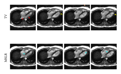
    Field-map-combined images for selected cardiac phases. The TV reconstructions have near-band-flow-related streaking artifacts (red arrows) and undersampling artifacts (peach arrow). In addition, some hyperintensities (yellow arrows) persist, possibly from using noisier field maps for phase-cycle combination. The MSLR reconstructions have smoother blood signal since flow artifacts were localized to near the bands, which were then removed by field-map combination. In addition, both blood-myocardium boundaries (cyan arrows) and background structures appear sharper.
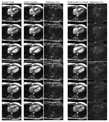
    Reconstructions from the inverse-gridded and retrospectively undersampled dataset. Six cardiac phases evenly spaced through the cardiac cycle are shown. The phase cycles were root-sum-of-squares combined. Multi-scale low rank results in sharper images and better removal of undersampling artifacts (e.g., in the background) than total variation.
  • Simplified Phase-Sensitive Inversion Recovery (PSIR) Reconstruction using Multi-dimensional Integration (MDI) for Elevated SNR
    Yichen Hu1 and Junpu Hu2
    1UIH America, Inc., Houston, TX, United States, 2United Imaging Healthcare, Shanghai, China
    We applied MDI algorithm to classic PSIR reconstruction for cardiac imaging and demonstrated the effectiveness of the approach for improved SNR. In comparison to the conventional reconstruction, the algorithm offers a simplified and fast pathway to achieve desired image contrast.
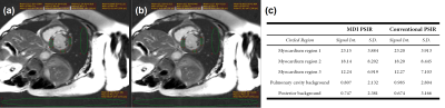
    Figure 2. Images for detecting myocardial infarction by (a) MDI PSIR and (b) the conventional PSIR reconstruction methods. (c) signal intensity analysis for the five representative regions (three in myocardium and two in the background) circled in (a) and correspondingly in (b).
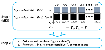
    Figure 1. Illustration of MDI PSIR reconstruction method
  • Single ProjectIon DrivEn Real-time (SPIDER) Multi-contrast MR Imaging Using Pre-learned Spatial Subspace
    Pei Han1,2, Junzhou Chen1,2, Fei Han3, Zhehao Hu1,2, Debiao Li1,2, Anthony G. Christodoulou1,2, and Zhaoyang Fan1,4
    1Biomedical Imaging Research Institute, Cedars-Sinai Medical Center, Los Angeles, CA, United States, 2Department of Bioengineering, UCLA, Los Angeles, CA, United States, 3Siemens Medical Solutions USA, Inc., Los Angeles, CA, United States, 4Departments of Radiology and Radiation Oncology, University of Southern California, Los Angeles, CA, United States
    A new technique called SPIDER is proposed for real-time multi-contrast 3D imaging. With the information learned and stored in the “Prep” scan, 3D multi-contrast images can be generated in the "Live" scan with simple matrix multiplication, which yielding a latency of 50ms or less.
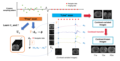
    Figure 1: The SPIDER framework.
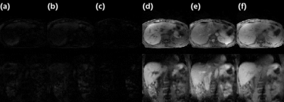
    Figure 3: Comparison of images from Multitasking reconstruction (reference) and the proposed SPIDER method. From left to right: (a) Reference from Multitasking reconstruction; (b) Contrast-variated SPIDER real-time images; (c) Difference between (a) and (b); (d-f) Contrast-frozen T1w, T2w, and PDw SPIDER real-time images.
  • Low-rank and Framelet Based Sparsity Decomposition for Reconstruction of Interventional MRI in Real Time
    Zhao He1, Ya-Nan Zhu2, Suhao Qiu1, Xiaoqun Zhang2, and Yuan Feng1
    1Institute for Medical Imaging Technology, School of Biomedical Engineering, Shanghai Jiao Tong University, Shanghai, China, 2School of Mathematical Sciences, MOE-LSC and Institute of Natural Sciences, Shanghai Jiao Tong University, Shanghai, China
    A low-rank and sparsity decomposition with framelet transform for spatial sparsity was proposed for reconstruction of interventional MR images. A group-based reconstruction showed that the proposed method can achieve an acceleration of 40 folds.
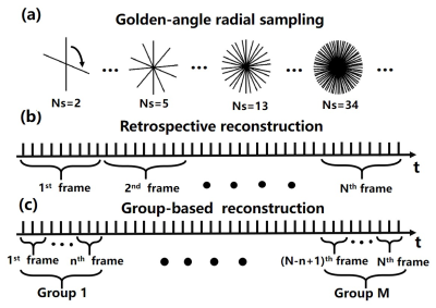
    Figure 1. Illustrations of the data acquisition and reconstruction scheme. (a) A continuous golden-angle radial sampling method was used for i-MRI in this study (golden angle = 111.25°). (b) Conventional dynamic image reconstruction based on a retrospective scheme. (c) The proposed group-based reconstruction method for real-time i-MRI reconstruction.
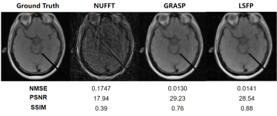
    Figure 2. A comparison of different algorithms. The ground truth is the 150th simulated brain intervention image. A group-based reconstruction strategy (10 spokes per frame, 5 frames per group, a total of 200 frames for 2000 spokes) was adopted for reconstruction using NUFFT, GRASP, and LSFP. The acceleration factor was about 40.
  • Reconstruction of Undersampled Dynamic MRI Data Using Truncated Nuclear Norm Minimization and Sparsity Constraints
    Runyu Yang1, Yuze Li1, and Huijun Chen1
    1Center for Biomedical Imaging Research, Department of Biomedical Engineering, Tsinghua University, Beijing, China
    Achieving high spatio-temporal resolutions is challenging in dynamic magnetic resonance imaging . It is effective to use low-rank prior and sparse prior  for dMRI reconstruction. We proposed a novel method used low rank  which utilize a nonconvex norm and sparse  for dMRI reconstruction.
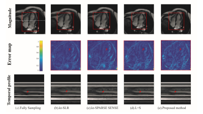
    FIG.2. Reconstruction results comparison for cardiac cine dataset with radial trajectory and r=2. From top to bottom: time-frame magnitude images, error map, x-t temporal profiles in white dotted line. The proposed method had lower artifacts for reconstruction of cardiac blood pools than the other methods by the arrows in the error map. By the arrows in the temporal profile, the methods to be compared present temporal blurring artifacts, which are effectively removed by the proposed mothed.
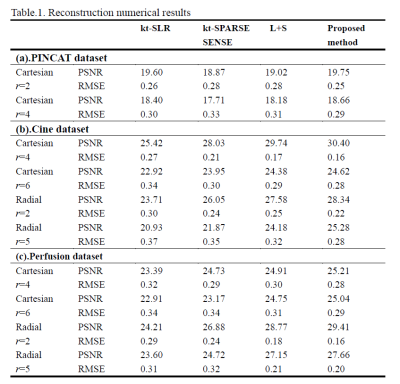
    Table.1. PSNR/RMSE comparison of various reconstruction methods on PINCAT dataset, Cine dataset and Perfusion dataset.
  • Time Domain Principal Component Analysis for Rapid, Real-Time MRI Reconstruction from Undersampled Data
    Mark Wright1, Bryson Dietz1, Jihyun Yun1,2, Eugene Yip2, B Gino Fallone1,2, and Keith Wachowicz1,2
    1Oncology, University of Alberta, Edmonton, AB, Canada, 2Medical Physics, Cross Cancer Institute, Edmonton, AB, Canada
    A real-time acceleration method using Principal Component Analysis (PCA) was developed for use on hybrid MR-radiotherapy machines. Good temporal-robustness was achieved at high frame-rate and low latency (less than 50ms), without the need for any complex coil geometries.
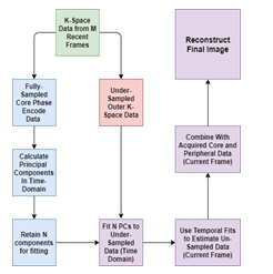
    Figure 2: Flow chart of the acceleration method. All calculations are done in the k-space domain.
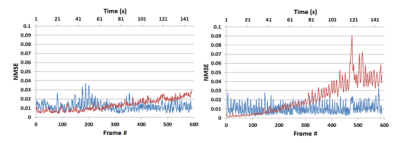
    Figure 3: A time evolution comparison of for two patients over 2.5 minutes using the spatial PCA method (red) and our proposed time-domain PCA method (blue) using normalised mean square error for an acceleration factor of 3.
  • Optimal Transport Based Convex Hybrid Image and Motion-Field Reconstruction
    Ingmar Middelhoff1, Matthias Schlögl2, Adrián Martín Fernández3, Silvio Fanzon4,5, Kristian Bredies4,5, and Rudolf Stollberger1,5
    1Institute of Medical Engineering, TU Graz, Graz, Austria, 2Solgenium OG, Linz, Austria, 3Department of Information and Communications Technologies, Pompeu Fabra University, Barcelona, Spain, 4Institute of Mathematics and Scientific Computing, NAWI Graz, University of Graz, Graz, Austria, 5BioTechMed-Graz, Graz, Austria
    An Optimal Transport based reconstruction for motion-afflicted data is tested which yields an image series and pixel-wise motion fields. Reconstructions based on simulated single-coil 4-fold undersampled k-space time-series data show good image quality.
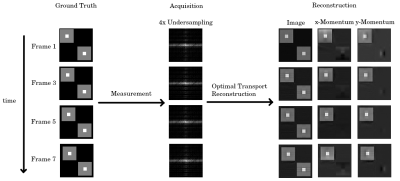
    A visualization of the reconstruction concept: A moving object is measured in a series of undersampled k-spaces. In this abstract, we simulated a 4-times acceleration per k-space with the 8 center k-space lines being measured additionally. The OT reconstruction allows not only to recover the image-series, but also the momentum fields of the object. For illustration purposes, only every second frame is shown.
    Zoom-in on the results from figure 2: The reconstructed image closely mirrors the ground truth. Despite every frame only having approximately one fourth of k-space sampled with a single coil, the brain could be reconstructed in detail with only the optimal transport regularization. The registered image is slightly blurry, showing potential problems with the motion field.
  • Accelerating gSlider-based Diffusion MRI: Phase constraints enable reduced RF encoding
    Yunsong Liu1, Kawin Setsompop2, and Justin P. Haldar1
    1Signal and Image Processing Institute, University of Southern California, Los Angeles, CA, United States, 2Department of Radiology, Stanford University, Stanford, CA, United States
    We investigate whether smooth-phase constraints can be used to reduce the required number of RF encodings in gSlider diffusion MRI.  Theoretical and simulation results demonstrate that, it can be done if optimized RF encodings are used.
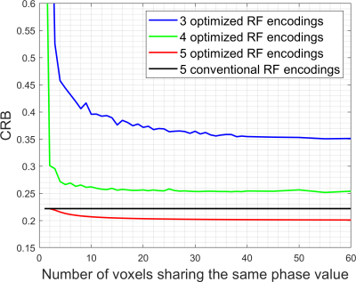
    Figure 2. CRBs for resolving 5 thin slices as a function of the number of voxels sharing the same phase value, plotted for several different RF encoding strategies. Note that without phase constraints, the CRB blows up to infinity whenever the number of RF encodings is smaller than the number of thin slices (5 in this case).
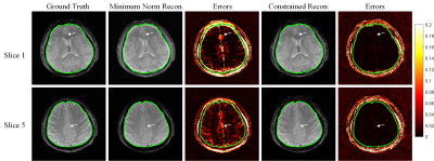
    Figure 3. Simulated reconstruction of 5 thin slices from the set of 4 optimized RF encodings, for both phase-constrained and minimum norm reconstruction. For ease of visualization, we only show two out of the five reconstructed slices for each case.
  • Improved Sampling for Distortionless Diffusion Weighted 2D Cartesian Multi-Shot Fast Spin Echo
    Philip Kenneth Lee1,2, Yuxin Hu1,2, Catherine Judith Moran2, Bruce Lewis Daniel2, and Brian Andrew Hargreaves1,2,3
    1Electrical Engineering, Stanford University, Stanford, CA, United States, 2Radiology, Stanford University, Stanford, CA, United States, 3Biomedical Engineering, Stanford University, Stanford, CA, United States
    Cartesian Fast Spin Echo can improve distortionless diffusion weighted imaging with multi-shot sampling patterns that complement low-rank compressed sensing reconstructions.
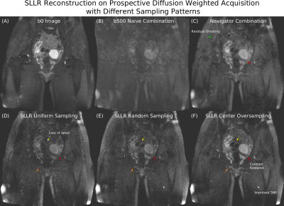
    Figure 5: (A) T2-weighted b0 image. (B) Naive combination of 12-shot data has severe multi-shot ghosting resulting in signal dropout. (C) Navigator shot combination has some residual ghosting (green). (D) SLLR reconstruction of uniformly sampled b500 data has loss of detail (yellow, orange), and reduced organ contrast (red). (E) Random sampling improves detail (yellow, orange). (F) Center oversampling further enhances detail (yellow, orange), restores organ contrast, matching the navigator combination (red), and improves SNR (white).
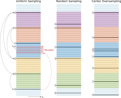
    Figure 1: Random sampling patterns were generated by dividing k-space into $$$ETL$$$ segments, represented by different colours. Each sample is randomly assigned to a shot. K-space is fully covered when shots are combined. For center oversampling, the center two ky lines were added to the beginning of a randomly generated shot. A low-resolution phase navigator was explicitly acquired with uniformly sampled data because the shot undersampling factor is too high for parallel imaging to recover phase maps.
  • Model-Based Iterative Reconstruction for Short-Axis Propeller EPI at 7T MRI
    Uten Yarach1,2, Frank Godenschweger3, Matt A Bernstein2, Myung-Ho In2, Itthi Chatnuntawech44, Kawin Setsompop5, Oliver Speck3, and Joshua Trzasko2
    1Radiologic Technology Department, Associated Medical Sciences, Chiang Mai University, Chinag Mai, Thailand, 2Department of Radiology, Mayo Clinic, Rochester, MN, USA, Rochester, MN, United States, 3Otto-von-Guericke University Magdeburg, Biomedical Magnetic Resonance, Magdeburg, Germany, 4National Nanotechnology Center (NANOTEC), National Science and Technology Development Agency (NSTDA), Bangkok, Thailand, 5Department of Radiology, Stanford University, Stanford, CA, United States
    Model-based iterative reconstruction can manage for off-resonance effect. This framework with locally-low-rank regularization enables high-resolution SAP-EPI images with minimizing blurring artifact. Moreover, no phase-calibration of different multi-blade directions is required.
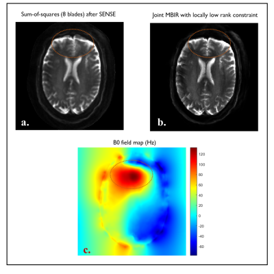
    Fig. 3. (a): Sum-of-squares image obtained from all individual blade images in 2a. (b): The image obtained by the proposed MBIR with virtual coil LLR. (c): B0 field map obtained from 6-echo fast gradient-echo images via fat-water separation. The red circles highlight severe geometric distortion (a) and its improvement with the proposed scheme (b) in the region with strong field inhomogeneity as shown in (c).
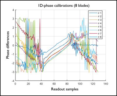
    Fig.1. 1D phase differences between positive and negative readout echoes (2x-oversampling not removed). Their offsets and slopes appear slightly different among the eight different blade angles.
  • On the possibility of reconstructing arbitrary FOVs using gradient waveforms with low-coherent aliasing properties
    Tobias Speidel1, Patrick Metze1, Kilian Stumpf1, Thomas Hüfken1, and Volker Rasche1
    1Internal Medicine II, Ulm University Hospital, Ulm, Germany
    The calculation of k-space trajectories in MRI usually involves prior knowledge of the FOV due to a minimum k-space sampling density. Based on a generalised form of the "Seiffert Spirals", this abstract describes an imaging modality that does not require prior commitment to an imaging FOV.
    Figure 1: 15 interleaves of the generated trajectory within normalised k-space.
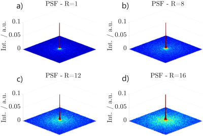
    Figure 2: Simulated PSFs in the $$$xy$$$-plane with $$$z=0$$$ of the presented approach according to the undersampling factors $$$R_1$$$ in a), $$$R_2$$$ in b), $$$R_3$$$ in c) and $$$R_4$$$ in d).
  • Learning a Preconditioner to Accelerate Compressed Sensing Reconstructions
    Kirsten Koolstra1 and Rob Remis2
    1Division of Image Processing, Leiden University Medical Center, Leiden, Netherlands, 2Circuits and Systems, Delft University of Technology, Delft, Netherlands
    In this work we design a preconditioner for compressed sensing reconstructions using a neural network. Results show that it is possible for a learned preconditioner to improve upon the performance of existing preconditioning techniques. 
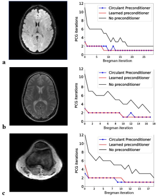
    Fig.2. Comparison of CG convergence with and without the learned and the circulant preconditioner. (a) Flair brain scan (128x128). The learned preconditioner reduces the number of iterations in CG by a factor 2.9. This is a slightly larger reduction compared to the factor of 2.7 obtained with the circulant preconditioner. (b) Similar results are obtained for a TSE scan with a twice as large matrix size compared to that in the training set. (c) The speed up is slightly lower in the knee (128x128), which is an anatomy that the network has not observed during training.
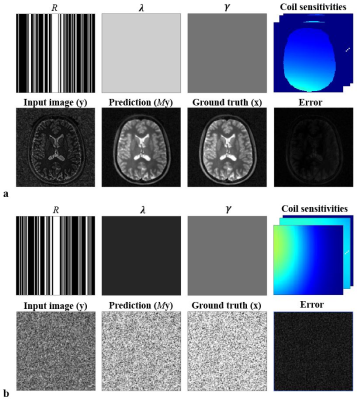
    Fig.1. The network’s input and output for two training examples. Each 37-channel input contains a sampling mask (R), regularization masks (λ and γ), 16 complex coil sensitivity maps and a complex image (y). Note that the complex channels are first split into real and imaginary components. The network’s prediction My is close to the ground truth, which is confirmed by the small error values both for the brain case (a) and for the noise case (b) (normalized norm < 0.16).
  • Robust and Computationally Efficient Missing Point and Phase Estimation for Zero Echo Time (ZTE) Sequences
    Curtis A Corum1,2, Abdul Haseeb Ahmed2, Mathews Jacob2, Vincent Magnotta2, and Stanley Kruger2
    1Champaign Imaging LLC, Shoreview, MN, United States, 2University of Iowa, Iowa City, IA, United States
    Here we modify and apply for the first time a robust and computationally efficient missing point and phase estimation algorithm originating in the solid state NMR community for zero echo time (ZTE) imaging sequences.
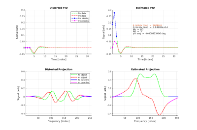
    Figure 1: Distorted FID, distorted Projection, corrected FID and corrected projection fro simulated hollow cube object.
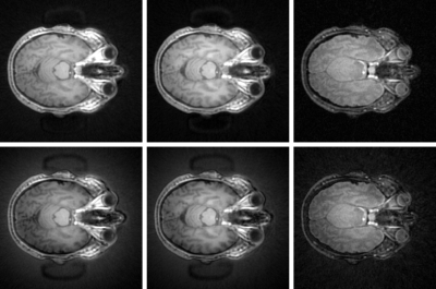
    Figure 2: Brain Images of Health Adult Subject.
    Bottom Row Left to Right: Un-corrected ZTE intrinsic contrast, IR-ZTE and T2-ZTE
    Top Row Left to Right: Corrected (Nd = 3) ZTE intrinsic contrast, IR-ZTE and T2-ZTE
  • Iterative Reconstruction for Enhanced Through-Plane Resolution T2-Weighted Spin-Echo Imaging of the Prostate
    Eric A Borisch1, Roger C Grimm1, Soudabeh Kargar2, Akira Kawashima3, Joshua D Trzasko1, and Stephen J Riederer1
    1Radiology, Mayo Clinic, Rochester, MN, United States, 2Radiology, University of Wisconsin-Madison, Madison, WI, United States, 3Radiology, Mayo Clinic, Phoenix, AZ, United States
    A sparsity-regularized forward model-based iterative reconstruction is presented improving the through-plane resolution and noise performance of the output images produced from a set of overlapping lower (thicker) resolution 2D acquisitions.
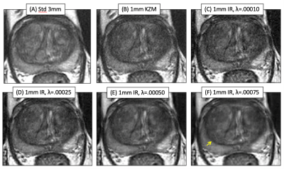
    Axial T2SE images of the prostate of one subject: (A) standard 3mm thick T2SE (acquired in 4:14) and (B-F) 1mm thick images. (B) was reconstructed using the linear KZM method[1], while (C-F) were generated with the new MBIR at the λ values shown. Note some improved sharpness of (B) vs. (A) owing to the thinner slice but an increased noise level. MBIR with increasing λ provides progressive reduction in noise in (C-F). However, use of λ=0.00075 caused undesirable blurring, e.g. between the peripheral and transition zones (F, arrow).
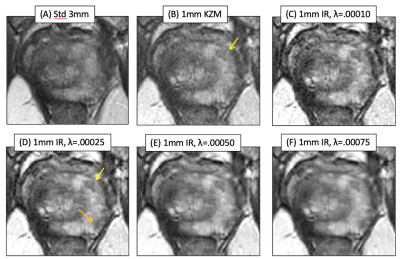
    Results from a second case. Fig. 2B vs. A better portrays a lesion (B, arrow), but MBIR provides further improved depiction (D, yellow arrow) and definition of a second lesion (orange arrow). Higher λ values again cause blur. Across the patient studies the radiologist’s perceived preference was for λ=0.00025.
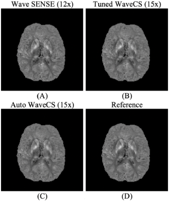
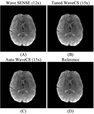
  • Automatic WaveCS reconstruction
    Gabriel Varela-Mattatall1,2 and Ravi S Menon1,2
    1Centre for Functional and Metabolic Mapping, Robarts Research Institute, Western University, London, ON, Canada, 2Department of Medical Biophysics, Schulich School of Medicine and Dentistry, Western University, London, ON, Canada
    We present an automatic, non-iterative, and prospective method to determine the regularization weighting for WaveCS reconstructions. The image quality from this reconstruction is on the same order as other reconstruction but without any tuning. 
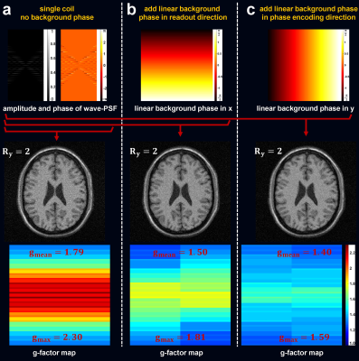
    Visualization of local phase images from the different reconstruction procedures. Panels (A) and (B) show the local phase from Bilgic et al7, while panel (C) shows the local phase obtained from our automatic WaveCS reconstruction procedure. Panel (D) shows the reference. In parenthesis it is shown the under-sampling factor for each reconstruction.
    Visualization of magnitude Images from the different reconstruction procedures. Panels (A) and (B) show the results from Bilgic et al7, while panel (C) shows the result from our automatic WaveCS reconstruction. Panel (D) shows the reference. In parenthesis it is shown the under-sampling factor for each reconstruction.
  • Jointly Reconstructed Undersampled Multiparameter MRI for Imaging Intratumoral Subpopulations
    Shraddha Pandey1,2, Arthur David Snider1, Wilfrido Moreno1, Harshan Ravi2, Ali Bilgin3, and Natarajan Raghunand2,4
    1Electrical Engineering, University of South Florida, Tampa, FL, United States, 2Cancer Physiology, Moffitt Cancer Center, Tampa, FL, United States, 3Departments of Medical Imaging, Biomedical Engineering, and Electrical & Computer Engineering, University of Arizona, Tucson, AZ, United States, 4Department of Oncologic Sciences, University of South Florida, Tampa, FL, United States
    A joint reconstruction framework is presented to concurrently reconstruct a series of complex MR images and their corresponding T1, T2 and T2* parameter maps. Tissue mapping and estimation of water and fat content within 4 objectively defined tissue types was possible using  18% k-space data. 
    Figure 1. k-space data obtained from the scanner is undersampled using the cartesian sampling mask. The Joint Reconstruction Algorithm is used to reconstruct the series of T1w images and their parameter maps. The process is repeated to reconstruct T2w/T2*w images and their parameter maps. Validation of the results is carried out by identifying the tissue types like muscle, fluid, tumor and adipose and using the rules on T1 and T2 maps. The T2*w images are subjected [1] to estimate PDFF, PDwF, & in the muscle, fluid, tumor and adipose tissue type.
    Figure 2. T1w reconstructed images for 2 mouse slices with mask of ~27%, ~36% k-space data are shown.The sampling masks are shown in col 1. The results for the Repetition times (TR) 0.4s and 5s are shown in col 2 & 3 for mouse 1 and col 4 & 5 for mouse 2. The 5th & 6th column shows the detailed version of the region highlighted in the red box.The MI value is computed for 4 undersampling masks 18%, 27%, 36%, & 52% shown on x-axis. The y-axis shows the MI value when the ground truth |u| are compared to the reconstructed |u|. The mean MI and standard error of the mean (S.E.M) is calculated over n = 30 mouse slices.
  • Automatic determination of the regularization weighting for wavelet-based compressed sensing MRI reconstructions
    Gabriel Varela-Mattatall1,2, Corey A Baron1,2, and Ravi S Menon1,2
    1Centre for Functional and Metabolic Mapping, Robarts Research Institute, Western University, London, ON, Canada, 2Department of Medical Biophysics, Schulich School of Medicine and Dentistry, Western University, London, ON, Canada
    Here we present an automatic, non-iterative, prospective, and fast approach to determine the regularization weighting, which enables wavelet-based compressed sensing MRI reconstructions.
    Knee images from the different strategies to define the regularization weighting. We compare (A) $$$ \lambda_{\textrm{Lcurve}} $$$ (14 iterations) and (B) $$$ \lambda_{\textrm{NMSE}} $$$ (14 iterations) strategies to (C) our approach, $$$ \lambda_{\textrm{auto}} $$$, using the 4th-level Daubechies mother wavelet (1 iteration). Panel (D) shows the reference and panel (E) shows quantitative results. Smaller values along each axis in (E) represent more accurate reconstructions.
    Visualization of magnitude and phase images from simulations. Panels (B) and (E) show the images from the reconstruction process using our approach for USF=5 and $$$\sigma=5$$$. Panels (A) and (D) show the reference. Panels (C) and (F) show the difference between both columns. All images are scaled according to their respective reference.
  • Improved CS-MRI using Hybrid Plug-and-Play Priors based Fast Composite Splitting Algorithm
    Qingyong Zhu1, Jing Cheng2, Zhuo-Xu Cui1, and Dong Liang1,2
    1Research Center for Medical AI, SIAT, Chinese Academy of Sciences, Shenzhen, China, 2Paul C. Lauterbur Research Center for Biomedical Imaging, SIAT, Chinese Academy of Sciences, Shenzhen, China
    The proposed hybrid Plug-and-Play priors (PnP) based on internal and external denoising can help improve the performance of fast composite splitting algorithm in MRI, and the hybrid PnP  is also easily incorporated in other algorithms such as ADMM and AMP.
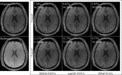
    Figure 1. The reconstruction results using all comparison methods at AF= 4, 5. The values at left-top of images are the corresponding RE($$$\%$$$) and PSNR(dB).
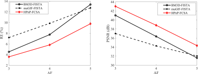
    Figure 2. The RE (left) and PSNR (right) plots of all reconstructions at various AFs (AF=3, 4, 5).
  • Fast Variable Density Poisson-Disc Sample Generation with Directional Variation for Compressed Sensing
    Nicholas Dwork1, Corey A. Baron2, Ethan M. I. Johnson3, Daniel O'Connor4, John M. Pauly5, and Peder E.Z. Larson1
    1Radiology and Biomedical Imaging, UCSF, San Francisco, CA, United States, 2Robarts Research, Western University, Ontario, ON, Canada, 3Biomedical Engineering, Northwestern University, Evanston, IL, United States, 4Mathematics and Statistics, University of San Francisco, San Francisco, CA, United States, 5Electrical Engineering, Stanford University, Stanford, CA, United States
    We have developed a fast algorithm to create a variable density poisson disc sampling pattern.
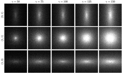
    Fig. 2: Variable density poisson disc sampling patterns for different values of $$$\gamma$$$ and different aspect ratios.
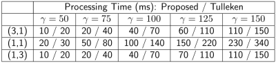
    Table 1: Run times for generating variable density poisson-disc sampling patterns with the proposed algorithm and the Tulleken algorithm. Time is reported in milliseconds. In all cases, the proposed algorithm is faster by $$$30-50\%$$$.
  • Automatic determination of the regularization weighting for low rank reconstruction problems
    Gabriel Varela-Mattatall1,2, Corey A Baron1,2, and Ravi S Menon1,2
    1Centre for Functional and Metabolic Mapping, Robarts Research Institute, Western University, London, ON, Canada, 2Department of Medical Biophysics, Schulich School of Medicine and Dentistry, Western University, London, ON, Canada
    We develop a general, non-iterative, fast, and automatic procedure to determine the regularization weighting for low-rank reconstruction problems. 
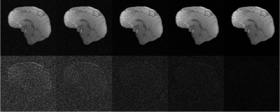
    Impact of the size of the dataset for automatic low rank reconstructions using SNR 5 and an under-sampling factor of 4x. The first row corresponds to the mean appearance of the reconstructions, $$$\hat{X}$$$, for 5,10,40,100, and 600 images (from left to right, respectively). The second row corresponds to the absolute difference with respect to the reference, $$$X$$$, as $$$5 \times |\hat{X}-X|$$$.
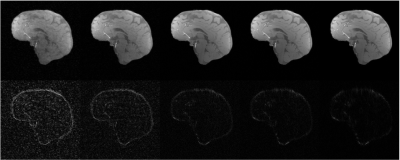
    Impact of the size of the dataset for automatic low rank reconstructions using SNR 30 and an under-sampling factor of 14x. The first row corresponds to the mean appearance of the reconstructions, $$$\hat{X}$$$, for 5,10,40,100, and 600 images (from left to right, respectively). The second row corresponds to the absolute difference with respect to the reference, $$$X$$$, as $$$5 \times |\hat{X}-X|$$$.