-
MRI Safety for Leave-on Powdered Hair Thickeners: Measurement of Deflection Force and MRI Artifact
Norio Hayashi1, Akio Ogura1, Atsuya Fuju2, Tomokazu Takeuchi3, Yusuke Sato4, Maiko Hashimoto5, and Masahiko Takahashi5
1Radiological Technology, Gunma Prefectural College of Health Sciences, Maebashi, Japan, 2Radiology, Kiryu Kosei General Hospital, Kiryu, Japan, 3Graduate School of Radiological Technology, Gunma Prefectural College of Health Sciences, Maebashi, Japan, 4Radiology, Gunma University Hospital, Maebashi, Japan, 5Isesaki Municipal Hospital, Isesaki, Japan
The purpose of this study was to evaluate
the attraction force and image artifacts of powdered hair thickeners.
Displacement force and MRI artifact was measured. Some of powdered hair
thickeners were highly susceptible to mechanical effects and will cause more
image artifacts.
Fig. 2 MRI T1-weighted images obtained by
the spin-echo sequence of the 1.5 Tesla MRI system.
-
Optimized 3D ultrashort TE protocol for lung imaging
CHIKARA NODA1, Chia Ying Liu2, Jason Ortman1, Bharath Venkatesh Ambale3, Webster Stayman4, Yoshimori Kassai5, and Joao A.C. Lima1
1Cardiology, Johns Hopkins University, Baltimore, MD, United States, 2Canon Medical Research, Cleveland, OH, United States, 3Radiology, Johns Hopkins University, Baltimore, MD, United States, 4Biomedical Engineering, Johns Hopkins University, Baltimore, MD, United States, 5Canon Medical Systems Corporation, Otawara, Japan
3D radial Ultrashort echo time sequence is a promising technique for lung imaging but the protocol has not been optimized. We examined the parameters including number of trajectories and flip angles using a lung phantom. In-vivo images were obtained and evaluated based on the parameters optimized in the phantom study. UTE acquisition in axial rather than coronal direction produced better quality of images. Breath holding acquisition may be an option in case patients cannot tolerate long scan time.
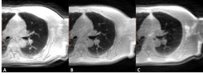
Figure 3. Lung images using ultrashort echo time sequence by different acquisitions. A) post reconstructed to axial by coronal acquisition with respiratory gating. The signal intensity was higher due to the longer acquisition time in this case. B) performed by axial acquisition with respiratory gating. Images were sharper in this orientation. C) performed by axial acquisition with 20 seconds breath holding.
-
Optimizing dark blood late gadolinium enhancement in cardiac magnetic resonance imaging.
Hannah Bergman1, Alaine Berry1, and Ben Statton1
1MRC, Imperial College, London, United Kingdom
Dark
blood late gadolinium enhancement imaging nulls both the blood pool and the
normal myocardium. Optimal image quality depends on a number of factors. This study sought
to determine whether gadolinium dose and delay time before imaging, affected
the nulling of the blood pool and myocardium.
-
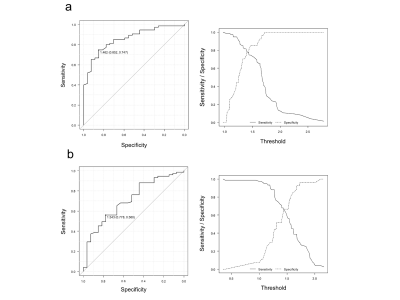
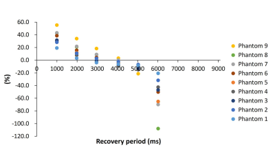
Quantitative evaluation of metal artifacts in 3-T MRI by using Blind/Referenceless Image Spatial Quality Evaluator (BRISQUE): a phantom study
Wakiko Tani1, Yutaka Katayama2, Tomoaki Yamakawa1, Shintaro Horii1, Ryuji Shimada1, Yuichiro Somiya1, and Akiko Kusaka1
1Center for Radiology and Radiation Oncology, Kobe University Hospital, Kobe, Japan, 2Division of Radiological Technology, Osaka City University Hospital, Osaka, Japan
A quantitative evaluation of metal artifacts was performed using Blind/Referenceless Image Spatial Quality Evaluator (BRISQUE), an objective evaluation, and compared with visual evaluation. BRISQUE was associated with visual evaluation and can quantitatively evaluate metal artifacts.
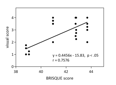
Figure 4. Correlations Between BRISQUE score and visual score
-
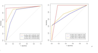
Compressed Sensing for Turbo Spin-echo Diffusion-weighted Imaging: Influence of Denoising Level on the Signal Intensity and Apparent Diffusion Coefficient Values in Cervical Cancer
Yuichiro Somiya1, Yoshiko Ueno2, Shintaro Horii1, Ryuji Shimada1, Keitaro Sofue2, Naoki Yoshida1, Wakiko Tani1, Akiko Kusaka1, and Takamichi Murakami2
1Center of Radiology and Radiation Oncology, kobe university hospital, kobe, Japan, 2Department of Radiology, Kobe University Graduate School of Medicine, kobe, Japan
This study evaluated the effect of denoising level on ADC values of diffusion weighted imaging using the single-shot TSE sequence with compressed sensing in cervical cancer. At each denoising level, the significant difference was observed in the ADC values between the cancer and normal cervix.
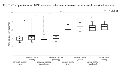
Figure3. Comparison of ADC values
between normal cervix and cervical cancer
-
Comparison of cardiac T1 mapping on a high-performance 0.55T scanner and a conventional 1.5T scanner
Christine Mancini1, W. Patricia Bandettini1, Peter Kellman1, Hui Xue1, and Adrienne E. Campbell-Washburn1
1NHLBI, National Institutes of Health, Bethesda, MD, United States
There is a good correlation between native T1 myocardial mapping and ECVs between a conventional 1.5T scanner and a high-performance 0.55T scanner.
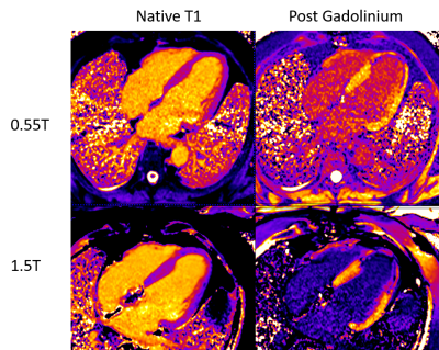
Pre and post MOLLI images from 0.55T and 1.5T in a patient with myocardial infarction
-
Effects of image quality deterioration and data shortage on automatic white matter bundle segmentation by diffusion magnetic resonance imaging
Yuichi Suzuki1, Tsuyoshi Ueyama1, Takahiro Iwasaki1, Jiro Sato1, Hideyuki Iwanaga1, and Osamu Abe1
1Department of Radiology, THe University of Tokyo Hospital, Tokyo, Japan
We investigated the effect of image quality
deterioration and data shortage of DWI on automatic
white matter bundle segmentation. There was almost no difference in bundle
segmentation ability between SMS factors of 2 and 3. For MPG, 16
axes provided results comparable to the GS data (64 axes).
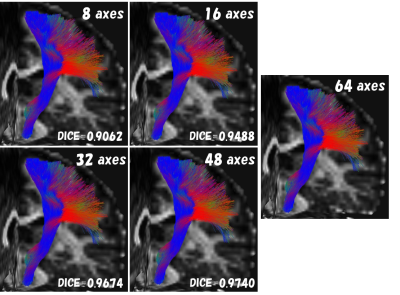
Fig.4. The left pyramidal tract (example)
images of MPG comparison
Fiber bundles different from anatomical
running were not visually observed under any conditions as well as SMS
comparison.
-
Optimization of Variable TI 4D ultrashort TE MR Angiography: A Numerical Simulation and Phantom Study
Toshiya Akatsu1, Haruyuki Fukuchi2,3,4, Kei Fukuzawa4, Nao Takano1, Yutaka Ikenouchi3, Michimasa Suzuki3, Kohji Kamagata3, Akihiko Wada3, Osamu Abe2, and Shigeki Aoki3
1Department of Radiology, Juntendo University Hospital, Tokyo, Japan, 2Department of Radiology, Graduate School of Medicine, University of Tokyo, Tokyo, Japan, 3Department of Radiology, Juntendo University Graduate School of Medicine, Tokyo, Japan, 4Department of Radiology, Toranomon Hospital, Tokyo, Japan
We
implemented the new Variable TI 4D UTE-MRA in order to improve the visibility
of hemodynamic flow. It was found that a 50 % reduction of TI steps increases 50 %
of signal from the flow volume without impairment of structural information in a
numerical simulation and a phantom study.
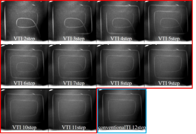
The maximum intensity
projection (MIP) of all TI phases in coronal plane were displayed. The signal
intensity from the tube was increased with less VTI steps. However less than VTI
5 step shows discontinuous of tube imaging because of the long shot intervals.
-
Assessing the reproducibility of semi-automated segmentation methods on post-operative T1 post contrast and FLAIR MRI of GBM.
Olga V Fadeeva Da Costa1,2, Shah Islam2,3, Mark M Boubnovski3, Eric Aboagye3, and Adam D Waldman4,5
1Department of Surgery & Cancer, Cancer Imaging Centre, Hammersmith Campus, Imperial College, London, United Kingdom, 2Imaging Department, MRI Unit, Hammersmith Hospital, Imperial College Healthcare NHS Trust, London, United Kingdom, 3Department Of Surgery & Cancer, Cancer Imaging Centre, Hammersmith Campus, Imperial College, London, United Kingdom, 4Department of Brain Sciences, Hammersmith Campus, Imperial College, London, United Kingdom, 5Centre for Clinical Brain Sciences, University of Edinburgh, Edinburgh, United Kingdom
The primary purpose of this study was to quantify the inter and intra reader reproducibility of segmentation of postoperative MRIs in patients with GBM. Accurate delineation and analysis of disease residuum is important for clinical trial endpoints and image biomarker development.
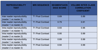
Table 1: Summary of reproducibility of segmentation voxels and their respective volumes
-
Osteoporotic vertebral fracture analysis with intravoxel incoherent motion: a preliminary study
Hiroyuki Takashima1,2, Rui Imamura1, Tsuneo Takebayashi3, Yasuhisa Abe3, Izaya Ogon2, Hiroshi Oguma3, Yoshihiro Akatsuka1, and Toshihiko Yamashita2
1Division of Radiology and Nuclear Medicine, Sapporo Medical University Hospital, Sapporo, Japan, 2Department of Orthopaedic Surgery, Sapporo Medical University School of Medicine, Sapporo, Japan, 3Department of Orthopaedic Surgery, Sapporo Maruyama Orthopedic Hospital, Sapporo, Japan
IVIM can assess
tissue water diffusivity and microcapillary perfusion and enable clear distinction
between VF and without fracture. This study provides the basic data for analyzing
VF using IVIIM.
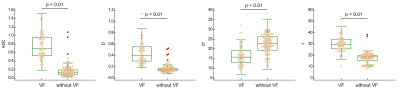
Fig. 3: Comparison between VF and without VF on each IVIM parameter
-
Optimizing a Motion Tracking Marker for Pediatric Patients
Kristina Mary Pelkola1,2, Onur Afacan1,2, Tess E. Wallace1,2, Pauline Connaughton1, Jenna McKay1, Joseph Zmuda1, Camilo Jaimes1, and Simon K. Warfield1,2
1Radiology, Boston Children's Hospital, Boston, MA, United States, 2Computational Radiology Laboratory, Boston Children's Hospital, Boston, MA, United States
Magnetic Resonance Imaging (MRI) can be challenging for pediatric
patients due to factors such as the large tunnel, imaging coil, and loud
gradient noises. This can spark anxiety and fear causing them to be uncooperative
and unable to hold still [1,2]. Due to these barriers, pediatric MRI exams can
be plagued with motion artifacts which result in poor diagnostic quality of the
images and make it challenging for Radiologists to interpret [3]. It is a
common practice to administer sedation or anesthesia to attempt to acquire
diagnostic images of pediatric patients. These methods are not only costly and
potentially harmful, but do not eliminate motion artifacts [4]. Exams performed
under sedation and anesthesia can still be affected from breathing and/or
uncontrollable muscle spasms. There is an unmet need for alternative methods to
enable diagnostic imaging exams in the presence of motion.
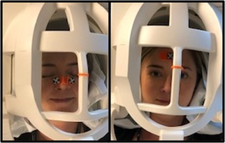
Figure 2: Adult volunteer displaying the
double winged marker, on the left, and the single flat marker, on the right in
a Siemens 64-channel head coil [7].
-
Deep Inspiration Breath-Hold Radiotherapy does not induce MRI detectable LV functional and structural changes in patients with left-sided breast cancer – A six months follow-up
Xin Dong1,2, Arnold Ng3,4, Sharon Watson5, Harish Sharma5, Graham Galloway1,2,6, and Margot Lehman5
1Queensland University of Technology, Brisbane, Australia, 2Translational Research Institute, Woolloongabba, Australia, 3Cardiology, Princess Alexandra Hospital, Brisbane, Australia, 4University of New South Wales, Sydney, Australia, 5Radiation Oncology, Princess Alexandra Hospital, Brisbane, Australia, 6The University of Queensland, St Lucia, Australia
Deep
inspiration breath-hold Radiotherapy for left-sided breast cancer does not induce MRI detectable LV functional
and structural changes in patients with left-sided breast cancer
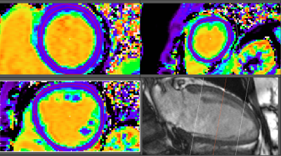
ECV maps with myocardium ROIs at the basal, mid, and apical
levels.
-
7T Knee MRI: Clinical Benefits and Challenges
Kevin Epperson1, Karla Epperson1, Garry Gold2, and Feliks Kogan3
1Radiology School of Medicine, Stanford University, Stanford, CA, United States, 2Radiology, Stanford Univeristy, Stanford, CA, United States, 3Radiology - SOM, Stanford Univeristy, Stanford, CA, United States
Chronic knee pain, invading osteoarthritis, and rising diagnostic costs continue to hinder diagnosis and treatment of this disease. Can the benefits of high field MR imaging such as 7T provide the needed resolution to present pathology as well as allow the quantitative results expected.
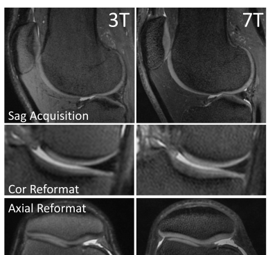
Figure 2. 3D PD Cones images acquired at 3T (left) and 7T (right) with equivalent parameters. 7T images shown considerably better tissue signal and contrast in the deep layers of patellar and tibial cartilage. Additionally, on coronal and axial reformats, 7T images show improved visualization of structures with reduced blurring.