Virtual Meetings Archive
The SMRT Virtual Meetings Archived has moved!
Visit the ISMRM Virtual Meeting Archives for all past SMRT meeting videos.
Watch past Virtual Meetings and learn from the convenience of your home, office, or anywhere around the world. Access is free for SMRT & ISMRM members!
Joint ISMRM High Field Study Group & ISMRT Virtual Meeting:
Challenges, Supports, and Networks for 7T Scanner Users
09 December 2021
Watch the recording here (Members only; no CE credit) Username: Your ID number Password: Your last name (Case-Sensitive)
Overview:
Challenges and experience of setting up a 7T service as a new user Pip Bridgen, (PgCert) Our 7T service is based within an inner-city London hospital, which allows us to offer a clinically focused service alongside research work in an ever-developing field. This presentation is an overview of the challenges we experienced whilst setting up a new 7T service, and the ways in which we overcame these. We focus on some of the processes and procedures implemented to achieve this and set out our plans going forward.
Team up at 7T Prof. Dr. Oliver Speck Establishing and running a 7T facility can be challenging. Once the operation is established, how can efforts between multiple centers be combined and a higher impact be generated? A few 7T research networks, including GUFI, UK7T and EUFIND, have addressed these questions and demonstrated added value in combined high field research efforts.
Moderators:
Alexander P. Lin, Ph.D. Rebecca Glarin, RT
Challenges and experience of setting up a 7T service as a new user
Pip Bridgen, (PgCert) 7T Lead Research Radiographer King’s College London London, UK
Team up at 7T
Oliver Speck, Ph.D. Otto-von-Guericke-University Magdeburg Magdeburg, Germany
Panel Discussion
Alexander P. Lin, Ph.D. Rebecca Glarin, RT
Joint ISMRM & ISMRT Virtual Meeting:
Preview of 2022 Joint Annual Meeting – London
12 November 2021
Watch the recording here (Members only; no CE credit) Username: Your ID number Password: Your last name (Case-Sensitive)
Overview:
ISMRM goes Hybrid! Steven P. Sourbron, Ph.D. After 2 years of virtual ISMRM annual meetings, we are now planning the first ever hybrid annual meeting of the ISMRM. While many of the details are still being worked out, we are committed to provide an interactive hybrid format. Those travelling to London will experience an ISMRM meeting much like in pre-pandemic times, with all the benefits of in-person interaction but with an additional option to interact with online attendees. Online attendees will be able to watch live streams of all sessions in London, and recordings will be available for those in more distant time zones, with increased interactivity than in previous online annual meetings. Importantly, online attendees will also be able to present their work to attendees in London and online. In this talk I will update on the current ideas for the meeting, not only on format but also on content including also the educational program and other special sessions under development.
MR education during the ISMRM annual meeting in 2022: What to expect Nivedita Agarwal, MD The ISMRM annual meeting in London will offer a total of 218 hours of education distributed from Saturday to Thursday designed by the AMPC. This also includes 28 hours of tutorials organised by eight different education tracks (Neuro, Body, Cross-Organ, MSK, Image acquisition, contrast mechanisms, Physics & Engineering and Transferable Skills). All education is planned onsite and all speakers have been requested to be onsite to allow for maximum interactivity between them and the participants. We also have planned a three day meeting within a meeting or a Special focus meeting (SFM) dedicated to cardiovascular MR research. This is specially designed for attendees (such as clinicians) who may not have time to attend a six-day meeting but want to get the most of the cutting-edge technologies centered around CMR.
First Hybrid SMRT (just renamed to ISMRT!) Annual Meeting Rhys Slough, MSc. Similar to ISMRM annual meeting, SMRT (renamed to ISMRT) annual meeting will go hybrid as well! The annual meeting will have a similar schedule as the meetings in pre-pandemic time with an interesting poster session on Friday afternoon, accredited theme-based education sessions for MR radiographers/technologists on both Saturday and Sunday, and accredited ISMRM sessions and ISMRM-ISMRT joint forum on Monday. We will cover different hot topics in the meeting including AI, high and low-field imaging and advanced practices in MR. We will continue the great Master Class tradition we started in the past two years. Attendees traveling to London will be able to have in-person interaction with gorgeous speakers and other attendees (both in-person and online). Attendees online will be able to view live presentations and join the Q & A and discussions virtually. We look forward to seeing you!
2022 ISMRT Annual Meeting will be fun! Huijun ‘Vicky’ Liao, MR-RT 2022 ISMRT Annual Meeting encourages interaction/discussion since it will be our first chance to see each other in-person after two years! Besides the hot topics mentioned by Rhys, we plan to have Q&A and panel/group discussions in the safety and clinical sessions where both in-person and online attendees can share their opinions and questions. We also encourage you to join our poster-tour on Friday to interact with our MR radiographer/technologist poster presenters and other MR colleagues around the world. There also will be Grab-n-Go Lunch served for ISMRT Business Meeting on Saturday which will give the most updated status of ISMRT and the Abstract/Proffered Papers Awards on Sunday. We will also be planning on our evening welcoming party which has been a tradition in the pre-pandemic times!
Moderators:
Nancy H Beluk (ISMRT Education Chair)
Anne Dorte Blankholm (ISMRT President)
ISMRM goes Hybrid!
Steven P. Sourbron, Ph.D. (ISMRM AMPC Chair) The University of Sheffield Sheffield, UK
MR education during the ISMRM annual meeting in 2022: What to expect
Nivedita Agarwal, M.D. (ISMRM AMPC Vice Chair) Pediatric Hospital in Lecco Milan, Italy
First Hybrid SMRT (just renamed to ISMRT!) Annual Meeting
Rhys Slough, M.Sc. (ISMRT AMPC Chair) Cambridge University Hospitals Cambridge, UK
2022 ISMRT Annual Meeting will be fun!
Huijun Vicky Liao, B.Sc., ARMRIT (ISMRT AMPC Vice Chair) Brigham and Women’s Hospital Boston, MA, USA
Joint ISMRM Interventional MR Study Group & SMRT Virtual Meeting: Interventional MR Gallery: MR-Guided Therapeutic Ultrasound for Essential Tremor, MR-Guided Cardiac Interventions, MR-Linac & MR Safety in Multimodality Operation Room
14 October 2021
Watch the recording here (Members only; no CE credit) Username: Your ID number Password: Your last name (Case-Sensitive)
Overview: MRI safety in a Multidisciplinary OR environment – Janice Fairhurst, R.T. (R) MR The MRI Safety challenges are inherent in a Multi-Modality Image Guided Operation Room. This talk will describe the potential hazards that exist in a multimodality imaging suite as it relates specifically to MRI safety and identify the key safety strategies that are used within the multidisciplinary iMRI environment to ensure safety.
Transcranial MR-guided Focused Ultrasound for Essential Tremor – Rachelle Bitton, Ph.D. MR guided Focused ultrasound is a noninvasive, therapeutic technology with the potential to improve the quality of life and decrease the cost of care for patients with essential tremor. This novel technology focuses multiple beams of ultrasound energy precisely and accurately on targets deep in the brain without damaging surrounding normal tissue. MR imaging is used for treatment planning and inter-procedural monitoring, providing images to pinpoint the area to treat and real time MR thermometry to monitor treatment progress. In this talk we explore the mechanism behind the technology, explore treatment protocols, show 3 year follow up results in patients, and talk about next steps for the technology.
Cardiac interventional MRI: current status and remaining technical challenges – Olivier Jaubert, Ph.D. This talk will give a brief overview of the cardiovascular applications of interventional MRI (iMRI). We will give a description of the clinical MRI scans routinely performed at our institution in pulmonary hypertension patients. Finally we will give some future directions being investigated to resolve the remaining challenges for better translation of cardiovascular iMRI in clinical practice.
Initial experience with the clinical implementation of quantitative MRI biomarkers in a 1.5T MR-Linac – Ergys Subashi, Ph.D. MR-guided systems, particularly the integrated MR-linac, provide a novel platform for the delivery of precision radiotherapy and may enable improvements in therapeutic response by increasing dose to the target while sparing organs at risk. The clinical implementation of these systems necessitates a review of current commissioning and quality assurance (QA) methods and, if needed, revisions to address differences with conventional MRI and radiotherapy machines. Integrated MR-linacs present with challenges that are not encountered when each component is considered separately. In this talk we will discuss our initial experience with the clinical implementation of quantitative MRI biomarkers in a 1.5T MR-Linac.
Moderators: Elena Kaye, Ph.D., Chair & Jurgen J. Fütterer, M.D., Ph.D., Vice Chair, Interventional MR Study Group
Accreditation:
The SMRT has designated this live activity for a maximum of 1.00 Category A Credit.
The ISMRM designates this live activity for a maximum of 1.0 AMA PRA Category 1 Credits™. Physicians should claim only the credit commensurate with the extent of their participation in the activity.
Objectives:
- Appreciate the academic and clinical potential of technologies in the field of iMRI;
- Identify the unique demands of iMRI; and
- Development iMRI technologies with this knowledge to better inform referring clinicians and the patients and/or their carers.
MRI safety in a Multidisciplinary OR environment
Janice Fairhurst, B.Sc.,R.T.(R)(MR) Brigham and Women’s Hospital Boston, MA, United States
Transcranial MR-guided Focused Ultrasound for Essential Tremor
Rachel R. Bitton, Ph.D. Stanford University Palo Alto, CA, United States
Cardiac interventional MRI: current status and remaining technical challenges
Olivier Jaubert, Ph.D. University College London London, United Kingdom
Initial experience with the clinical implementation of quantitative MRI biomarkers in a 1.5T MR-Linac
Ergys David Subashi, Ph.D. Memorial Sloan Kettering Cancer Center New York, NY, United States
SMRT Virtual Meeting: From Birth to Death: Non-Oncological Applications of MR Spectroscopy in the Brain
09 September 2021
Watch the recording here (Members only; no CE credit) Username: Your ID number Password: Your last name (Case-Sensitive)
Overview:
Dr. Stefan Bluml will describe MRS acquisition and technical aspects in a clinical environment, metabolic maturation from birth to adolescence, birth asphyxia and localized temperature measurement.
Dr. Alex Lin will present MRS diagnosis of metabolic disorders and anoxia in the ER, traumatic brain injury, psychiatric disorders and neurodegenerative diseases.
Objectives:
- Understand the requirements for the use of MRS in a clinical environment;
- Appreciate the evolving metabolic features of the brain and the need for standardized acquisition methods and appropriate control data;
- Recognize main features of neonatal hypoxic/ischemic brain injury;
- Understand the usefulness of MRS for measuring regional brain temperatures in newborns undergoing therapeutic hypothermia treatment;
- Learn how MRS can be used to impact patient management;
- Compare and contrast the spectroscopic changes that occur from acute to chronic and severe to subconcussive brain injury;
- Describe the metabolic changes that occur in schizophrenia, bipolar disorder, and depression; and
- Understand how spectroscopy can be used to diagnose Alzheimer’s disease and mild cognitive impairment.
Moderators:
Huijun ‘Vicky’ Liao, B.Sc., ARMRIT George Bouzalis, (MR)
Accreditation:
The SMRT has designated this live activity for a maximum of 1.00 Category A Credit.
The ISMRM designates this live activity for a maximum of 1.0 AMA PRA Category 1 Credits™. Physicians should claim only the credit commensurate with the extent of their participation in the activity.
MR Spectroscopy of Pediatric Brain Diseases
Stefan Bluml, Ph.D. Children’s Hospital LA Los Angeles, CA, United States
MR Spectroscopy of Adult Brain Diseases
Alexander Lin, Ph.D. Brigham & Women’s Hospital/Harvard Medical School Boston, MA, United States
Joint ISMRM PET-MRI Study Group & SMRT Virtual Meeting: PET-MRI Acquisition/Reconstruction Harmonization
26 August 2021
Watch the recording here (Members only; no CE credit) Username: Your ID number Password: Your last name (Case-Sensitive)
Overview:
Presentation 1: Dementia Platform UK PET-MR Harmonisation Study In the UK a network of 8 PET-MR scanners has been set up for dementia scanning, 3 Siemens mMR and 5 GE Signa scanners. However for future clinical trials, it is important to reduce variability of measurements through the harmonisation of scanning protocols, and to characterise the repeatability and reproducibility of these measurements. Following the design of harmonised protocols, a travelling volunteer test-retest study is currently being conducted using the amyloid radiotracers, [18F]flutemetamol and [18F]florbetaben. The presentation will describe the study design, the many challenges that had to be met, and hopefully some preliminary results from the [18F]flutemetamol arm of the study which finished on the 26th July 2021.
Presentation 2: Harmonization of PET in simultaneous PET/MR and Path to Qualification The talk will present in one part an approach and its results to perform Harmonization of PET in simultaneous PET/MR in which we use NEMA phantom data acquired in a standard and controlled manner at two sites equipped with a Siemens mMR and a GE Signa PET/MR scanner. PET data were reconstructed by systematically varying image reconstruction parameters of number of iteration and filtration level and the best agreement between the reconstruction were found by evaluating the root mean square difference (RMSD) in contrast recovery coefficients (CRC). In a second part of the talk, an approach will be presented as a possible path to site qualifications of PET/MR scanners for multicenter brain imaging studies without the use of phantom to validate the accuracy of attenuation correction.
Moderators:
Udunna Anazodo, Ph.D. Kylie Walters, B.Appl.Sc.,M.H.Sc.(MRS)(MRI)
Objectives:
- Appreciate the academic and clinical potential of acquisition and data harmonization in PET-MRI;
- Identify the unique demands of PET-MRI acquisition and reconstruction harmonization; and
- Develop PET-MR protocols or processes with this knowledge to better inform referring clinicians and the patients and/or their carers.
Dementia Platform UK PET-MR Harmonisation Study
Julian Matthews, Ph.D. University of Manchester Manchester, United Kingdom
Harmonization of PET in Simultaneous PET/MR and Path to Qualification
Richard Laforest, Ph.D. Washington University School of Medicine St. Louis, MO, United States
MR Safety Week Virtual Meeting: MR Safety in Physics, Physiology and Practice Plus Practical Advice on Active Implants
29 June 2021
Watch the recording here (Members only; no CE credit) Username: Your ID number Password: Your last name (Case-Sensitive)
Overview:
In the presentation of ‘Understanding MRI safety: Physics, Physiology and Practice’, the interaction between the physical principles of the three types of magnetic fields in the MRI environment, viz., the static magnetic field, time-varying spatial field gradient, and radio-frequency magnetic fields, and their interactions with the biologic tissue will be highlighted. This understanding can guide the development of policies and procedures that can help create a safe MR environment for patients, staff, and support personnel. From this presentation, the audience will understand the characteristics of the three main magnetic fields in the MR environment and a framework for creating MRI safe policies and procedures.
The presentation of ‘Active Implant MRI Safety’ will provide a review of the process to clear active implants, with a focus on pacemakers. The audience will have an understanding of how to analyze the vendor documentation to identify what is relevant for the technologist to ensure that examinations are performed safely.
Moderators:
Sonja K. Boiteaux, MS, RT(R)(MR), MRSO, CHC Brandy Willis, MBA, RT(R)(MR)
Presentations:
Understanding MRI Safety: Physics, Physiology and Practice
Raja Muthupillai, PhD, DABR, DABMP, MRSE Live Healthy Imaging Bellaire, TX, United States
Active Implant MRI Safety
Seferino Romo, AAS, RT(R)(MR)(ARRT) Memorial Hermann Hospital – Texas Medical Center Houston, TX, United States
SMRT Virtual Meeting in Collaboration with the ISMRM Pediatric MR Study Group: Pediatric Diffusion Weighted MRI: Applications and Opportunities
17 June 2021
Watch the recording here (Members only; no CE credit) Username: Your ID number Password: Your last name (Case-Sensitive)
Overview:
From an academic perspective, Diffusion Weighted Imaging (DWI) MR is being used to inform and develop some neonatal human connectome projects, but DWI of the connectivity within the brain is itself is a growing and evolving field. Multi-disciplinary teams are therefore required to develop the acquire, process, analyze and interpret the data, because of the many and varied challenges in this field such as subject size, safety, and motion. The development, and importantly the application of the knowledge from this field is leading to refined clinical diagnosis, but this is dependent on applied parameters and radiological knowledge. This virtual meeting will discuss the development and use of advanced academic applications, through to the clinical applications and real world use of DWI MR today and into the near future.
Objectives:
- Appreciate the academic and clinical potential of DWI in mapping human cerebral connectivity.
- Identify the unique demands of the pediatric or neonatal DWI image acquisition, analysis, and application.
- Development clinical DWI imaging protocols or processes with this knowledge to better inform referring clinicians and the patients and/or their carers.
Moderators:
Michael Kean, R.T. FSMRT Minhui Ouyang, Ph.D.
Accreditation:
The SMRT has designated this live activity for a maximum of 1.00 Category A Credit.
The ISMRM designates this live activity for a maximum of 1.0 AMA PRA Category 1 Credits™. Physicians should claim only the credit commensurate with the extent of their participation in the activity.
Clinical Applications of Pediatric Brain Diffusion Weighted Imaging
Yi Li, M.D. University of California, San Francisco San Francisco, CA, United States
Challenges and Opportunities in Perinatal Diffusion MRI
Jacques-Donald Tournier, Ph.D. King’s College London London, England, United Kingdom
SMRT Virtual Meeting: A “How-To” Guide to Presenting & Moderating at a Scientific Meeting
22 April 2021
Watch the recording here (Members only; no CE credit) Username: Your ID number Password: Your last name (Case-Sensitive)
Overview: Every year, the ISMRM and SMRT have a scientific meeting which is almost certain to metamorphize into a varied format of “face to face” blended with “virtual” forms of address, presentation, and moderation. The techniques of presenting are strongly aligned with techniques of education and teaching, and these techniques are best developed with both meeting formats in mind, with a foundation in quality educational material and delivery. A skilled educator and presenter is ideal to give members a lecture on goals to be aimed for and hopefully achieved during their time on the real or virtual stage. Similarly, moderating in this mixed format environment is a skill to be used to guide the meeting, and extract the most information from the presentation and presenters. Moderating in its own way, can go someway towards making or breaking a meeting.
A large number of ISMRM and SMRT members are presenting at the upcoming meeting, and another significant member cohort will be moderating the sessions during this meeting. This virtual meeting will come 3-4 weeks before the ISMRM/SMRT annual meeting and the timing of this virtual meeting would be perfect to address this much needed skillset.
Moderator: Shawna Farquharson, Ph.D.
Accreditation: Credits are not available for this event.
This virtual meeting is a general instruction session and is not intended for a specific meeting or event, but could be helpful to moderators and speakers of the 2021 ISMRM & SMRT Virtual Meeting & Exhibition.
A Guide to Presenting at a Scientific Meeting
Thijs Dhollander, Ph.D. Murdoch Children’s Research Institute Melbourne, VIC, Australia
A Guide to Moderating at a Scientific Meeting
Sonja Boiteaux M.Sc. University of Texas MD Anderson Cancer Center Houston, TX, USA
Joint SMRT & ISMRM MR of Cancer Study Group Virtual Meeting: Imaging in Neuro-Oncology
08 April 2021
Watch the recording here (Members only; no CE credit) Username: Your ID number Password: Your last name (Case-Sensitive)
Overview: There is broad recognition that cancer imaging is best performed with standardized quality imaging protocols to enable serialized assessment of disease and treatment progression. That is a worthy goal in itself. However, there is a constant growth and body of work in the research domain which is yet to progress to routine clinical imaging practice. There is a recognized gap in application and translation of contemporary work and research in many imaging fields, including cancer. This meeting will explore current, and some emerging imaging techniques in the field of cancer, as well as the important process of translating contemporary imaging research into routine clinical practice.
Objectives:
- Recognize the benefits of quality routine imaging practice;
- Appreciate the process of applying new and emerging techniques into quality routine imaging; and
- Appreciate and apply the techniques to translating contemporary research into application.
Accreditation: The Society for MR Radiographers & Technologists (SMRT), A Section of the ISMRM, has designated this live activity for a maximum of 1.00 Category A Credit. The International Society for Magnetic Resonance in Medicine designates this live activity for a maximum of 1.0 AMA PRA Category 1 Credits™. Physicians should claim only the credit commensurate with the extent of their participation in the activity.
Moderators: Brandy Willis, M.B.A.,R.T.(R)(MR) Fulvio Zaccagna, M.D.
Imaging Challenges in Neuro-Oncology: Current Clinical Pathways and New Therapies
Adam D. Waldman, M.D., Ph.D. University of Edinburgh Edinburgh, Scotland, United Kingdom
Accreditation: 0.50 CE/CPD
Translating Research into Clinical Practise
Rhys A. Slough, M.Sc. (Med. Img. – MRI) Cambridge University Hospital Cambridge, England, United Kingdom
Accreditation: 0.50 CE/CPD
The SMRT is recognized by the American Registry of Radiologic Technologists (ARRT) as a Recognized Continuing Education Evaluation Mechanism (RCEEM).
SMRT Virtual Meeting: MRI Safety: Scanning of Active Implants
25 March 2021
Watch the recording here (Members only; no CE credit) Username: Your ID number Password: Your last name (Case-Sensitive)
Overview: This meeting presents information to ensure the safe scanning of patients with active implants, namely, neuromodulation systems. The lectures will provide an explanation of the MRI issues that impact neuromodulation systems and will provide several examples of how best to manage patients with these devices.
Objectives:
- Understand the MRI issues for active implants;
- Understand the imaging parameters that are applied to MR Conditional, active implants; and
- Understand the MRI labeling information for neuromodulation systems.
Accreditation: The Society for MR Radiographers & Technologists (SMRT), A Section of the ISMRM, has designated this live activity for a maximum of 1.00 Category A Credit. The International Society for Magnetic Resonance in Medicine designates this live activity for a maximum of 1.0 AMA PRA Category 1 Credits™. Physicians should claim only the credit commensurate with the extent of their participation in the activity.
Moderators: Anne Dorte Blankholm, Ph.D., M.Sc., Radiographer, MRSO Titti Owman, R.N.(R)(CT)(MR)FSMRT
MRI Issues for Active Implants: Neuromodulation Systems, Part 1
Frank Shellock, Ph.D., FACR, FISMRM, FACC Adjunct Clinical Professor of Radiology and Medicine Keck School of Medicine, University of Southern California Playa del Rey, CA, USA
Accreditation: 0.50 CE/CPD
MRI Issues for Active Implants: Neuromodulation Systems, Part 2
Ilse Joubert Patterson, B.Rad.(MR) Addenbrooke’s Hospital Cambridge, England, United Kingdom
Accreditation: 0.50 CE/CPD
The SMRT is recognized by the American Registry of Radiologic Technologists (ARRT) as a Recognized Continuing Education Evaluation Mechanism (RCEEM).
SMRT Virtual Meeting: Fetal MRI: Why, When, Where & How Do We Do It?
11 March 2021
Watch the recording here (Members only; no CE credit) Username: Your ID number Password: Your last name (Case-Sensitive)
Overview: Why Fetal MRI? This is an expanding area of interest and usefulness that has moved from research to clinical practice with no training programs for radiographers or radiologists. All have had to learn on the job with limited guidance to provide a service driven by clinician or patient requests. It’s still limited in availability but due in part to social media and the internet, patients now request it. Providers are playing catch up, made harder in countries that rely on an insurance system for healthcare.
Aligned tightly with fetal MRI is the fetal or neonatal postmortem service which further expands the application of this field into clinical governance and also brings significant learning resources.
Objectives:
- Identify whether or not their site is an appropriate site for a fetal MRI service;
- Identify goals and targets for the development of a formalized fetal MRI service; and
- Identify the clinical and technical needs in building, as well as formalizing that fetal MRI service in what might be a sensitive and difficult context.
Accreditation: The Society for MR Radiographers & Technologists (SMRT), A Section of the ISMRM, has designated this live activity for a maximum of 1.00 Category A Credit. The International Society for Magnetic Resonance in Medicine designates this live activity for a maximum of 1.0 AMA PRA Category 1 Credits™. Physicians should claim only the credit commensurate with the extent of their participation in the activity.
Moderators: Martin Sherriff, B.Appl.Sc.,MRT(MR) Kristina Pelkola, B.Sc.,R.T.(R)(MR)
Fetal MRI: Why, When & Where?
Elspeth H. Whitby, M.B.,Ch.B. The University of Sheffield Sheffield, UK
Accreditation: 0.50 CE/CPD
Fetal MRI: How? A Radiographer’s Guide
Jeff Chen, Grad.Dip. MRI, MRSO Monash Children’s Hospital Melbourne, VIC, Australia
Accreditation: 0.50 CE/CPD
The SMRT is recognized by the American Registry of Radiologic Technologists (ARRT) as a Recognized Continuing Education Evaluation Mechanism (RCEEM).
SMRT Virtual Meeting in Collaboration with the ISMRM MR in Radiation Therapy Study Group: MRI and Its Role in Radiation Therapy Planning
04 February 2021
Watch the recording here (Members only; no CE credit) Username: Your ID number Password: Your last name (Case-Sensitive)
Overview: The role of MRI in radiation therapy is not like that of diagnostic medical imaging, medical research, or any other traditional or conventional context. Diagnostic imaging prioritizes morphology and signal enabled tissue typing. While lesion size, shape, and signal are key goals in this typical environment, the exact location of that lesion in space is not. Radiation therapy planning has very different goals in MRI to what most of us are used to. The diagnosis has already been made, and is no longer required. What is required is the ability to accurately, precisely, and repeatedly predict the exact location in space, of the gross tumour volume so as to prescribe a precise external beam of radiation. This ability is enabled only by equally precise distortion correction methods, reproducible patient positioning, and appropriate signal choice and parameters for gross tumour volume identification. This in turn creates a broad but heavy demand for quality assurance not previously seen in the MRI environment, such that MRI guided radiation therapy planning would not succeed without it.
Objectives:
- Recognize the quality assurance demands in a radiation therapy planning MRI department;
- Implement processes or people to develop, test, and validate the spatial accuracy of the MRI system for this context; and
- Identify the sequences and parameters best used to meet the new goals of MRI in the context of radiation therapy planning.
Accreditation: The Society for MR Radiographers & Technologists (SMRT), A Section of the ISMRM, has designated this live activity for a maximum of 1.00 Category A Credit. The International Society for Magnetic Resonance in Medicine designates this live activity for a maximum of 1.0 AMA PRA Category 1 Credits™. Physicians should claim only the credit commensurate with the extent of their participation in the activity.
Moderators: Cynthia Eccles D.Phil Glenn Cahoon, MSc., FSMRT
Quality Assurance for MRI in Radiation Therapy Planning
Maria Schmidt, Ph.D. Royal Marsden NHS Foundation Trust Sutton, United Kingdom
Accreditation: 0.50 CE/CPD
The Goals for MR Imaging in Radiation Therapy Planning
Kate Skehan B. Med. Rad. Sc Calvary Mater Hospital Newcastle, NSW, Australia
Accreditation: 0.50 CE/CPD
The SMRT is recognized by the American Registry of Radiologic Technologists (ARRT) as a Recognized Continuing Education Evaluation Mechanism (RCEEM).
SMRT Virtual Meeting in Collaboration with the ISMRM Diffusion Study Group: Diffusion MRI of the Spine: What the Clinician Wants & How Optimization Can Deliver It
10 December 2020
Watch the recording here (Members only; no CE credit) Username: Your ID number Password: Your last name (Case-Sensitive)
Moderators: Shawna Farquharson, Ph.D., and Kurt Schilling, Ph.D.
Overview:
This virtual meeting will consist of two speakers and experts in the field of Diffusion MRI, particularly Diffusion MRI of the spine. The session will start with Dr Francesca Bagnato, a neurology certified physician from the Vanderbuilt Medical Centre, speaking on Diffusion MRI of the spine, with a focus on what the clinician wants to see.
The second speaker is Mr Ben Kennedy from QScan Radiology Clinics. Ben is the MRI senior radiographer for QScan and has for many years taken a particular interest in sequence optimization. This includes general DWI techniques, and in particular, DWI of the spine.
The session will conclude with a live discussion and question and answer session of around 10-15 minutes, where registrants can ask the experts in a semi-public forum, questions pertaining to DWI clinical demands and utilization, as well as sequence techniques and optimization.
Objectives:
- Identify imaging objectives when applying Diffusion MRI to the spine;
- Recognize the unique technical challenges when performing DWI of the spine; and
- Apply teaching points to their own sequences for greater optimization and utility of DWI in the spine.
Presentation 1:
 Diffusion MRI of the Spine: What the Clinician Wants
Francesca Bagnato Ph.D. Vanderbilt University Medical Centre Nashville, TN, USA
Presentation 2:
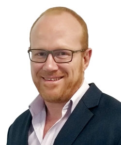 Diffusion MRI of the Spine: Sequence Optimisation
Ben Kennedy, M.Sc., B.Appl.Sc.(MRI) QScan Radiology Clinics Brisbane, QLD, Australia
SMRT & ISMRM Virtual Meeting: Top Tips for your Upcoming Abstract Submissions
19 November 2020
Watch the recording here (Members only; no CE credit) Username: Your ID number Password: Your last name (Case-Sensitive)
Moderators: Sheryl L. Foster, M.H.Sc.(MRS)(MRI) & Nick J Palmer, PG.Dip. MRI
Overview: This will be an information and question & answer virtual meeting for ISMRM and SMRT members. It will serve as a guide on successfully writing and submitting abstracts for ISMRM and SMRT meetings, whether they be virtual or face to face. Goals will include resolving any ambiguity or misunderstanding of the points the abstract reviewing committee are looking for when assessing, and help eliminate any confusion over the submission requirements as well as the nomenclature of the submission steps.
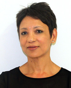 Tips and Tricks for Creating an SMRT Award Winning Abstract
Petronella Samuels, B.Sc. University Of Cape Town Cape Town, South Africa
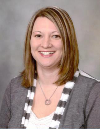 What SMRT Abstract Reviewers Look For in an Abstract
Erin Gray, M.H.A., R.T.(R)(MR) Mayo Clinic Rochester, MN, USA
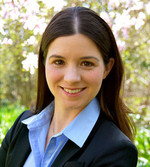 What ISMRM Abstract Reviewers Look For in an Abstract
Nicole E. Seiberlich, Ph.D. University of Michigan Ann Arbor, MI, USA
SMRT Virtual Meeting in Collaboration with the ISMRM MR of Cancer Study Group: Habitat Imaging With a Particular Emphasis on Validation of MRI Biomarkers for Imaging Tumor Heterogeneity
11 June 2020
Watch the recording here (Members only; no CE credit) Username: Your ID number Password: Your last name (Case-Sensitive)
Moderators: Janine M. Lupo, Ph.D. & Rhys A. Slough, M.Sc. (Medical Imaging – MRI)
Overview:
This virtual event will include two 30 minutes live presentations from experts in the field of Oncology Imaging. The session will open with a live presentation from Professor Evis Sala (MD, PhD, FRCR) an academic Radiologist with a special interest in Cancer Imaging. Dr Sala will be presenting on “Habitat imaging for unravelling tumour heterogeneity in ovarian and kidney.”
The second live presentation will be given by Dr Natarajan Raghunand (PhD), an Associate Professor of Radiology and Director of Translational Cancer Imaging in the College of Medicine at the University of Arizona Tucson who will be presenting on “Imaging tumor heterogeneity: tackling the inverse problem”
The live presentations will be followed by time for discussion – to provide the audience of MDs, PhDs, MRI Technologists and Radiographers with the opportunity to “Ask the Experts” questions about the challenges they encounter, techniques they use and the experience they have when using MRI for Cancer imaging.
Objectives:
- Describe the basis for habitat imaging and its value over current medical imaging models;
- Review the organ systems currently being validated for biomarkers and methods under investigation; and
- Explain implementation and its effect on current imaging sequences and post-processing.
Presentation 1:
 Habitat Imaging for Unravelling Tumour Heterogeneity in Ovarian and Kidney Cancers
Evis Sala, M.D.,Ph.D.
University of Cambridge
Cambridge, United Kingdom
Overview: The proposed session will highlight the role of MRI based habitat imaging in unravelling tumour heterogeneity in ovarian and kidney cancers and its implications in clinical trials.
Objectives:
- Understand the importance of tumour heterogeneity on patient outcome;
- Learn the concept of habitat imaging; and
- Understand the need for biological validation of tumour habitats.
Presentation 2:
 Multispectral Analysis of mpMRI Images to Identify Tumor Sub-Populations
Natarajan Raghunand, Ph.D.
H. Lee Moffitt Cancer Center
Tampa, FL, United States
Overview: This presentation will discuss some methods for reproducibly generating multispectral clusters on multiparameter MRI images to visualize tumor heterogeneity as a map of tumor sub-populations or “habitats”.
Objectives:
- Understand the value of acquiring spatially co-registered multiparameter MRI images for imaging tumor habitats;
- Understand the need for image intensity calibration for reproducible habitat imaging; and
- Understand some applications of habitat imaging such as tumor response assessment.
SMRT Virtual Meeting in Collaboration with the ISMRM Pediatric MR Study Group: Pediatric MR Imaging Techniques and Caveats and Insight into Imaging the Neonatal Brain
28 May 2020
Watch the recording here (Members only; no CE credit) Username: Your ID number Password: Your last name (Case-Sensitive)
Moderators: Glenn Cahoon, M.Appl.Sc., FSMRT, SMRT Representative, Pediatric MR Study Group Duan Xu, Ph.D., Chair: Pediatric MR study group
Overview:
This virtual event will include two 30 minute live presentations from experts in the field of Pediatric MR. The session will open with a live presentation from Nancy Hill Beluk (USA) a MRI Radiographer from the Pediatric Imaging Research Center, Children’s Hospital of Pittsburgh (UPMC), Pennsylvania. Nancy will be presenting on techniques and caveats in Pediatric MR Imaging which she has developed or experienced over her years of working with children.
The second live presentation will be given by Jorge Humberto Davila Acosta (Canada) from the Children’s Hospital of Eastern Ontario. One of Dr Davila areas of interests include pediatric neuroradiology and will be speaking on MR Imaging the neonatal brain.
The live presentations will be followed by time for discussion – to provide the audience of MDs, PhD, MRI Technologists and Radiographers with the opportunity to ‘Ask the Experts’ questions about the challenges they encounter, techniques they use and the experience they have when imaging pediatrics.
Presentation 1:
 Neonatal Brain MRI: Why the Technique Parameters Differ from Adults?
Jorge Humberto Davila Acosta, M.D.
Children’s Hospital of Eastern Ontario
Ottawa, ON, Canada
Accreditation: 0.50 CE/CPD
Overview: The proposed session will highlight the brain maturation changes happening from the third trimester of fetal life to adulthood and will will provide an overview of the specific parameters that should be optimized in neonatal and infantile brain MRI imaging.
Objectives:
- Understand the brain maturation process;
- Understand why the need of special considerations for conventional brain MRI in neonates; and
- Understand why the need of special considerations for non conventional brain MRI in neonates.
Presentation 2:
 Pediatric MR Imaging Techniques and Caveats
Nancy Hill Beluk, R.T.(R)
UPMC Children’s Hospital of Pittsburgh
Pittsburgh, PA, USA
Accreditation: 0.50 CE/CPD
Overview: The session will highlight different techniques for successful non-sedation MRI scans in the pediatric population and provide insights into specific safety considerations.
Objectives:
- Understand the differences of the various pediatric age ranges;
- Understand screening procedures, preparation needs and how to limit motion; and
- Understand specific safety concerns for this unique population.
ISMRM & SMRT Joint Virtual Meeting: COVID-19: Perspectives from the MRI Front Line
CE credits coming soon!
Your login to view these videos is:
Username: [Your Email Address]
Password: [Your Member Number]
Broadcast 1
15 April 2020
Moderators: Shawna Farquharson (AUS) & Larry Wald (USA)
Broadcast 2
18 April 2020
Moderators: Shawna Farquharson (AUS) & Pek-Lan Khong (HK)
Organizers:
Kirsty Campbell SMRT VMPC Chair New Zealand
Shawna Farquharson SMRT President Australia
Presentations:
- COVID-19 Emergency: The Italian Experience • Dr. Nivedita Agarwal, Italy (ISMRM)
- COVID-19 Emergency: A Clinical Perspective from Korea • Dr. Jongmin Lee (ISMRM)
- A Managers Perspective on Staffing and Operational Priorities • Chris Kokkinos, Australia (SMRT)
- The Importance and Practicalities of PPE in MRI • Rhys Slough, UK (SMRT)
- Strategies to Prevent the Intra-Departmental Spread of COVID-19 in Radiology • Dr. Yong Geng Goh, Singapore
- Q&A and Panel Discussion
Accreditation:
The Society for MR Radiographers & Technologists (SMRT), A Section of the International Society for Magnetic Resonance in Medicine has designated this live activity for a maximum of 1.00 Category A Credit.
The SMRT is recognized by the American Registry of Radiologic Technologists (ARRT) as a Recognized Continuing Education Evaluation Mechanism (RCEEM).
SMRT Virtual Meeting: MR Safety
27 February 2020
Watch the recording here (Members only; no CE credit) Username: Your ID number Password: Your last name (Case-Sensitive)
Members can earn CE credit for watching this video in the SMRT eLearning Center! Log in, click the eLearning Center button, followed by the MR Learning Labs option.
Moderators: Kate Negus, B.Appl.Sc.,RMIT(MR) & Laura Vasquez. Ph.D., RVT, R.T.(R)(MR), MRSO
Overview: This virtual event will include two 30 minute live presentations from experts in the field of MR Safety. The session will open with a live presentation from Dr Donald McRobbie Ph.D, (Australia) co-author of the popular MR physics textbook MRI from Picture to Proton. Dr McRobbie is a registered diagnostic imaging medical physicist, MRI physicist and MR safety expert.
The second live presentation will be given by Dr Michael Steckner Ph.D, M.D, (USA), who is an experienced Senior Research Manager with a demonstrated history of working in the medical device industry. Skilled in medical devices, biotechnology, biomedical engineering, product development, and research and development.
The two presentations will be followed by a question and answer session.
Objectives:
- Identify and understand risks related to the MR safety of implants and devices;
- Identify areas of MR Safety not commonly known;
 Title: Six Things You Didn’t Know About MR Safety
Speaker: Donald W. McRobbie, Ph.D.
University of Adelaide, School of Physical Sciences
Adelaide, SA, Australia
 Title: Implants and the MR Worker
Speaker: Michael Steckner, Ph.D, M.D.
Canon Medical Research USA, Inc.
Beachwood, OH, United States
ISMRM & SMRT Virtual Meeting: Women in Leadership: Insights from our ISMRM & SMRT MR Community Leaders
03 December 2019 at 11:30 PST
Learn from our women in leadership and hear them describe their journeys, greatest accomplishments and advice on how to further your professional career.
Watch the recording here (Members only; no CE credit) Username: Your ID number Password: Your last name (Case-Sensitive)
Karla L. Miller, Ph.D. University of Oxford United Kingdom
Shawna Farquharson, M.Sc.(R) The Florey Institute of Neuroscience Australia
Roberta A. Kravitz ISMRM Executive Director United States
Pia C. Maly Sundgren, M.D., Ph.D. Lund University Sweden
Moderators: Margaret Hall-Craggs, M.D., Women in ISMRM Chair, and Chris Kokkinos, B.Appl.Sc., Pg.Cert.(MRI), Past SMRT President
SMRT Virtual Meeting: The Principles, Challenges & Applications of Diffusion Weighted Imaging in the Body
21 November 2019
Watch the recording here (Members only; no CE credit) Username: Your ID number Password: Your last name (Case-Sensitive)
Members can earn CE credit for watching this video in the SMRT eLearning Center! Log in, click the eLearning Center button, followed by the MR Learning Labs option.
Moderators: Liana G Sanches-Rocha, M.Sc.(MR)(R) and Kurt G Schilling, Ph.D.
Overview: This virtual event will include two 30 minutes live presentations from experts in the field of MR Diffusion. The session will open with a live presentation from Michael Kean (AUS), who is a very experienced and respected MR Radiographer interested in MR Diffusion and will provide an expert’s explanation on “the principals, concepts and technical challenges of acquiring diffusion weighted imaging in the body.”
The second live presentation will be given by Dr Eric E Sigmund (PhD), an Associate Professor at NYU Langone Health, who will present on “DWI in the body: applications and challenges for oncology imaging.”
The live presentations will be followed by time for discussion – to provide the audience of MDs, PhDs, MRI Technologists and Radiographers with the opportunity to “Ask the Experts” questions about the challenges they encounter, techniques they use and the experience they have when acquiring diffusion weighted imaging in the body.
Objectives:
- Explain the basic principles of Diffusion-Weighted Imaging;
- Recognize the main technical challenges of acquiring Diffusion-Weighted Imaging in the body; and
- Understand why Diffusion-Weighted Imaging is used in the oncology protocols.
 Title: Diffusion Imaging of the Body: Concepts and Technical Challenges
Speaker: Michael Kean R.T., FSMRT
Royal Children’s Hospital
Melbourne, VIC, Australia
Accreditation: 0.50 CE/CPD
Overview: The presentation will address the technical concepts and challenges associated with acquiring DWI of the abdomen. The concepts to be addressed will include acquisition strategies, fat suppression and its impact on image quality and artefact reduction.
Objectives:
- Be familiar with the impact of different acquisition strategies on image quality;
- Understand the impact on sequence parameters on image quality; and
- Recognise artefacts and potential solutions.
 Title: DWI in the Body: Applications and Challenges for Oncology Imaging
Speaker: Eric P Sigmund Ph.D.
New York University, Department of Radiology, Langone Medical Center
New York, NY, United States
Accreditation: 0.50 CE/CPD
Overview: This talk will discuss the tools of acquisition and analysis applied to the imaging of cancer in the body with several variants of diffusion-weighted MRI. These include elements to ensure good image quality at the organ level, as well as capture the specific complexity of tumors and their host tissue the microscopic level. Both of these priorities involve approaches uniquely suited for body diffusion imaging.
Objectives:
- Understand unique challenges and solutions for good image quality for body DWI of cancer;
- Understand the applications of DWI techniques beyond apparent diffusion coefficient (ADC) (IVIM, DTI, DKI) in the body for both cancerous tissue and the surrounding host organs; and
- Understand examples of advanced modeling of cancerous tissue for further specificity of body DWI.
SMRT Virtual Meeting: How to Deal With Arrhythmia in CMR Imaging
17 October 2019
Watch the recording here (Members only; no CE credit) Username: Your ID number Password: Your last name (Case-Sensitive)
Members can earn CE credit for watching this video in the SMRT eLearning Center! Log in, click the eLearning Center button, followed by the MR Learning Labs option.
Moderators: Rhys Slough, BSc (Hons) Pg Dip (MRI), and Ben Statton, M.Sc., B.Appl.Sc.(MedRad)
 Title: Cardiac MR Imaging in Patients with Arrhythmias
Speaker:Peter Kellman, Ph.D.
National Heart, Lung and Blood Institute
Bethesda, MD, USA
Accreditation: 0.50 CE/CPD
Overview: The proposed session will focus on methods for cardiac magnetic resonance imaging (CMR) in patients with arrhythmias. CMR methods using breath-held, gated, segmented imaging protocols are vulnerable to artifacts in the presence of significant RR-variation. Real-time and/or single shot methods can be used to mitigate these artifacts. The presentation will cover cine, perfusion, and late enhancement imaging.
Objectives:
- Understand the mechanism that causes artifacts in segmented imaging protocols;
- Understand methods using arrhythmia rejection; and
- Understand the benefits of single shot and/or real-time cine imaging.
 Title: Cardiac MR Imaging in Patients with Arrhythmias: The Technologists Perspective: Tips, Tricks, and Possible Solutions
Speaker: Jane M Francis DCR(R), DNM, FSCMR
Barts Heart Centre
London, England, UK
Accreditation: 0.50 CE/CPD
Overview: This session will focus on recognising normal cardiac rhythms, including sinus bradycardia and tachycardia and common arrhythmias, distinguishing them from gradient induced artefact and provide some tips on how to improve image quality particularly cine imaging with different MR sequences.
Objectives:
- Recognise sinus rhythym and how to optimise for fast and slow rates;
- Recognise common cardiac arrhythymias and distinguish them from gradient induced artefact; and
- How to optimise ECG triggering in dealing with cardiac arrhythymias and sequence choice.
The Fundamentals & a Clinical Perspective of Current & Emerging Applications of 4D Flow Imaging Virtual Meeting in Collaboration with the ISMRM MR Flow & Motion Study Group
19 September 2019
Watch the recording here (Members only; no CE credit) Username: Your ID number Password: Your last name (Case-Sensitive)
Members can earn CE credit for watching this video in the SMRT eLearning Center! Log in, click the eLearning Center button, followed by the MR Learning Labs option.
 Title: The Fundamentals of 4D Flow Imaging
Speaker: Oliver Wieben, Ph.D.
University of Wisconsin, Madison
Madison, WI, USA
Accreditation: 0.50 CE/CPD credit
Moderator: Kevin Michael Johnson, Ph.D., MR Flow & Motion Study Group Chair
Overview: This presentation will review the basic concepts of 4D Flow MRI including data acquisition, post processing, visualization, common artefacts and pitfalls.
Objectives:
- Understand the principles on how velocity sensitive MRI data are acquired;
- Understand the benefits, pitfalls, and future potentials of 4D Flow MRI; and
- Understand basic principles of hemodynamic analysis possible with 4D Flow MRI.
 Title: A Clinical Perspective of Current & Emerging Applications of 4D Flow MRI
Speaker: Nicholas S. Burris, M.D.
University of Michigan
Ann Arbor, MI, USA
Accreditation: 0.50 CE/CPD credit
Moderator: Shawna L. Farquharson, M.Sc.(R), MR Flow & Motion Study Group SMRT Representative
Overview: This presentation will highlight a variety of emerging clinical applications of 4D Flow MRI, will review some of the current barriers to clinical adoption, and suggest potential future pathways towards routine clinical use of 4D Flow techniques.
Objectives:
- Understand the advantages and disadvantages of 4D Flow MRI for assessment of clinical patients.
- Understand some basic technical challenges and considerations when acquiring and analyzing 4D Flow MRI data in patients.
- Review some potential applications for clinical 4D Flow MRI.
MRI Safety: Implant Labeling & its Practical Applications Virtual Meeting in Collaboration with the ISMRM MR Safety Study Group
22 August 2019 Organizers: Kirsty Campbell & Vera Kimbrell
Watch the recording here (Members only; no CE credit) Username: Your ID number Password: Your last name (Case-Sensitive)
Members can earn CE credit for watching this video in the SMRT eLearning Center! Log in, click the eLearning Center button, followed by the MR Learning Labs option.
 Title: Implants and Other Devices: MRI Safety Testing and Labeling Guidelines
Speaker: Terry Woods, Ph.D.
Food and Drug Administration, Center for Devices & Rad. Health
Silver Spring, MD, USA
Accreditation: 0.5 CE/CPD credit
Moderator: Titti Owman, R.N.(R)(CT)(MR), FSMRT
 Title: Interpreting and Applying MRI Labelling Information for Implants & Devices
Speaker: Frank G. Shellock, Ph.D., FACR, FACC, FISMRM
Director, MRI Safety
USC Stevens Neuroimaging and Informatics Institute
Los Angeles, CA, USA
Accreditation: 0.5 CE/CPD credit
Moderator: Anne Marie Sawyer, B.S., R.T.(R)(MR), FSMRT
Q&A Session: Ask The Experts
30 minutes (maximum) for questions from the virtual meeting audience.
Overview: This virtual event will include two 30 minute live presentations from experts in the field of MR Safety. The session will open with a live presentation from Terry Woods, Ph.D. The session will highlight MRI related safety concerns produced by implants and other devices, provide an overview of standards and guidelines for assessing MRI safety, and describe standard MRI safety labeling guidelines.
The second live presentation will be given by Frank Shellock, Ph.D., who is a physiologist with more than 30 years of experience conducting laboratory and clinical investigations in the field of magnetic resonance imaging. Dr. Shellock will present on interpreting the standards and guidelines presented by Dr. Woods and how to apply MRI Labeling Information for Implants & Devices.
Objectives:
- Identify and understand risks related to the MR safety of implants and devices based on their intended location in the MR environment, electrical activity, and implant status;
- Describe standards and guidelines that relate to MR safety of implants and other medical devices; and
- Recognize and understand MRI safety labeling for medical devices, including the standard MR safety terms: MR Safe, MR Conditional, and MR Unsafe.
The How & Why of MR Spectroscopy Virtual Meeting in Collaboration with the ISMRM MR Spectroscopy Study Group
18 July 2019 Organizers: Kirsty Campbell & Huijun Vicky Liao
Watch the recording here (Members only; no CE credit) Username: Your ID number Password: Your last name (Case-Sensitive)
Members can earn CE credit for watching this video in the SMRT eLearning Center! Log in, click the eLearning Center button, followed by the MR Learning Labs option.
 Title: The “How to” of MRS: Things to Consider When Planning Acquisition
Speaker: Caroline (Lindy) Rae, Ph.D.
Neuroscience Research Australia
Melbourne, VIC, Australia
Accreditation: 0.5 CE/CPD credit
Moderator: Michael Kean, (RT)FSMRT
Overview: The proposed session will discuss the range of different approaches to single voxel MRS and highlight their purposes and limitations. It will canvass things to consider when planning MRS acquisitions and what, realistically, can be achieved with the method.
Objectives:
- Understand the different acquisition options for acquiring MR spectra
- Understand which acquisition options may be optimal for what you want to measure
- Understand the practical issues you need to consider when setting up a MRS acquisition
 Title: The Clinical Applications of MR Spectroscopy
Speaker: Alexander P. Lin, Ph.D.
Brigham and Women’s Hospital
Boston, MA, USA
Accreditation: 0.5 CE/CPD credit
Moderator: Huijun Vicky Liao, B.Sc., ARMRIT SMRT Representative, MR Spectroscopy Study Group
Overview: The goal of this presentation will be to provide technologists with practical knowledge of how magnetic resonance spectroscopy is utilized for diagnosis and treatment monitoring across a broad range of clinical diseases.
Objectives:
- Understand how MR spectroscopy is utilized in different pathologies such as cancer, neurological disorders, metabolic disorders, and non-neuro application in both pediatrics and adults.
- Understand how to acquire spectroscopy for these different diseases by reviewing different protocols for each condition.
- Learn how to provide quality control on MRS data and recognize when real-time changes need to be made to ensure accuracy and excellence.
Challenges, Advances & Applications of MR Spinal Cord Imaging Virtual Meeting in Collaboration with the ISMRM White Matter Study Group
20 June 2019 Organizers: Kirsty Campbell & Helle Simonsen
Watch the recording here (Members only; no CE credit) Username: Your ID number Password: Your last name (Case-Sensitive)
Members can earn CE credit for watching this video in the SMRT eLearning Center! Log in, click the eLearning Center button, followed by the MR Learning Labs option.
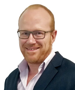 Speaker 1: Ben Kennedy, B.Appl.Sc.,Mst(MRI)
Title: MRI of the Spine at 3T
Overview: The proposed session will look at historical challenges of MRI of the spine at 3T, addressing many of these challenges with technology advancements and parameter optimization and novel imaging that 3T SNR allows us to take advantage of.
Objectives:
- Understanding challenges of 3T physics vs large FOV spine imaging
- Current technology making clinical 3T of the spine a new gold standard
- Parameter optimization for best standards of Spine imaging at 3T
Moderator: Hugo Vrenken, Ph.D.
Note for ARRT R.T.s: This talk was previously accredited for CE credit in May 2019 at the SMRT Annual Meeting in Montreal, QC, Canada. It is your responsibility to maintain a record of previously submitted and credited live or self-learning CE activities. Duplicate credit for the same activity is not permitted in the same biennium.
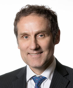 Speaker 2: Klaus Schmierer, M.D., Ph.D.
Title: Spinal Cord MRI in the Diagnosis & Monitoring of Multiple Sclerosis: A Clinical Perspective
Objectives:
- Understand that MS is a common and complex disease;
- Understand the importance of correlating MRI findings with the underlying pathology; and
- Understand the challenge of assessing specific tissue features other than myelin in the spinal cord
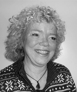 Moderator: Helle J. Simonsen, (MRT)FSMRT
SMRT Representative, White Matter Study Group
SMRT Virtual Meeting: MRI Safety: Risk Prevention & Creating an MRI Safety Culture
26 April 2019
Watch the recording here (Members only; no CE credit) Username: Your ID number Password: Your last name (Case-Sensitive)
Members can earn CE credit for watching this video in the SMRT eLearning Center! Log in, click the eLearning Center button, followed by the MR Learning Labs option.
MRI Safety is universal. There are no geographic boundaries, cultural preferences, or religious exemptions. The danger is real for everyone.
Accreditation: 1.0 CE/CPD credit
Target Audience: MRI Technologists, Physicians, Nurses, MRI Support personnel
Registration Fee: Free for members; US$25 for non-members
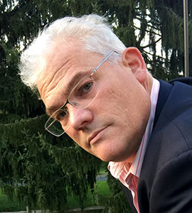 Speaker 1:
John C. Posh, B.S.,R.T., (R)(MR)
Title:
Preventing MRI Accidents: Best Practice Recommendations to Minimize Risk
Objectives:
- Understand the concept of risk and how it applies to MRI;
- Understand how risk is managed in general and the impact it has on MRI operations; and
- Learn the types of accidents that happen in MRI and the countermeasures for preventing them.
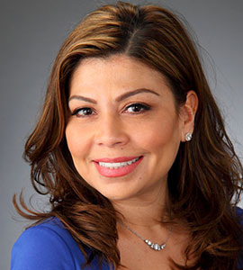 Speaker 2:
Laura P. Vasquez, PhD, RVT, RT(R)(MR), MRSO
Title:
Creating an MRI Safety Culture and Performance Improvement Program
Objectives:
- Deliver recommendations for implementation of an institutional interprofessional MRI safety committee;
- Provide suggestions for facilitation of leadership engagement and administrative accountability; and
- Describe ways to foster a culture of education, standardization, quality and safety.
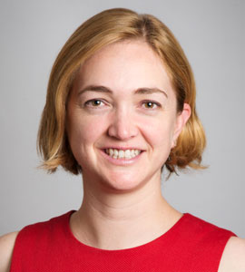
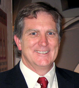 Moderators:
Claire B. Mulcahy, M.MRT., B.Appl.Sc.
Greg C. Brown, A.Dip.Rad.Tech.
SMRT/CAMRT Virtual Meeting: Accelerated MRI Using Compressed Sensing
Tuesday, 05 March 2019
Members can earn CE credit for watching this video in the SMRT eLearning Center! Log in, click the eLearning Center button, followed by the MR Learning Labs option.
Watch this webinar to gain basic concepts of Nyquist sampling, aliasing, and parallel imaging (SENSE and SMASH). Understand the application of compressed sensing (CS) for MRI data acquisition and learn how this newer method can accelerate imaging without the need for Nyquist sampling. Walk away understanding the principles of the CS approach along with a host of clinical applications now available from MRI vendors.
 Speaker:
Michael Noseworthy, Ph.D.
 Moderator:
James J. Stuppino, B.S., R.T.(R)(MR)
Accreditation: 0.25 CE/CPD credit (not available for credit for archive viewers)
Registration Fee: CLOSED (was: Free for members; $15 for non-members)
Objectives:
- Refresh the understanding of undersampling and its consequences with MRI;
- Introduce how undersampling using compressed sensing works;
- Understand how compressed sensing can be applied using clinical examples.
Target Audience: MRI Radiographers, Technologists and Radiologists
Thursday, 21 February 2019
Watch the recording here (Members only; no CE credit) Username: Your ID number Password: Your last name (Case-Sensitive)
Members can earn CE credit for watching this video in the SMRT eLearning Center! Log in, click the eLearning Center button, followed by the MR Learning Labs option.
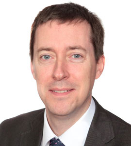 Speaker 1:
Declan O’Regan, FRCP, FRCR, Ph.D.
What Clinicians Need from Cardiac MRI
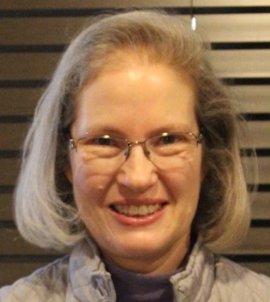 Speaker 2:
Cindy Comeau, B.S., RT(N)(MR), FSMRT, FSCMR
Getting Started: The Core Cardiac MRI Exam
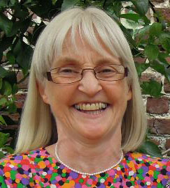
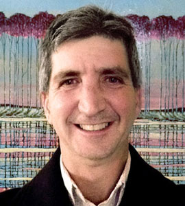 Moderators:
Muriel Cockburn, DCR, B.Sc.(Hons), FSMRT
Marty Sherriff, B.App.Sc., R.T. (MR)
Accreditation: 1.0 CE/CPD credit (not available for credit for archive viewers)
Registration Fee: CLOSED (was: Free for members; $25 for non-members)
Objectives:
- Understand the common indications for CMR and the information clinicians require.
- Learn how different CMR sequences are used to answer clinical questions.
- Appreciate the appearances of common pathologies on CMR.
Target Audience: MRI Radiographers, Technologists and Radiologists
Improving Throughput Without Compromising Patient Care & Creating Value in MR
Friday, 18 January 2019
Watch the recording here (Members only; no CE credit) Username: Your ID number Password: Your last name (Case-Sensitive)
Members can earn CE credit for watching this video in the SMRT eLearning Center! Log in, click the eLearning Center button, followed by the MR Learning Labs option.
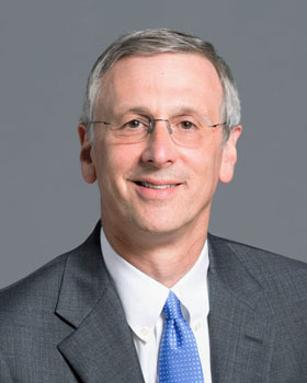
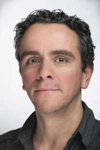 Presenters:
Michael P. Recht, M.D.
Chris Kokkinos, B.Appl.Sc., Pg.Cert.(MRI)
Target Audience: MRI Radiographers and Technologists, Radiologists
Accreditation: 1.25 CE/CPD credits
Registration Fee: CLOSED (was: Free for members; $25 for non-members)
Talk 1: Value in MR: What Is It and How Do We Create It? Presenter: Michael P. Recht, M.D.
Talk 2: When Throughput Affects Patient Care: How to Improve Your Numbers Without Compromising Care Presenter: Chris Kokkinos, B.Appl.Sc., Pg.Cert.(MRI)
Moderators: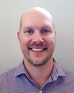
 Kirsty Campbell, NDMDI, PG Dip (MRI)
Joe Joslin, B.S., R.T.(MR)(R)
Duration: 1 hour 15 minutes, with questions at the end
Objectives:
- How to improve patient throughput;
- How value can be created and operationalized in MR; and
- How to use data to improve the MRI department.
RF Safety in MRI: An MR Technologist’s Perspective Wednesday, 14 November 2018
Members can earn CE credit for watching this video in the SMRT eLearning Center! Log in, click the eLearning Center button, followed by the MR Learning Labs option.
 Presenter: Vera Kimbrell, BSRT, (R)(MR), FSMRT
Vera Kimbrell is a MR technologist/radiographer from Boston, Ma. USA. She works at the Brigham and Women’s Hospital in the role of clinical educator for MRI. She has been in MR for over 30 years and an educator for the majority of that time. Currently the Chair of the SMRT Safety committee, Vera has lectured and taught MR safety internationally.
Target Audience: MR Technologists, Radiographers, Students, Clinicians & Early Career Scientists
Accreditation: 0.50 CE/CPD credits
Registration Fee: CLOSED (was: Free for members; $15 for non-members)
 Moderator: James J. Stuppino, B.S., R.T.(R)(MR)
Time: There will be two presentations of this webinar. Please only register for one.
- 09:00 PST / 12:00 EST / 20:00 UTC
- 12:00 PST / 15:00 EST / 23:00 UTC
Duration: 30 minutes
Objectives:
- Explain the safety related issues related to Radiofrequency Magnetic Fields (RF)
- Define devices and practices that contribute to RF burns
- Describe workflows to mitigate hazards related to RF
- Review accidents and explore preventative solutions
Diffusion Imaging & Fiber Tractography: Tips & Tricks for Clinical Use
Wednesday, 24 October 2018
Watch the recording here (Members only; no CE credit) Username: Your ID number Password: Your last name (Case-Sensitive)
Members can earn CE credit for watching this video in the SMRT eLearning Center! Log in, click the eLearning Center button, followed by the MR Learning Labs option.
Moderators: Ben Statton, M.Sc., B.Appl.Sc. (London, England) and Ileana Jelescu, Ph.D. (Lausanne, Switzerland)
Target Audience: MR Technologists, Radiographers, Students, Clinicians & Early Career Scientists
Accreditation: 1.0 CE/CME/CPD credits
Time: 07:00 PDT; 16:00 The Netherlands; 01:00 Melbourne, Australia
Duration: 1 hour
Fee: Free for members; $25 for non-members
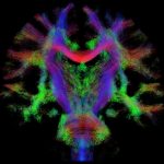
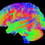
Overview: This session provides a focused review suitable for a broad audience on one widely-used or emerging area of Neuroimaging. This presentation is targeted to radiographers, technologists, researchers and clinicians interested in understanding and developing diffusion tractography MRI methods for neurosurgical planning. Core techniques and their shortcomings will be discussed in the context of practical clinical cases.
Speaker 1: Shawna Farquharson, M.Sc.(R) (Melbourne, VIC, Australia)
Speaker 2: Alexander Leemans, Ph.D. (Utrecht, Netherlands)
SMRT/CAMRT Virtual Meeting: MRI Contrast Agents: Safety Issues and Practice
Wednesday, 25 July 2018
Members can earn CE credit for watching this video in the SMRT eLearning Center! Log in, click the eLearning Center button, followed by the MR Learning Labs option.
Speaker: Lawrence Tanenbaum, M.D.
Moderator: Jim Stuppino, B.S., R.T.(R)(MR)
Target Audience: MR Technologists, Radiographers, Students, Clinicians & Early Career Scientists
Accreditation: 1.0 CE/CPD credits
Learning objectives:
- Describe the different gadolinium contrast agents available, including their chelate structure, stability and retention.
- Describe the link between gadolinium contrast agents and Nephrogenic Systemic Fibrosis (NSF).
- Review current literature and regulation related to the safe and effective use of gadolinium contrast agents.
- Identify strategies for discussing contrast media with patients.


 Download the flyer