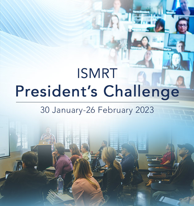
2023 ISMRT President’s Challenge
30 January – 26 February 2023
 Are you up for a challenge? Then why not join us by participating in this month-long event bringing world-class MR education to your local area? The call for an “Expression of Interest” to host an ISMRT President’s Challenge event is NOW OPEN!
So, what is an ISMRT President’s Challenge event? As a member of the ISMRT, you register to host an event, choose a package, and organize a 1-hour meeting with your colleagues. We have done all the upfront work of creating several different packages that you can choose from. Each package includes a PowerPoint presentation to support you in leading the meeting. Embedded in the slides are videos on the topic of choice. Details on providing attendees certificates of attendance or CE/CPD credit will also be included. It’s that simple!
Can this event be hosted virtually, or does it have to be in person? The choice is yours. We understand that not everyone will be able to host an in-person event due to current restrictions. If you have a way to host a virtual event (such as having your own personal Zoom, AdobeConnect, etc., account), you can participate. We only ask that you limit attendees to those in your local area. (Please note that if you do hold a meeting virtually, you will be expected to confirm each person’s attendance at the beginning, middle, and end of the meeting in order to provide CE or CPD).
This is a great way to highlight the advantages of being an ISMRT member and to convey to others the importance of MR education for radiographers and technologists. If the attendees find the content valuable and are interested in membership, they will have the opportunity to join. This is also a chance for you to share knowledge with your colleagues by offering them free CE/CPD. A win-win!
We know that times have been challenging, and it is more important than ever to come together to share our knowledge. We challenge you to organize your event, spread the word, and have fun networking and presenting an MR education topic with your colleagues!
ISMRT President’s Challenge
Event Host Application
Educational Package Options
Click the package titles below to view more details about each package, including presentations, organizers and more.
Presentation 1:
MR Imaging of Brachial Plexus
Kimberly K. Amrami, M.D.
Presentation 2:
Advances in Spinal Cord MRI at 3T
Seth Smith, Ph.D.
Course Organiser:
Barbara Pirgousis (AUS), ISMRT Online Learning Committee member, ISMRT Australian Chapter Secretary
Course Summary:
The two presentations chosen provide an overview of the challenges when imaging the brachial plexus and the spine at 3T. Each presentation provides the technologist/radiographer with clinical examples, including how advances in MRI technology assist with optimising image and diagnostic quality. MR Imaging of the Brachial Plexus, presented by Kimberly K. Amrami, M.D., of the Mayo Clinic, acknowledges the difficulty with imaging the brachial plexus and the importance of performing post-gadolinium imaging, supported with case studies. Advances in Spinal Cord MRI at 3T, presented by Seth Smith, Ph.D., of Vanderbilt University Medical Center, highlights the challenges posed by 3T in relation to optimising contrast in the spinal cord. The presentation then outlines examples of advanced techniques including DWI/DTI, CSET, fMRI, qMT of the spinal cord.
Presentation 1:
Neonatal MR Imaging: Expanding the Diversity of Imaging Through Innovation
Michael Kean, R.T., FSMRT
Presentation 2:
Pediatric Body MR Imaging: Challenges & Strategies for Success
Hansel J. Otero, M.D.
Course Organiser:
Glenn Cahoon (AUS), ISMRT President-elect & Education Co-Lead
Course Summary:
These presentations were chosen with the goal of providing information to MRI radiographers/technologists who are currently or soon to be working at a paediatric facility or have an interest in paediatric MRI. Michael Kean, a highly experienced MR radiographer working at the Royal Children’s Hospital in Melbourne, Australia, discusses the common paediatric brain tumours and the techniques, benefits, and challenges of MR imaging in this patient group. Dr. Hansel Otero, is the director of body imaging at the Children’s National Medical Center in Washington, D.C., USA, focuses on the challenges and strategies for successful body imaging in paediatric patients. Both presenters have first-hand knowledge of day-to-day operations in busy paediatric centres, and the presentations provide information and demonstrations on how to succeed in acquiring MR images on our younger population.
Presentation 1:
Quantitative MRI in MSK: Beyond Tissue Morphology
Emily McWalter, Ph.D.
Presentation 2:
Imaging Biomechanics of Whole Joints
David R. Wilson, D.Phil.
Course Organiser:
Debra Patterson, M.S., R.T. (R)(MR)(CT) (USA), ISMRT Online Learning Committee Chair
Course Summary:
The following presentations provide MRI radiographers/technologists with an overview of how new techniques in MRI can be used to examine and diagnose pathology and function in MSK. Emily McWalter and David Wilson are both mechanical engineers and have spent years conducting research to explore the relationship between biomechanics and MRI. These presentations specifically focus on imaging techniques used for dynamic imaging of joints and illustrate how MRI can generate quantitative maps to improve diagnosis of musculoskeletal diseases.
Presentation 1:
MR Physics & Techniques for Clinicians - Artifacts to Artefacts: Causes & Cures from a Clinical Perspective
Walter F. Block, Ph.D.
Presentation 2:
Introduction to Hardware
Martin J. Graves, Ph.D.
Course Organiser:
Sonja K. Boiteaux, MSRS, RT(R)(MR), MRSO, MCHC (USA), 2022 ISMRT President
Course Summary:
These presentations provide MR radiographers/technologists with information on fundamental principles of MR physics. The presentations are well-suited for those who want to take a look “under the hood” or beyond the gantry of your MR system. Dr. Walter Block—MR physicist, Professor of Biomedical Engineering at University of Wisconsin - Madison reviews artifacts in MRI and Dr. Martin Graves of the University of Cambridge describes the main hardware components of an MRI system and explains how they contribute to image formation and discusses their limitations.
Presentation 1:
ISMRT Video Safety Series, Part 1
William Faulkner, B.S.,R.T.(R)(MR)(CT), FSMRT, MRSO (MRSC™)
Presentation 2:
ISMRT Video Safety Series, Part 2
William Faulkner, B.S.,R.T.(R)(MR)(CT), FSMRT, MRSO (MRSC™)
Course Organiser:
Haidee Paterson (UAE), member, ISMRT Online Learning & ISMRT Marketing Committees
Course Summary:
The following presentations provide MRI radiographers/technologists with an insight into some of the elements that create the backbone of MRI safety. These two presentations are ideal for those who would like a greater understanding of the different magnetic fields present in an MRI environment and the subsequent safety issues that can arise from these inherent fields. William Falkner, a radiologic technologist with advanced certification in MRI and CT, explains the static and gradient magnetic fields in Part 1, and addresses the radiofrequency field and a quench in Part 2.
Presentation 1:
Diffusion in the Body: Diffusion-Weighted MR Imaging of the Extrahepatic Abdomen & Pelvis
Russell N. Low, M.D.
Presentation 2:
Pancreas & Biliary: Tailored Techniques for Biliary & Pancreatic Imaging
Mustafa Shadi R. Bashir, M.D.
Course Organiser:
Lori Nugent, ISMRT Online Learning Committee member
Course Summary:
The following two MR body presentations provide medical imaging technologists and MR radiographers with learning content related to the clinical applications of DWI in the abdomen and pelvis and strategies for pancreatic and biliary imaging.
Dr. Russel Low—a board-certified diagnostic radiology specialist practicing at Balboa Nephrology and affiliated with Sharp Memorial Hospital in San Diego, CA—discusses the clinical applications of DWI in the abdomen and pelvis and demonstrates the ways in which DWI is beneficial in providing qualitative and quantitative information and in detecting and characterizing lesions. He also details the effectiveness in DWI in both monitoring treatment as well as the patient’s responses to the therapy.
Dr. Mustafa Bashir of Duke University Medical Center describes the diagnostic information that is obtained through the use of MRCP techniques, T1 pre- and post-dynamic CE imaging, T2 pathology weighting, and DWI to diagnose pancreas and biliary disease. He also discusses when the use of MRI is the gold standard for detecting pathology in these structures.
Presentation 1:
Imaging Cells with Nanoparticles
Paula Foster, Ph.D.
Presentation 2:
Interventional: Prostate, Liver & Kidney Interventions
Kemal Tuncali, M.D.
Course Organiser:
Anne Dorte Blankholm, Ph.D., MSc., Radiographer, ISMRT Past President & Education Co-Lead
Course Summary:
These two presentations provide information to radiographers/MR technologists who are interested in hot topics in MR. Both presentations give a look into things that may affect the future work life of radiographers/MR technologists—and maybe some already have. In the first presentation, Paula Foster, Ph.D.—medical biophysics professor at Western University and Cellular and Molecular Imaging Research Program lead at Robarts Research Institute in London, ON, Canada—discusses how nanoparticles in MR can be used to measure both disease and treatment. The second presentation, given by Kemal Tuncali, M.D., Interventional MRI director at Brigham and Women’s Hospital, Harvard Medical School, presents some challenges and achievements in interventions of the prostate, liver, and kidney, including cryoablation and MR-guided prostate biopsy through the perineum
