BY YOGESH MARIAPPAN
As the field of MRI is slowly but surely moving from qualitative to quantitative, and from imaging structures to imaging other properties, the choice of this month’s Highlights article is apt: the research group led by Ingolf Sack has been at the forefront of both of these aspects with their work on MR Elastography (MRE). This is a quantitative imaging technique capable of measuring the mechanical properties of tissues of interest and is available from all the major vendors. The current clinical application is focused on liver where the stiffness (measured in kilopascals) is used for fibrosis assessment. In this article they have provided recent results from their “In vivo wideband multi-frequency work” on liver and brain.
MRMH: Tell us about yourselves and your academic journey so far.
Florian: I did my Bachelor’s and Master’s degrees in medical informatics, worked in the field of radiation therapy for the German Cancer Research Center in Heidelberg, Germany, and then at Massachusetts General Hospital in Boston. Later I found my true calling and have been doing my doctoral thesis work with the Elastography group since 2013. My future plan is to work in industry, but as of now I am not sure where. (Talent HR in industries: are you reading?)
Ingolf: I have always been interested in the physical and chemical aspects of biological tissues. I started as a chemist from which I came to analytical chemistry, to spectrometry, to MR imaging and now finally to MR Elastography. I started working with Elastography when it was in its infancy and have seen it grow leaps and bounds for the last 16 years.
MRMH: Could you please provide us a glimpse of your academic institute Charité, especially from the MR Research perspective?
Ingolf: Charité is one of the biggest university hospitals in Europe, which provides us the opportunity to have many clinical collaborators constantly pushing us to develop methods for detecting diseases better and quicker. The hospital itself is spread over three campuses in Berlin with multiple research-dedicated MR scanners. Our group works mainly on basic research, solving equations, but our collaborators provide the motivation and bring our methods to the clinic.
MRMH: Elastography and MR Elastography in particular are still considered novel. Could you comment on the application, awareness and acceptance?
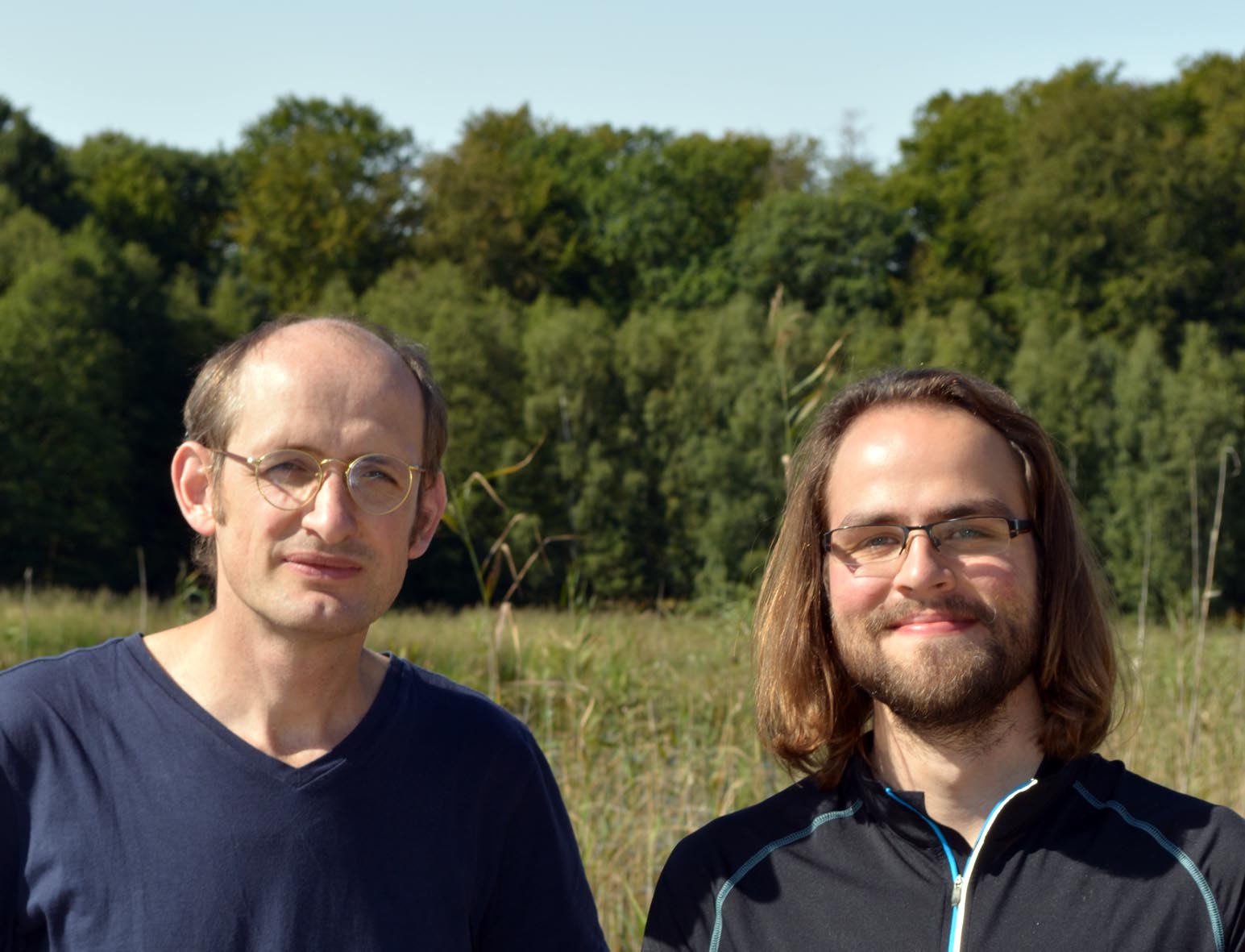
Florian exploring a glacier on his travels.
Florian: MRE is quite an interesting field, currently being used for the staging of liver fibrosis clinically. And it is being actively investigated for brain and tumor characterization applications. In most of these cases, I believe that it will be beneficial to use the wideband approach elaborated in our article.
Ingolf: Elastography has penetrated deeper in the field of ultrasound, where it is implemented in all commercially available Ultrasound systems. Elasticity imaging is not only a diagnostic imaging technique, it is a new approach to study the biophysics of tissues. Once we can better solve the Inverse problem (calculating the tissue mechanical properties from the acquired Ultrasound/MRI images), I think there will be widespread applications. While MR Elastography is also available from all the big vendors, it is currently used only for additional investigation for cases that are suspicious. The clinicians typically have an idea of the tissue mechanical properties from ultrasound, but then turn to MRE for providing high resolution images.
MRMH: Could you please provide a brief overview of the paper?
Florian: Our goal was to develop the MRE method for time-harmonic multi frequency elastography, specifically using very low frequencies below 25 hertz. We developed a new modeling framework and a fast imaging sequence to achieve this in clinically acceptable scan times. We tested our technique first in gel samples, and then in human brain and liver. We were able to create very high resolution stiffness elastograms using our wideband multi frequency approach.
Ingolf: Brain is surprisingly soft at low frequencies; so while we did expect to measure low elasticity values, we obtained values almost half of what we had expected. We were initially skeptical of the accuracy of these results. But then we went to literature, and we found our values fit perfectly. In the liver, we have low SNR with long echo times, and at low frequency elastograms suffer from noise. This is even worse in fibrosis where the stiffness increases. In the brain, it is the other way around, brain gets softer with most brain disease processes. This softness in the brain enables the use of low frequency vibrations and helps our inversion algorithm. Low frequency vibrations are better transmitted in the body. In fact these waves do actually travel through the entire body, so one can do MRE with one actuator without strong attenuation. This is a big advantage with the lower frequencies compared to conventional MRE.
MRMH: So maybe in the future one can get whole body MR Elastography under five minutes…
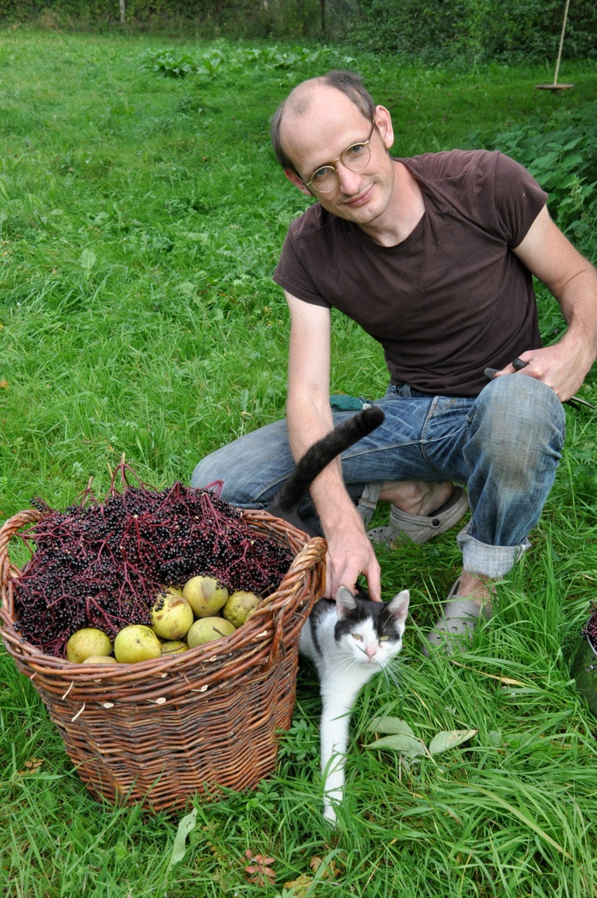
Ingolf harvesting fruits from his bountiful garden.
Ingolf: [laughs] Yes, that is the idea, we are not interested in low hanging fruits. Getting the high hanging fruits is the motivation.
MRMH: Could you comment on the safety of these vibrations?
Ingolf: Vibrations coming from the scanner during normal MR Imaging are sometimes higher in amplitude than the low frequency vibrations we apply for MRE. That is one of the advantages of low frequency: safety, homogeneous penetration, and the sensitivity to solid-fluid interactions.
MRMH: What is one key concept you would like the community to remember after reading your paper?
Florian: Low frequency MRE is feasible, and is good for the future of MRE research, as other mechanical properties like poroelasticity come into play.
Ingolf: With the low frequency vibrations, the safety is greater and tolerance for the technique is higher. When we tell the patients that we don’t touch their head but we do brain MRE, that helps.
MRMH: What do you do when you are not in the lab?
Florian: I like bicycles which I ride every day, I enjoy cooking, and traveling to places that are peaceful and breathtaking.
Ingolf: Florian, you have a life after the lab [laughing]?. For me, I spend time with family, kids, and I love my garden.
That wraps up our discussion with Florian and Ingolf. This group is working on a wide range of applications (fibrosis, cancer, diagnostic MRE for tissue culture analysis), organs (liver, brain, breast) and frequencies (from a few Hertz to a few kilo Hertz). Thus, when Ingolf, being an avid gardener, says that they are heading for the high hanging fruit, we cannot wait to find out what’s on their menu next.