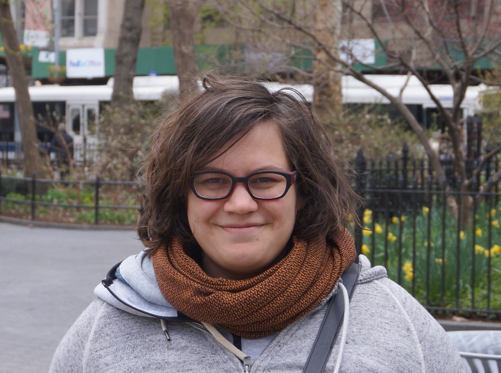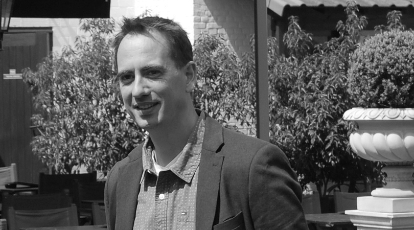

Gwendolyn Van Steenkiste is currently a doctoral student at the University of Antwerp in Belgium, working on super-resolution reconstruction (SRR). Gwendolyn’s paper entitled “Super-resolution reconstruction of diffusion parameters from diffusion-weighted images with different slice orientations,” was selected as Editor’s Pick for the month of January. We contacted Gwendolyn and her supervisor, Dr. Jan Sijbers to discuss the details of the paper.
MRMH: Gwendolyn, how did you get interested in MRI?
Gwendolyn: I have a strong clinical influence from my family who are dentists, doctors and pharmacists. So when I finished studying engineering in applied physics as an undergraduate, I became very interested in MRI, which has clinical impact but is still physics based.
MRMH: Can you tell us in plain language the main points of your paper?
Gwendolyn: Whenever you want to increase the spatial resolution of an image, there is either an increase in acquisition time or a decrease in signal to noise ratio (SNR). Our paper showed that it is possible to use the same acquisition time and achieve greater spatial resolution. Additionally we showed that the diffusion model can be incorporated into the super resolution reconstruction process, thus limiting the propagation of errors in the pipeline. Specifically with our approach, you can have greater parameter selection, while sampling Q space more optimally and incorporating the motion correction.
 Jan: As Gwendolyn said, this method allows for higher resolution and greater freedomwith the acquisition parameters. We achieve this by acquiring a set of low-resolution diffusion images with high SNR, and incorporating well-chosen orientations. By optimizing the scanning parameters, you can break the traditional trade-off between acquisition time, SNR, and spatial resolution.
MRMH: Are the voxels you acquire isotropic?
Gwendolyn: No, the idea is that you acquire images with high in-plane, but low through-plane resolution. The thick slices are acquired at different angles, so that if you project in k-space you create a circle in 2D, or a cylinder in 3D. From this you can reconstruct higher resolution images.
MRMH: In short can you describe the signal-generating model?
Gwendolyn: The signal-generating model explains what happens in the scanner. First we model the motion (patient movement and table vibration), then we incorporate the geometry (slice orientation), and also the voxel size (downsampling). This is all modeled by an affine transformation, and it is followed by filtering that accounts for the incomplete k-space coverage and the imperfect slice selection.
Jan: The signal-generating model is very important in forward modeling. It allows to forecast how low resolution images (acquired at a specific orientation) would look like, given an estimate of the high-resolution DTI image. This simulated low resolution image can then be compared to actually measured low resolution images. The difference measures how close your assumed high-resolution DTI image is to the true (unknown) high resolution DTI image.
MRMH: What are the benefits of using the SRR method? How do you see this translating into future research?
Gwendolyn: Well, SRR could be an alternative to the strong gradients of the Connectom scanners. In regular clinical research, it will result in shorter acquisition times, which will in turn produce images with fewer motion artifacts. As for research on SRR, there is still lots to do, on improving the modeling, but also on applying to different modalities, such as T1 mapping and perfusion.
Jan: Our ultimate goal is for SRR to optimize each single k-space point for the best parameter maps in a range of protocols (diffusion, T1 mapping, perfusion).
MRMH: What has been the biggest challenge of this project?
Gwendolyn: Our protocol is very different from what MR operators usually use, so you need to have good communication if you want somebody else to acquire your data. Basically you need to convince them to let go of what they know and stick with the weird slice orientations and the unconventional acquisition strategy. As we progress, with the more complex diffusion models, I think the difficulty will lie in the computational complexity of the fitting.
MRM: What was your eureka moment?
Gwendolyn: When I finally got the acquisition set up on point and I visually saw an enhancement in the spatial resolution of the estimated DTI parameters. Another moment was when I was simulating motion in my diffusion data and realized that including a variety in the q-space sampling resulted in a better estimation of the diffusion parameters!
MRM: Jan, what is life like at the University of Antwerp?
Jan: The University is comprised of three campuses, and our campus is located outside the city. It primarily hosts life sciences, it is green, has top research infrastructure, and the food is great.
MRMH: What comes next for you, Gwendolyn?
Gwendolyn: I am in my final year of my PhD, and am hoping to continue with MRI research focusing on super resolution methods. I would like to go abroad to explore different opportunities and viewpoints.
Jan: Indeed, going abroad during or after your PhD is for sure an enriching experience. Within the Vision Lab, we will continue exploring new avenues in the area of quantitative magnetic resonance imaging and hope that our collaboration with Gwendolyn will last for many years.
*Interview conducted by Karolina Urban and Nikola Stikov.
