
BY BO ZHU & NIKOLA STIKOV
For the first Editor’s pick of June, Dr. Yunkou Wu and Prof. Dean Sherry from the University of Texas Southwestern Medical Center have developed a novel paraCEST agent seeking to push the envelope of robust pH quantification in-vivo. We spoke with them over Skype to discuss this new technique and its implications for the CEST field, as well as the broader MR community.
MRMH: What is your background and how did you get involved with MRI research?
Yunkou: I obtained my PhD in Chemistry, with a background in organic synthesis. I started to do research on developing MR contrast agents as a post-doc, but things really picked up when I moved to Dr. Dean Sherry’s group at UT Southwestern Medical Center, where I became more familiar with operating MRI scanners and understanding more fundamental MR Physics.
Dean: My scientific interests in this area go way back – I was likely doing NMR before either of you were born! I’ve had a long history of working on lanthanides, studying everything from ligand synthesis, to water coordination numbers, to NMR Dispersion analyses of gadolinium relaxation, to measuring water exchange rates. We had used gadolinium as a “relaxation reagent” to study protein structure before the discovery of MRI by Paul Lauterbur so we immediately recognized the potential of gadolinium as an imaging contrast agent. Paul’s discovery really jump-started my interest in the field of MR contrast agent development.
MRMH: What were the motivations for this work, and could you give a brief summary of the paper?
Yunkou: We are interested in pH imaging because dysregulation of pH is associated with many diseases, like cancer or renal tubular acidosis, so developing a simple way to image pH in vivo is a big component of our MR imaging research.

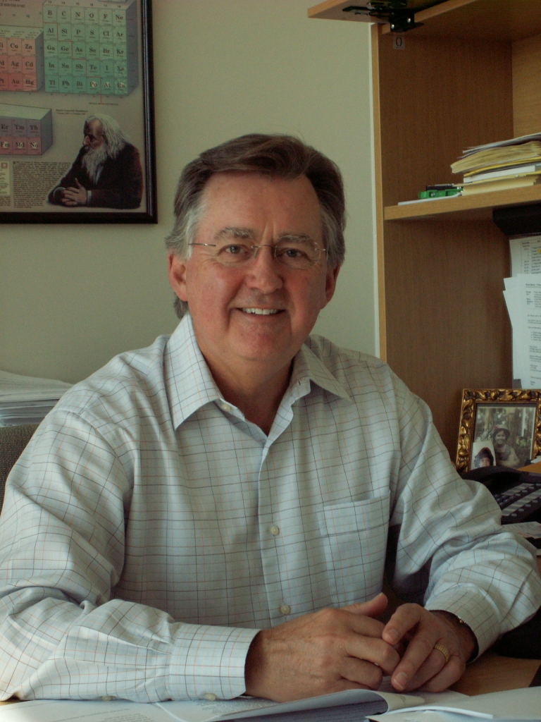
Dean Sherry.
Dean: When Yunkou came to my lab, he was clever enough to realize that instead of altering the intensity of the paraCEST signal with changes in pH, you might be able to alter the frequency of the signal instead. After he developed this current agent, we then began thinking about the best way to detect it in vivo to get a direct readout of pH based upon frequency rather than intensity. That’s the fundamental idea behind this latest paper in Magnetic Resonance in Medicine.
MRMH: What are some foreseeable advantages of this technique in the context of other pH-sensitive CEST approaches?
Dean: The frequency of many paraCEST signals can be quite large – in this case, the paraCEST signal is around 50 ppm downfield of water. DiaCEST agents, on the other hand, have exchange peaks typically no more than ~5-6 ppm from water so suffer from interference from hundreds of other endogenously exchanging species. This makes it more difficult to separate the CEST signal you wish to detect from all the others.
Yunkou: Also, the conventional way to obtain a pH value from CEST agents is by a ratiometric analysis, where the agents have two chemically distinct exchanging protons exchanging with bulk water at two different frequencies, and you make a ratiometric plot of the pH based on the ratio of CEST intensities at these two frequencies. The problem is that this ends up being very sensitive to solvent or tissue composition, which you would have to correct for with prior knowledge which isn’t always available. Our agent allows us to simply follow the frequency of the CEST peak as a function of pH, which we have observed to be a more stable method, because the pH calibration curve is independent of the solution system.
MRMH: How flexible would you consider paraCEST to be as a chemical platform for sensing new biological phenomena?
Dean: The nice thing about paraCEST systems is that they are paramagnetic and sensitive to many physiological and biochemical phenomena such as temperature, oxygen levels, pH, endogenous metal ions, specific metabolites, etc… There are different lanthanides you can use as well, so imagine giving a cocktail of molecules – one reading out pH, one reading out tissue redox, and one reading out calcium ion concentration with each activated at a different, specific frequency. That’s the direction we’d like to take.

Dean Sherry and Yunkou Wu relaxing with colleagues.
MRMH: And for this paper, why did you choose to image kidneys?
Dean: One of the difficulties with imaging pH in vivo is that there isn’t a gold standard. You can use a tumor model and get a pH readout but the question always arises, does this readout truly reflect the correct tissue pH? At least in well-functioning kidney, the pH gradient has been established by others using invasive microelectrodes to map out the pH gradient across the kidney. So if we can do this non-invasively using a paraCEST agent, we would be able to compare our results with the established gold-standard microelectrode measurement.
MRMH: Where do you see this research going into the future?
Yunkou: We would like to use this sensor for tumor imaging, as pH is associated with various aspects of tumor physiology. However, the concentration of CEST agents like ours accumulated in tumors is too low to be detected reliably. We are currently working on ways to improve the sensitivity of these agents, for instance by introducing them into polymers and nanoparticle systems.
Dean: Also, we’re gradually learning how to make these agents less sensitive to the presence of hundreds of other exchanging species in tissues, which would dramatically improve sensitivity, perhaps up to 10-fold. That would allow us to keep the agent concentration low and well within acceptable toxicity limitations.
