 BY Blake Dewey

Jun Chen
I recently had the pleasure of chatting with Drs. Jun Chen and Jiang Du about their recent MRM manuscript entitled “Measurement of Bound and Pore Water T1 Relaxation Times in Cortical Bone Using Three-Dimensional Ultrashort Echo Time Cones Sequences”. We came together over three distinct time zones, stretching from Peking, China to San Diego, California and finally to me right outside of Washington, D.C, where thanks to a strong internet connection we discussed the ups and downs of ultrashort echo time (UTE) imaging. UTE is a method for direct imaging of tissues that have short transverse relaxation times by shortening the delay between excitation and readout. For example, in this paper, the echo time for UTE imaging was only 8 μs, compared to a traditional gradient echo readout, that would have a minimum of 2-5 ms, depending on the sequence. Jun and Jiang, along with their colleagues in the U.S. and China, have been working to apply UTE sequences to explore the T1 properties of cortical bone and give clinically relevant information on components of the cortical bone structures not easily investigated with conventional radiological techniques.
Blake: Jun, how did you get started working in MR research?
Jun: I started working in the field of MR in 2007 when I began my Ph.D. studies. Back then our lab focused on mental diseases such as schizophrenia and major depression. As a noninvasive method, MRI, especially fMRI can detect patients’ brain activation. This “abnormal” brain activation may be a potential endophenotype which can be used as a classification standard to homogenize patients with various presentations. From then on, I have been focusing on MR imaging and sequence development.
Blake: And then how did you start this long-distance collaboration that we are highlighting?
Jun: After I got my Ph.D. degree, I started working in the department of Orthopedics in Peking Union Medical College Hospital, where we were looking for ways to evaluate the properties of bone. Dr. Du is well known in this field and one of my colleagues once worked in his lab as a visiting scholar. That is how I established contact with Dr. Du and began our collaboration. To further study UTE sequences, I moved to San Diego and worked in Dr. Du’s lab as a post-doc for two years.
Blake: Jiang, what brought you to MR research?
Jiang: My background is in physics, actually nuclear physics, and then I switched to magnetic materials, so it is related to MRI to some degree. Then I went to UW Madison for Medical Physics and started in MRI for my Ph.D. My first project was actually in MR Angiography. Then I joined the UCSD team, I was one of the first recruits of Prof. Graeme Bydder for his UTE program.
Blake: It seems like a long road to get here, but also very applicable to the current work that you do. Let’s move on to the research at hand. Jiang, how could you provide some background on this work?
Jiang: With this technique, we are trying to quantify bone water, specifically the T1 relaxation times. Previously, people have used UTE to image bone and we have demonstrated, probably several years ago, that UTE sequences can detect both pore water and bound water on clinical MR scanners. Since then people have become very interested in quantifying the absolute volume of bound water, which is a biomarker for the collagen matrix, and pore water, which provides information about cortical porosity. To try to quantify bound and pore water accurately, we need accurate T1 information of both water components.
Blake: What strengths does this sequence have? Does it have a specific clinical impact that you are looking for?
Jun: UTE has ultrashort echo time, so this sequence can detect the bone structure better than normal sequences because bone has very short T2. It can also separate free water from bound water. It is relatively fast and can be used in clinic. It also tells us more about the bone function, instead of just the structure.
Jiang: A strength of the technique is the ability to measure T1s of both pore water and bound water. Those measurements can be clinically useful. For example, pore water T1 may be related to cortical porosity. Bigger pore leads to a longer pore water T1. Bound water, on the other hand, may be related to the organic matrix, so for the first time we can clinically evaluate the organic matrix part of cortical bone. The current gold standard, DEXA (Dual-Energy X-ray Absorptiometry), can only access the mineral component which takes about 40% of bone by volume. This means that with DEXA, 60% of the information is ignored because of technical limitations. With UTE, we get back the missing information about cortical porosity (pore water) and the organic matrix (bound water), therefore UTE may allow for more accurate evaluation of bone quantity and quality.

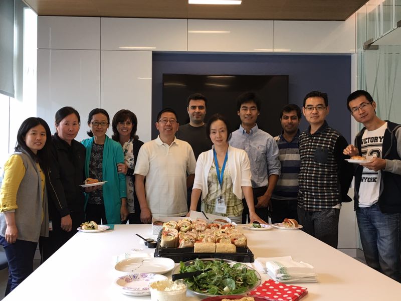
From Left to Right: Shujuan Fan, Yinghua Zhao, Xin Cheng, Lori Hamill, Jiang Du, Saeed Jerban, Rose Luo, Aaron Zhu, Amin Nazaran, Xing Lv and Yanjun Ma’ Regards!
Blake: You mentioned osteoporosis in your manuscript as a clinical target. Is this the main patient population that you will be investigating?
Jiang: Osteoporosis is one application. Not only that, we can apply this to any bone disease: Paget’s disease, osteomalacia, and even fractures. We can use this technique for diagnosis and treatment monitoring of any of these diseases.
Blake: What was the largest difficulty in implementing this method?
Jiang: Eddy currents are one major limitation for all non-Cartesian based techniques that are not commonly used on clinical MR scanners. Each scanner may have different sensitivity to eddy currents, so we need to calibrate the gradient system accurately and make UTE imaging more reliable. I think this is by far the biggest challenge when it comes to clinical application.
Jun: In China, almost all MRI machines are in hospitals and the users are mostly clinicians. When we want to implement the UTE sequence, we need the engineer’s help. There are not enough engineers to help everyone. Also, if we want to use the sequence we have to buy the license to use the sequence and that can be expensive.
Blake: What is the next step for this research? What is on the horizon?
Jun: When we use this technique in volunteers, the measurement is not always stable, so we need to optimize the imaging parameters. Second, we need to reduce the imaging time. This is very important in the clinic, as the patient cannot stay still for a long time.
Jiang: One day, it would be great to have dedicated bone imagers. It could be on the desktop, be low cost and could be used for imaging of bone in fingers or wrists. This could include a whole package, not just T1s of bound and pore water, but T2s, bound and pore water concentrations, collagen backbone protons including their fractions and exchange rates with water (via UTE magnetization transfer and signal modeling), and bone mineral (via UTE quantitative susceptibility mapping or UTE-QSM).
