We are pleased to present the list of the five most-cited Magnetic Resonance in Medicine articles from 2014:
1. Uecker, M., Lai, P., Murphy, M. J., Virtue, P., Elad, M., Pauly, J. M., Vasanawala, S. S. and Lustig, M. (2014), ESPIRiT—an eigenvalue approach to autocalibrating parallel MRI: Where SENSE meets GRAPPA. Magn Reson Med, 71: 990–1001. doi: 10.1002/mrm.24751
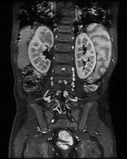
Combined parallel imaging compressed sensing reconstruction of an abdomen using l1-ESPIRiT as implemented in the BART toolbox. The scan was accelerated seven-fold using variable-density Poisson-disc sampling.
This article bridges the gap between the two main approaches for parallel imaging (SENSE and GRAPPA) allowing the reconstruction of images from undersampled multicoil data. SENSE explicitly uses coil sensitivities, and GRAPPA makes use of learned correlations in k-space. In this work, Martin Uecker, Peng Lai and colleagues present a new autocalibration technique combining the extended reconstruction of SENSE with GRAPPA-like robustness to errors. A matlab demo, implementation, and reproduction of the results in the paper can be found here.
2. Chow, K., Flewitt, J. A., Green, J. D., Pagano, J. J., Friedrich, M. G. and Thompson, R. B. (2014), Saturation recovery single-shot acquisition (SASHA) for myocardial T1 mapping. Magn Reson Med, 71: 2082–2095. doi: 10.1002/mrm.24878

In this work, Kelvin Chow and colleagues proposed a new approach for in vivo cardiac T1 mapping, consisting of a saturation recovery single-shot acquisition (SASHA) with 10 electrocardiogram-triggered single-shot balanced steady-state free precession images acquired in a breath-hold. Their work was validated through Bloch equation simulations, Monte Carlo simulations, and phantom experiments and illustrated in healthy controls and patients with heart failure
3. Feng, L., Grimm, R., Block, K. T., Chandarana, H., Kim, S., Xu, J., Axel, L., Sodickson, D. K. and Otazo, R. (2014), Golden-angle radial sparse parallel MRI: Combination of compressed sensing, parallel imaging, and golden-angle radial sampling for fast and flexible dynamic volumetric MRI. Magn Reson Med, 72: 707–717. doi: 10.1002/mrm.24980
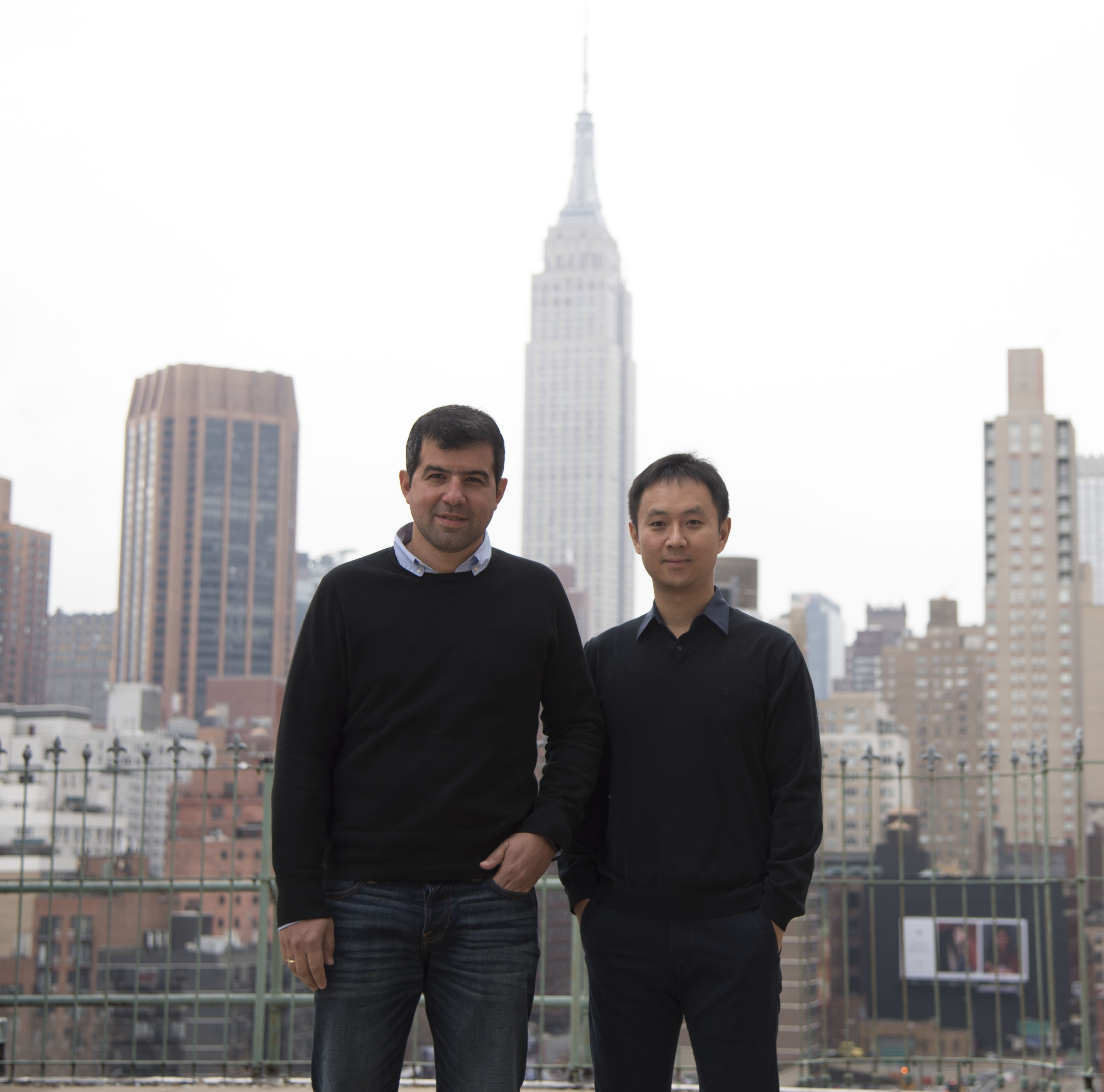
Li Feng(right) and Ricardo Otazo (left)
This work aimed to develop a fast and flexible free-breathing dynamic volumetric MRI technique, called iterative Golden-angle RAdial Sparse Parallel MRI (iGRASP). Feng and colleagues combined compressed sensing, parallel imaging, and golden-angle radial sampling to improve clinical studies that require robustness to motion and simultaneous high spatial and temporal resolution. iGRASP has been shown to achieve higher acceleration capability than either parallel imaging or coil-by-coil compressed sensing alone. The technique was used in clinical applications, such as free-breathing dynamic contrast-enhanced imaging in the abdomen of both adult and pediatric patients, and in the breast and neck of adult patients.
4. Kogan, F., Haris, M., Singh, A., Cai, K., Debrosse, C., Nanga, R. P. R., Hariharan, H. and Reddy, R. (2014), Method for high-resolution imaging of creatine in vivo using chemical exchange saturation transfer. Magn Reson Med, 71: 164–172. doi: 10.1002/mrm.24641
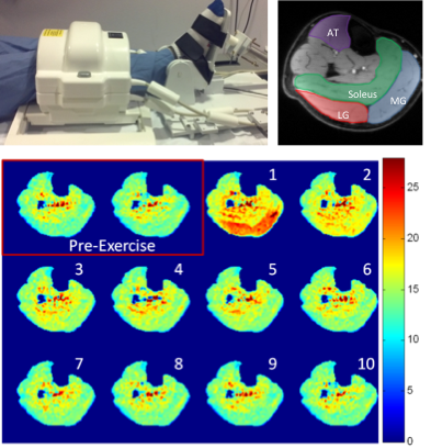
In this work, Feliks Kogan and colleagues developed a chemical exchange saturation transfer (CEST) based technique for in vivo measurement of free Creatine (Cr) changes for spatial and temporal mapping of muscle energetics. The method was validated against 31P MRS before and after a plantar flexion exercise protocol. This approach may provide new insights into muscle energetics and can serve as a tool for the diagnosis and treatment of muscle disorders as well as secondary complications associated with heart failure, renal failure, and peripheral vascular disease.
5. Shajan, G., Kozlov, M., Hoffmann, J., Turner, R., Scheffler, K. and Pohmann, R. (2014), A 16-channel dual-row transmit array in combination with a 31-element receive array for human brain imaging at 9.4 T. Magn. Reson. Med., 71: 870–879. doi:10.1002/mrm.24726

In this article, Shajan and colleagues explored the advantages inherent in transmit and receive arrays and combined them to image the human brain at 9.4T. Arranging transmit array elements in multiple rows provides an additional degree of freedom to correct for B1+ field inhomogeneities and to achieve whole-brain excitation at ultrahigh field strengths. Receive arrays shaped to the contours of the anatomy increase the signal-to-noise ratio of the image. Using a combination of the two in the brain achieved a two-fold increase in signal-to-noise ratio when compared with a 16-channel elliptic microstrip transceiver array.