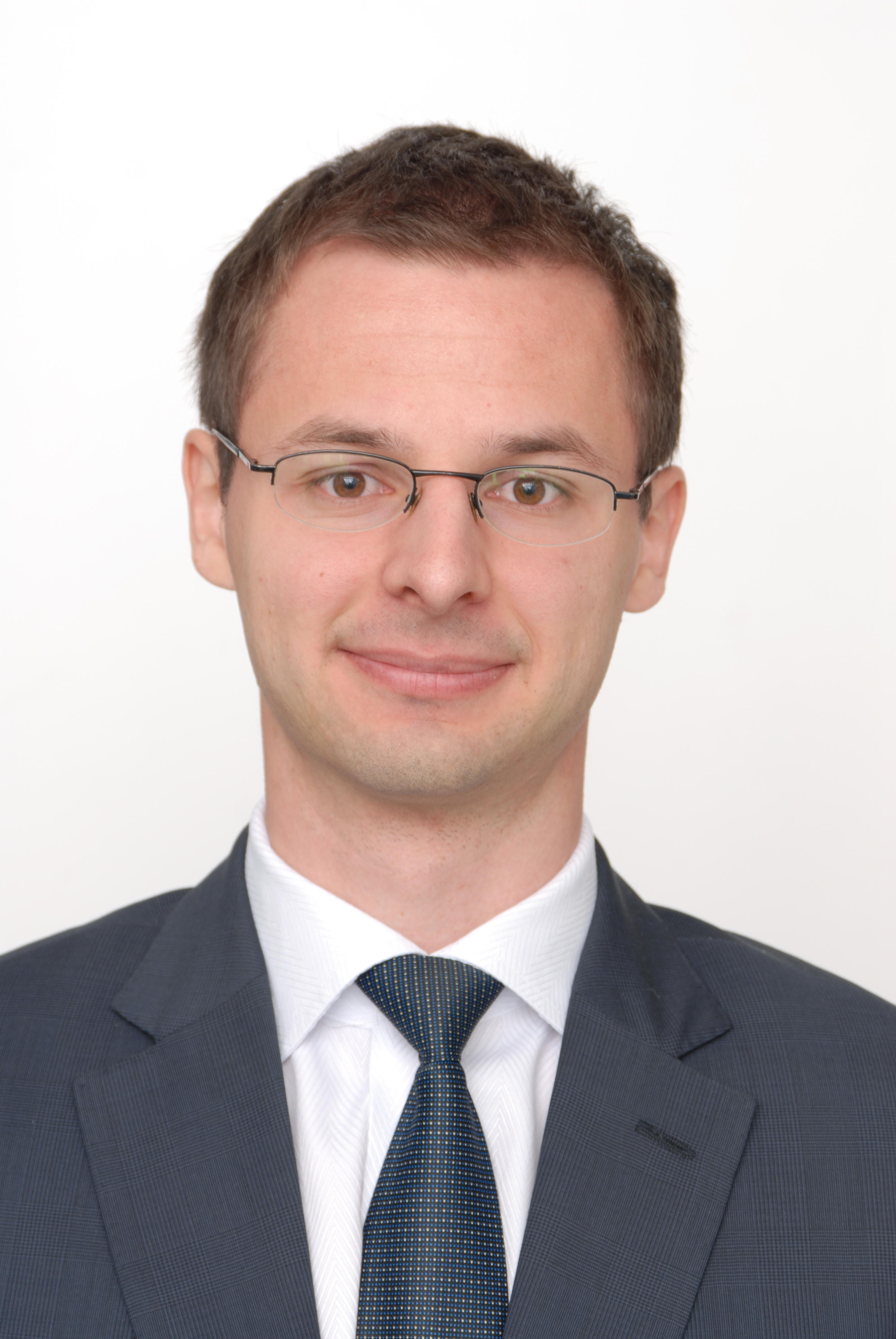

Both of our October Editor’s picks are on prostate imaging, and the first one comes from the University of Turku in Finland. Jussi Toivonen recently published a paper on diffusion imaging of prostate, and we invited him and senior author Ivan Jambor to tell us about their work.
MRMH: Thank you for accepting our invitation, can you please tell us a bit about yourselves and your background?
Jussi: I have a Master’s degree in Computer Science that I obtained in 2007 and I am now a PhD student at the University of Turku. I worked as a programmer for five years, then decided to go back to academia to pursue a PhD degree
Ivan: I am a research fellow at the University of Turku and currently in the transitional phase of being a PhD student to be a supervisor of PhD students. Jussi is one of my first PhD students. My main research interests are DWI and various spin locking methods.
MRMH: Your work is on using diffusion imaging to characterize prostate cancer. Could you please summarize your paper?
Jussi: We scanned 50 patients twice with diffusion magnetic resonance performed using b values in the range of 0 to 2000 s/mm2. Rectangular ROIs were placed on trace images in cancer and healthy tissue. Then, we fitted four different diffusion models/function, tested their performance in prostate cancer detection and characterization, and evaluated the repeatability of the fitted parameters.
Ivan: There are two main aspects of the paper: modeling and clinical application. On the modeling end, the question is how good the diffusion model is for representing the MRI signal. On the clinical end, can clinicians use this, meaning how good the model is for cancer detection and characterization? We found that these more complex models fit the signal better by having more free parameters, but did not outperform the simpler monoexponential model in terms of cancer detection and characterization.
MRMH: So were you surprised, in a way, that simpler is better?
Ivan: More disappointed than surprised really, because the theory and the fitting work so well. But I am sure that clinicians are happy. Of course, this is just one step towards better prostate cancer DWI signal characterization obtained using high b values.
MRMH: Why did you choose prostate cancer and could we apply these methods to different types of cancer?
Ivan: The reason we are investigating prostate cancer is because it is a very common cancer with wide range of cancer aggressions. Thus, it’s not anymore about detecting prostate cancer but more about differentiating cancers which need active treatment from those which are better to be left on active surveillance. But of course, the models/functions can be applied to any cancer and we would like to apply these methods to other cancer types such as glioma.
MRMH: How easy is it to use your software? In particular, for clinicians to use it?
Jussi: At the moment it would be a bit challenging since we are modifying the code quite a bit and the documentation is lagging behind. But ultimately we would like to produce simple instructions in order for other people to reproduce what we have done. The software is freely available, but I am still working on its documentation for easier use.
MRMH: Where do you want to take this? Can we use your method to characterize tissue microstructure?
Jussi: Physiological interpretation of the signal is a challenge. Some research groups are doing experiments on high-field MRI but it is far from clinical applications.
Ivan: Our group is pushing towards semi-automatic quantification in order to take away this burden from the clinicians. We are also trying to move away from ROI fitting to voxel-wise fitting.
MRMH: Thank you for your time, we look forward to hearing more from you in the coming years!
Ivan: This is a very nice initiative. It helps open up the science, especially a complicated field like MR physics.
*Interview conducted by Olivier Comtois, Benjamin De Leener, and Nikola Stikov.

